Introduction
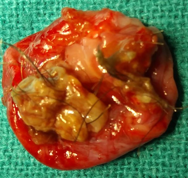
A dermoid cyst is a benign cutaneous developmental anomaly that arises from the entrapment of ectodermal elements along the lines of embryonic closure.[1][2] These benign tumors are lined by stratified squamous epithelium with mature skin appendages found on their wall and their lumens filled with keratin and hair.[3] Dermoid cysts are considered to be congenital, but not all of them are diagnosed at birth.[3][4][5] Only about 40% of dermoid cysts are diagnosed at birth, while about 60% of the dermoid cysts are diagnosed by five years of age.[3] The dermoid cysts usually present within the first year of life and grow slowly.[1] Dermoid cysts occur most commonly on the head and neck, with 84% of these cysts occurring in this region.[4]
Etiology
Register For Free And Read The Full Article
Search engine and full access to all medical articles
10 free questions in your specialty
Free CME/CE Activities
Free daily question in your email
Save favorite articles to your dashboard
Emails offering discounts
Learn more about a Subscription to StatPearls Point-of-Care
Etiology
The etiology of dermoid cysts remains mostly unknown. The cause of this congenital developmental anomaly has yet to be determined.[3][4] Dermoid cysts are true hamartomas. These occur when skin and skin structures become trapped during fetal development. Prior et al. did not find any correlation between the localization of the dermoid cyst and sex, histology, or age of the patient.[4]
Epidemiology
Dermoid cysts are among the most common pediatric skull tumors.[4][3] Dermoid cysts account for about 15.4%-58.5% of all scalp and skull masses in pediatric patients.[4] Dermoid cysts are usually congenital, with about 70% of cases discovered in children five years old or younger.[3][6] Cases of dermoid cysts discovered in adulthood have also been reported.[3][4] Pollard et al. found a slight predominance of dermoid cysts in girls.[5] However, this significant predominance has not been seen in other case series.[3] No racial predilection is apparent; however, most cases are described in Whites.
Pathophysiology
Dermoid cysts result from an abnormal alteration in fetal development.[2][4] They occur due to the abnormal sequestration and inclusion of the surface ectoderm along the lines of skin fusion during embryologic development.[4][2][7] Due to this abnormality, a dermoid cyst can usually be found along cranial sutures or the anterior fontanelle.[4][2]
Histopathology
Dermoid cyst on histology shows a well-defined wall lined by stratified squamous epithelium and a lumen that may be filled with mature adnexal structures of mesodermal origin, such as hair follicles and shafts, sebaceous and eccrine glands.[2][4][8]
History and Physical
In the majority of cases, dermoid cysts occur in the head and neck region, although they may be found anywhere on the body.[4][3] In the head and neck region, dermoid cysts can most commonly be seen in the frontal, occipital, and supraorbital areas, with the outer third of the eyebrow being the most frequently affected region.[2][4] An eyelid dermoid cyst attached to a tarsus may present as a firmly adherent non-tender upper eyelid nodule.[9]
A lower lid dermoid cyst may be evident as a painless, gradually enlarging swelling of the lower lid.[10] Dermoid cysts in the medial canthal area may present as masses adherent to lacrimal canaliculi.[11]
Dermoid cysts usually occur as solitary lesions; however, multiple concurrent dermoid cysts have also been reported.[2] Dermoid cysts typically present as a pale, flesh-colored, pearly, dome-shaped, firm, deep-seated, subcutaneous nodule.[1][2][3][12] They are usually asymptomatic, non-pulsatile, and non-compressible.[1][2]
Hair protruding from a dermoid cyst punctum is pathognomic for dermoid cysts.[13] Of note, midline dermoid cyst may present as a pit that secreted sebaceous material that can become intermittently inflamed and infected.[3][13]
Evaluation
Dermoid cysts have the potential to grow over time and extend intracranially or intraspinally.[1][3] Due to this potential, one should consider radiological studies before biopsy or manipulation, especially of a lesion that is midline or on the scalp.[3][1] Aspiration or biopsies of dermoid cysts have the potential to cause infection, further leading to osteomyelitis, meningitis, or cerebral abscess.[3][4][14] Other possible complications include bony erosions, eyelid displacement, and intracranial extension.[3][15]
Midline dermoid cysts have the highest association with cranial or spinal dysraphism or have an intracranial extension.[3] Nasal dermoid cysts are the most frequent midline congenital nasal malformations.[3] Studies have shown that there is a 10-45% incidence of intracranial extension in patients with a nasal dermoid cyst.[1] When undergoing neuroimaging, MRI is the preferred means of revealing evidence of intracranial or intraspinal extension.[1][3] Studies showed a higher association between a dermoid cyst located in the frontal and pterional regions and bony erosion.[4][16] If bony erosion is suspected, CT imaging is better at delineating these bony changes.[3][4] In some instances, high-resolution ultrasonography may help reveal a deep component.[3]
Dermoid cysts under an ultrasound will show a well-defined homogenous and hypoechoic cystic lesion.[2] Fistulography was done preoperatively in some cases to rule out the involvement of a deep tract in a dermoid cyst.[3] Dacryocystography was also performed in some atypical dermoid cysts cases.[3] Furthermore, consultation with a neurosurgeon is highly recommended for dermoid cyst complicated by intracranial or intraspinal extension.[1][3]
Treatment / Management
Dermoid cysts usually tend to grow slowly, further having the potential to cause bony deformities, intracranial extension, or intraspinal extension.[1][4] The presence of intracranial extension or intraspinal extension can further lead to meningitis or develop into an abscess.[1][3] A small, asymptomatic dermoid cyst may not necessitate immediate excision as it can be stable for years or even regress.[3][17] However, because most dermoid cysts grow over time, complete surgical excision without disruption of the cyst wall by an experienced surgeon is recommended before the development of such complications.[3][4](B2)
Early resection may also avoid extensive surgery and a shorter skin incision, further resulting in an improved cosmetic outcome.[3][4] An additional advantage of surgical excision is the possibility of obtaining a histologic diagnosis due to the rare possibility of a malignant tumor presenting as a solitary lump in the head and neck region of a child like a dermoid cyst.[4] The most dermoid cysts can be removed using a direct approach with careful dissection at the site where the cyst adheres to the bone.[3] If the cyst wall ruptures at the time of surgical removal, then remnant tissue should be removed using curettage and copious irrigation.[3](B2)
If the cyst wall has adhered to vital structures, a partial excision may be performed.[3] Recurrences of dermoid cyst have been seen in cases of incomplete excision.[3] Another benefit of early removal of dermoid cysts is a higher chance of obtaining a complete excision without disruption of the cyst wall, a factor associated with a reduced risk of recurrence.[3][4] For small dermoid cysts, endoscopic surgery is a novel approach for removal.[3] In cases of a dermoid cyst with intracranial extension, a craniotomy may still be required.[3](B2)
Differential Diagnosis
Dermoid cysts are rare, but all nodular cystlike lesions are included in the differential diagnoses of a patient presenting with a subcutaneous nodule, especially in the head and neck or midline region.[1][2][3] Following are the differentials of a dermoid cyst:
- Epidermoid cyst
- Glioma
- Encephalocele
- Juvenile xanthogranuloma
- Lipoma
- Pilar cysts
- Meningioma
- Neurofibroma
- Teratoma
- Rhabdomyosarcoma
- Olfactory neuroblastoma
- Lymphoma
- Subcutaneous abscess
- Facial trauma
- Trichilemmal cyst
- Pilomatrixoma
- Lymphatic malformation
- Thyroglossal duct cyst
Prognosis
The overall prognosis for patients with a dermoid cyst is good, especially when there is no intracranial or intraspinal extension.[3] Although histologically benign, dermoid cysts may grow and erode the skull, further being potentially susceptible to the epidural extension.[4] When there is intracranial or, intraspinal extension overall prognosis is still good if there is proper, timely surgical intervention.[3][4] In rare instances, when dermoid cysts become symptomatic due to local mass, effect, rupture, infection, or even in rare cases, cause brain compression prognosis can be poor.[3][4]
Complications
There are no complications for dermoid cysts that don't have an intracranial or intraspinal extension.[3][4] Dermoid cysts that have intracranial or intraspinal extension may lead to meningitis, abscess, or cause local mass effect.[3][1][4] Aspiration and biopsies of dermoid cysts have the potential to cause infection, further leading to osteomyelitis, meningitis, or cerebral abscess.[1][3]
Other possible complications include bony erosions, eyelid displacement, and intracranial extension.[3] Malignant transformation is a rare complication that may occur in patients with long-standing dermoid cysts. Carcinomatous transformation to squamous cell carcinoma is described in sublingual, ovarian, and intra-abdominal dermoid cysts.
Deterrence and Patient Education
A dermoid cyst is a benign cutaneous developmental anomaly that usually presents in the head and neck regions in pediatric patients.[1] Due to its tendency for growth and possible complications, early surgical intervention is recommended.[3]
Enhancing Healthcare Team Outcomes
An interprofessional team that provides a holistic and integrated approach to diagnosing and treating dermoid cysts can help achieve the best possible outcomes. Health care staff of primary care and emergency departments play a vital role in diagnosing and referring patients with head and neck subcutaneous nodules to dermatology or head and neck surgery. This will aid in better patient satisfaction, quality of life, proper care, and decrease the chance of complications.
Collaboration shared decision making and communication are crucial elements for a good outcome. The interprofessional care provided to the patient must use an integrated care pathway combined with an evidence-based approach to planning and evaluation of all joint activities. The earlier signs and symptoms of dermoid cysts are identified; the better is the patient outcome, satisfaction, and prognosis.
Media
References
Julapalli MR, Cohen BA, Hollier LH, Metry DW. Congenital, ill-defined, yellowish plaque: the nasal dermoid. Pediatric dermatology. 2006 Nov-Dec:23(6):556-9 [PubMed PMID: 17155997]
Level 3 (low-level) evidenceNakajima K, Korekawa A, Nakano H, Sawamura D. Subcutaneous dermoid cysts on the eyebrow and neck. Pediatric dermatology. 2019 Nov:36(6):999-1001. doi: 10.1111/pde.13976. Epub 2019 Aug 14 [PubMed PMID: 31414508]
Orozco-Covarrubias L, Lara-Carpio R, Saez-De-Ocariz M, Duran-McKinster C, Palacios-Lopez C, Ruiz-Maldonado R. Dermoid cysts: a report of 75 pediatric patients. Pediatric dermatology. 2013 Nov-Dec:30(6):706-11. doi: 10.1111/pde.12080. Epub 2013 Mar 14 [PubMed PMID: 23488469]
Level 2 (mid-level) evidencePrior A, Anania P, Pacetti M, Secci F, Ravegnani M, Pavanello M, Piatelli G, Cama A, Consales A. Dermoid and Epidermoid Cysts of Scalp: Case Series of 234 Consecutive Patients. World neurosurgery. 2018 Dec:120():119-124. doi: 10.1016/j.wneu.2018.08.197. Epub 2018 Sep 3 [PubMed PMID: 30189303]
Level 2 (mid-level) evidencePollard ZF, Calhoun J. Deep orbital dermoid with draining sinus. American journal of ophthalmology. 1975 Feb:79(2):310-3 [PubMed PMID: 1115198]
McAvoy JM, Zuckerbraun L. Dermoid cysts of the head and neck in children. Archives of otolaryngology (Chicago, Ill. : 1960). 1976 Sep:102(9):529-31 [PubMed PMID: 962697]
Sorenson EP, Powel JE, Rozzelle CJ, Tubbs RS, Loukas M. Scalp dermoids: a review of their anatomy, diagnosis, and treatment. Child's nervous system : ChNS : official journal of the International Society for Pediatric Neurosurgery. 2013 Mar:29(3):375-80. doi: 10.1007/s00381-012-1946-y. Epub 2012 Nov 21 [PubMed PMID: 23180312]
Reissis D, Pfaff MJ, Patel A, Steinbacher DM. Craniofacial dermoid cysts: histological analysis and inter-site comparison. The Yale journal of biology and medicine. 2014 Sep:87(3):349-57 [PubMed PMID: 25191150]
Koreen IV, Kahana A, Gausas RE, Potter HD, Lemke BN, Elner VM. Tarsal dermoid cyst: clinical presentation and treatment. Ophthalmic plastic and reconstructive surgery. 2009 Mar-Apr:25(2):146-7. doi: 10.1097/IOP.0b013e31819aae6e. Epub [PubMed PMID: 19300165]
Level 3 (low-level) evidenceGonsalves SR, Lobo GJ, Mendonca N. Dermoid cyst: an unusual location. BMJ case reports. 2013 Nov 8:2013():. doi: 10.1136/bcr-2013-200686. Epub 2013 Nov 8 [PubMed PMID: 24214152]
Level 3 (low-level) evidenceKim NJ, Choung HK, Khwarg SI. Management of dermoid tumor in the medial canthal area. Korean journal of ophthalmology : KJO. 2009 Sep:23(3):204-6. doi: 10.3341/kjo.2009.23.3.204. Epub 2009 Sep 8 [PubMed PMID: 19794949]
Level 3 (low-level) evidenceBrownstein MH, Helwig EB. Subcutaneous dermoid cysts. Archives of dermatology. 1973 Feb:107(2):237-9 [PubMed PMID: 4685580]
Wardinsky TD, Pagon RA, Kropp RJ, Hayden PW, Clarren SK. Nasal dermoid sinus cysts: association with intracranial extension and multiple malformations. The Cleft palate-craniofacial journal : official publication of the American Cleft Palate-Craniofacial Association. 1991 Jan:28(1):87-95 [PubMed PMID: 2004099]
Level 2 (mid-level) evidenceYavuzer R, Bier U, Jackson IT. Be careful: it might be a nasal dermoid cyst. Plastic and reconstructive surgery. 1999 Jun:103(7):2082-3 [PubMed PMID: 10359279]
Level 3 (low-level) evidencePensler JM, Bauer BS, Naidich TP. Craniofacial dermoids. Plastic and reconstructive surgery. 1988 Dec:82(6):953-8 [PubMed PMID: 3200958]
Pryor SG, Lewis JE, Weaver AL, Orvidas LJ. Pediatric dermoid cysts of the head and neck. Otolaryngology--head and neck surgery : official journal of American Academy of Otolaryngology-Head and Neck Surgery. 2005 Jun:132(6):938-42 [PubMed PMID: 15944568]
Shields JA, Shields CL. Orbital cysts of childhood--classification, clinical features, and management. Survey of ophthalmology. 2004 May-Jun:49(3):281-99 [PubMed PMID: 15110666]
Level 3 (low-level) evidence