Introduction
The brain receives vascular supply from a network of arteries that anastomose to form the circle of Willis. Because the brain has a constant high metabolic demand and no energy supply of its own, it requires a significant blood supply, consuming 15% of total cardiac output; any blockage of blood flow leads to severe damage and a host of neurological pathologies (see Figure. Diagram of the Brain Blood Circulation).[1]
The brain is supplied by the internal carotid arteries (ICAs), which branch from the common carotid arteries, and the vertebral arteries, which branch from the subclavian. The ICA gives rise to the anterior cerebral artery (ACA) and the middle cerebral artery (MCA). The 2 vertebral arteries unite to form the basilar artery, terminating in the 2 posterior cerebral arteries (PCA). The circle of Willis is the combination of these anterior and posterior divisions (see Image. Outer Surface of the Cerebral Hemisphere).[2]
Structure and Function
Register For Free And Read The Full Article
 Search engine and full access to all medical articles
- 10 free questions in your specialty
- Free CME/CE Activities
- Free daily question in your email
- Save favorite articles to your dashboard
- Emails offering discounts
Learn more about a Subscription to StatPearls Point-of-Care
Structure and Function
The internal carotid arteries penetrate the temporal bone through the carotid canal before giving off branches supplying the eyes via the ophthalmic artery and its daughter, the central retinal artery.[3] It then gives off branches supplying the hypothalamus regions via the posterior communicating arteries, the areas surrounding the globus pallidus, and the amygdala via the anterior choroidal arteries, and the medial surfaces of the anterior segment of the brain via the anterior cerebral artery.[4][5] The 2 anterior cerebral arteries then connect by the anterior communicating artery (ACoA), the junction with which is a frequent location of berry aneurysms.[6] Finally, the internal carotid arteries end in the middle cerebral arteries, which, together with the anterior cerebral arteries, represent its terminal branches and supply a wide territory of the lateral aspects of the cerebral hemispheres (see Image. Arterial Circulation of the Brain). These broad territories include the centers controlling speech production: Broca and Wernicke areas.[7] The lenticulostriate arteries, which branch from the proximal part of the middle cerebral arteries, further serve the deeper structures of this region.
Several small branches of the ACA branch at or near its junction with the ACoA. These include the recurrent artery of Heubner (RAH), the orbitofrontal artery, and the frontopolar artery. Identification of these arteries is essential, particularly the RAH, as their location places them at risk of injury in surgery for aneurysms and tumors.[8] The RAH is the largest medial lenticulostriate arteries, which also branch from the ACA and serve the basal ganglia with lateral branches from the MCA. In addition to those lenticulostriate arteries, the MCA also gives off the polar and anterior temporal arteries and the uncal artery, followed by numerous cortical branches that come off the distal part of the MCA.[9]
The vertebral arteries arise from the corresponding subclavian arteries and course via the transverse foramina of the cervical spine into the foramen magnum.[10] Before joining to form the basilar artery, they give off the posterior inferior cerebellar arteries and the anterior spinal arteries. Following the joining, the basilar artery gives off the paramedian pontine arteries, labyrinthine arteries, the long circumferential anterior inferior cerebellar arteries, and superior cerebellar arteries. Cranial nerve III passes between the superior cerebellar and posterior cerebral arteries as it exits the midbrain.[11] The anterior inferior cerebellar arteries supply various brainstem territories and pontine cranial nerve nuclei. Finally, the basilar artery bifurcates into its 2 terminal branches, the posterior cerebral arteries, which supply the midbrain and much of the posterior brain, including the visual cortex and related centers in the occipital and temporal lobes (see Figure. Diagram of the Posterior Cerebral Artery and its Branches). They also give off the medial and lateral posterior choroidal arteries supplying deeper structures and cortical branches, including the splenial artery.[12]
Occlusion of each arterial territory causes damage to different functional areas of the brain. As a result, each branch has a unique constellation of symptoms that may present when affected.
Embryology
The proximal ICA originates from the third aortic arch, and the distal portions come from the dorsal aorta. At roughly 28 to 30 days, the ICA has fully divided into cranial and caudal segments. The cranial segment includes the primitive olfactory artery (POA) that eventually forms the anterior choroidal and MCA. Together with the median artery of the corpus callosum (MACC), the POA is involved as part of the normal development of the ACA. Failure of the MACC to regress can lead to a variant called azygos ACA, described below. The MCA develops later, beginning around 32 to 40 days, and develops together with the cerebral hemispheres. Initially, numerous anastomoses exist between the carotid and vertebrobasilar systems, most of which regress. Occasionally, the hypoglossal artery or the trigeminal artery may persist into adulthood. The PCA develops from the caudal segments of the ICA; it is initially a continuation of the posterior communicating artery, which regresses in most individuals.[13]
Blood Supply and Lymphatics
Vasa vasorum, the microscopic vessel networks supplying large blood vessels, are rarely found in the brain and, when present, are usually related to intracranial cardiovascular pathologies such as atherosclerosis. Rather than through vasa vasorum, intracranial vessels likely get their blood supplied by simple diffusion with the cerebrospinal fluid (CSF).[14]
The brain does not have a lymphatic system as peripheral tissues do; instead, it has a similar system by which CSF enters the brain parenchyma, known as the glymphatic system. CSF initially travels through the pial arteries into the perivascular Virchow-Robin space, entering brain cells via aquaporin channels found on astrocytes. Eventually, it exits into lymphatics running along the spinal and cranial nerves and through the arachnoid granulations.[15]
Physiologic Variants
The ACA is divided into 5 segments, denoted A1 through A5. Its distal part runs along the corpus callosum, sometimes called the pericallosal artery. The largest branch of this pericallosal segment is the callosomarginal artery, which can be present in more than half of individuals. Most branches of the ACA serve the cortex branch either from the callosomarginal artery or, if it is not present, directly from the pericallosal artery. There are numerous additional variants, of which significant examples are the azygos ACA, in which the A2 segments of the 2 ACAs fuse before dividing again; bihemispheric, in which the A2 segment of 1 ACA diminishes and the other divides to feed both hemispheres; and medial ACA, in which a third ACA appears and feeds the distal territories.[16] Also, many individuals have fenestration of the anterior communicating artery, which can mimic an aneurysm.[13]
The MCA is divided into 4 parts: M1, from the ICA bifurcation through M4 to the lateral cerebral cortex. Corresponding with their relative locations, they are alternatively called the sphenoidal, insular, opercular, and cortical segments, respectively.[9] There may be a duplicate or accessory MCA in rare cases.[13]
The PCA is divided into 4 sections: P1, P2A (anterior), P (posterior), P3, and P4. The anastomosis with the posterior communicating artery separates P1 and P2.[17] As mentioned above, the PCA derives from the caudal segments of the ICA that form the posterior communicating artery. In many patients, the posterior communicating artery does not regress to a normal degree and remains a significant contributor to the PCA; the term for this is a "fetal" PCA.[13]
Surgical Considerations
As mentioned above, knowing cerebral branches and variants during surgery for intracranial aneurysms and neoplasms is vital. For example, avoiding occlusion of the fetal PCA is important, as it provides much of the posterior circulation during treating aneurysms of the posterior communicating artery.[13]
Clinical Significance
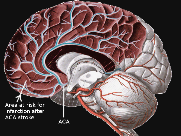
Occlusion of the cerebral arteries can lead to stroke, leading to significant morbidity and mortality (see Image. Anterior Cerebral Artery Stroke). Understanding cerebral arterial anatomy is also significant in diagnosing and managing intracranial aneurysms and tumors.
Ischemic occlusion of branches of the ICA results in anterior circulation strokes. Occlusion of cortical (pial) branches of the anterior circulation results in cortical stroke syndromes with clinical findings of aphasia, apraxia, cortical sensory impairment such as neglect or agraphesthesia, and contralateral hemiparesis. Occlusion of deep perforating branches, most commonly the lenticulostriate arteries, results in lacunar infarcts with the most common type of pure motor hemiparesis. Cortical infarction of the posterior cerebral artery results in homonymous hemianopia. Occlusion of the perforators from the PCA (thalamic perforators) may result in the lacunar syndrome of pure sensory hemianesthesia.
The ophthalmic artery arises from the ICA. A transient ischemic attack presenting with transient monocular blindness indicates a transient ischemic attack (TIA) in the ipsilateral ICA territory. The imaging modality of choice is a carotid duplex ultrasound or computed tomogram (CT) angiogram.
The anterior circulation supplies 80% of the brain, and the posterior circulation 20% of the brain. Logically, 80% of strokes occur in the anterior and 20% in the posterior circulation.
Moyamoya disease is an occlusive condition resulting from bilateral blockage of the terminal ICA and the development of abnormal collateral vessels in the basal ganglia and internal capsule region. These tiny collaterals give rise to the radiological appearance of a "puff of smoke," which is the literal meaning of Moyamoya in Japanese. It is a major cause of both ischemic and hemorrhagic strokes in childhood and early adulthood and is often treated with surgical bypass to revascularize the MCA distribution.[18]
Media
(Click Image to Enlarge)
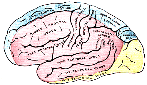
Outer Surface of the Cerebral Hemisphere. The outer surface of the cerebral hemisphere shows areas supplied by cerebral arteries. The blue areas are supplied by the anterior cerebral artery, the pink areas by the middle cerebral artery, and the yellow areas by the posterior cerebral artery.
Henry Vandyke Carter, Public Domain, via Wikimedia Commons
(Click Image to Enlarge)
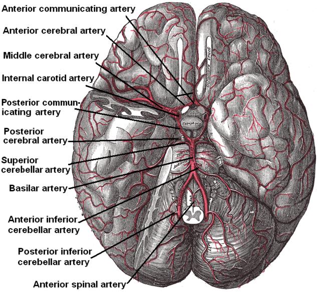
Arterial Circulation of the Brain. This inferior view shows the circle of Willis at the base of the brain formed by the anterior communicating, anterior cerebral, middle cerebral, internal carotid, posterior communicating, posterior cerebral, and basilar arteries. The temporal pole of the cerebrum and a portion of the cerebellar hemisphere have been removed on the right side. Other arteries in this illustration include the superior cerebellar, anterior inferior cerebellar, posterior inferior cerebellar, and anterior spinal arteries.
Henry Vandyke Carter, Public Domain, via Wikimedia Commons
(Click Image to Enlarge)
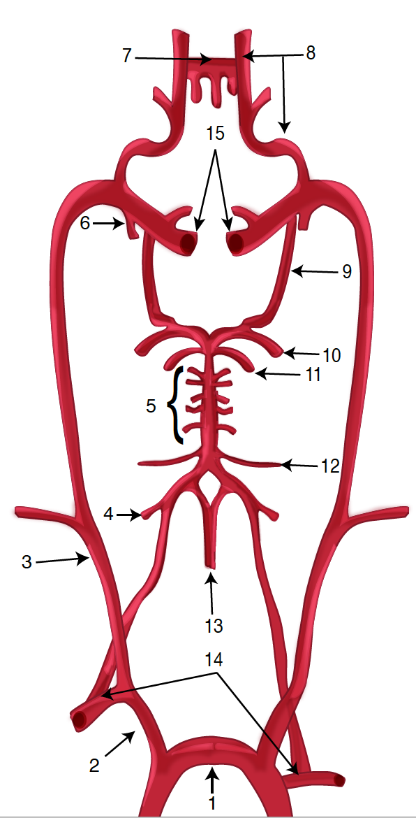
Diagram of the Brain Blood Circulation. Each number corresponds to the following neuroanatomy: 1) aortic arch; 2) brachiocephalic artery; 3) common carotid artery; 4) posterior inferior cerebellar artery; 5) pontine arteries; 6) anterior choroidal artery; 7) anterior communicating artery; 8) anterior cerebral artery; 9) posterior communicating artery; 10) posterior cerebral artery; 11) superior cerebellar artery; 12) anterior inferior cerebellar artery; 13) anterior spinal artery; 14) arches of vertebral arteries; and 15) internal carotid arteries.
Contributed by O Kuybu, MD
(Click Image to Enlarge)

Diagram of the Posterior Cerebral Artery and its Branches. Each number corresponds to the following neuroanatomy: 1) basilar artery; 2) superior cerebellar artery; 3) posterior cerebral artery; 4) thalamic subthalamic arteries; 5) posterior communicating artery; 6) internal carotid artery; 7) polar artery of thalamus; 8) posterior choroidal artery; 9) thalamogeniculate artery; 10) anterior inferior temporal artery; 11) posterior inferior temporal artery; 12) occipitotemporal artery; 13) calcarine arteries; and 14) occipitoparietal artery.
Contributed by O Kuybu, MD
(Click Image to Enlarge)
Anterior Cerebral Artery Stroke
Contributed by S Bhimji, MD
References
Xing CY, Tarumi T, Liu J, Zhang Y, Turner M, Riley J, Tinajero CD, Yuan LJ, Zhang R. Distribution of cardiac output to the brain across the adult lifespan. Journal of cerebral blood flow and metabolism : official journal of the International Society of Cerebral Blood Flow and Metabolism. 2017 Aug:37(8):2848-2856. doi: 10.1177/0271678X16676826. Epub 2016 Jan 1 [PubMed PMID: 27789785]
Takakuwa T, Koike T, Muranaka T, Uwabe C, Yamada S. Formation of the circle of Willis during human embryonic development. Congenital anomalies. 2016 Sep:56(5):233-6. doi: 10.1111/cga.12165. Epub [PubMed PMID: 27037515]
Toma N. Anatomy of the Ophthalmic Artery: Embryological Consideration. Neurologia medico-chirurgica. 2016 Oct 15:56(10):585-591 [PubMed PMID: 27298261]
Djulejić V, Marinković S, Georgievski B, Stijak L, Aksić M, Puškaš L, Milić I. Clinical significance of blood supply to the internal capsule and basal ganglia. Journal of clinical neuroscience : official journal of the Neurosurgical Society of Australasia. 2016 Mar:25():19-26. doi: 10.1016/j.jocn.2015.04.034. Epub 2015 Nov 16 [PubMed PMID: 26596401]
Javed K, Das JM. Neuroanatomy, Anterior Choroidal Arteries. StatPearls. 2024 Jan:(): [PubMed PMID: 30844216]
Brown RD Jr, Broderick JP. Unruptured intracranial aneurysms: epidemiology, natural history, management options, and familial screening. The Lancet. Neurology. 2014 Apr:13(4):393-404. doi: 10.1016/S1474-4422(14)70015-8. Epub [PubMed PMID: 24646873]
Eslinger PJ, Damasio AR. Age and type of aphasia in patients with stroke. Journal of neurology, neurosurgery, and psychiatry. 1981 May:44(5):377-81 [PubMed PMID: 7264683]
Avci E, Fossett D, Aslan M, Attar A, Egemen N. Branches of the anterior cerebral artery near the anterior communicating artery complex: an anatomic study and surgical perspective. Neurologia medico-chirurgica. 2003 Jul:43(7):329-33; discussion 333 [PubMed PMID: 12924591]
Level 3 (low-level) evidencePai SB, Varma RG, Kulkarni RN. Microsurgical anatomy of the middle cerebral artery. Neurology India. 2005 Jun:53(2):186-90 [PubMed PMID: 16010057]
Eskander MS, Drew JM, Aubin ME, Marvin J, Franklin PD, Eck JC, Patel N, Boyle K, Connolly PJ. Vertebral artery anatomy: a review of two hundred fifty magnetic resonance imaging scans. Spine. 2010 Nov 1:35(23):2035-40. doi: 10.1097/BRS.0b013e3181c9f3d4. Epub [PubMed PMID: 20938397]
Level 2 (mid-level) evidenceVitošević Z, Marinković S, Cetković M, Stimec B, Todorović V, Kanjuh V, Milisavljević M. Intramesencephalic course of the oculomotor nerve fibers: microanatomy and possible clinical significance. Anatomical science international. 2013 Mar:88(2):70-82. doi: 10.1007/s12565-012-0166-6. Epub 2012 Dec 15 [PubMed PMID: 23242853]
Pai BS, Varma RG, Kulkarni RN, Nirmala S, Manjunath LC, Rakshith S. Microsurgical anatomy of the posterior circulation. Neurology India. 2007 Jan-Mar:55(1):31-41 [PubMed PMID: 17272897]
Okahara M, Kiyosue H, Mori H, Tanoue S, Sainou M, Nagatomi H. Anatomic variations of the cerebral arteries and their embryology: a pictorial review. European radiology. 2002 Oct:12(10):2548-61 [PubMed PMID: 12271398]
Portanova A, Hakakian N, Mikulis DJ, Virmani R, Abdalla WM, Wasserman BA. Intracranial vasa vasorum: insights and implications for imaging. Radiology. 2013 Jun:267(3):667-79. doi: 10.1148/radiol.13112310. Epub [PubMed PMID: 23704290]
Jessen NA, Munk AS, Lundgaard I, Nedergaard M. The Glymphatic System: A Beginner's Guide. Neurochemical research. 2015 Dec:40(12):2583-99. doi: 10.1007/s11064-015-1581-6. Epub 2015 May 7 [PubMed PMID: 25947369]
Cilliers K, Page BJ. Review of the Anatomy of the Distal Anterior Cerebral Artery and Its Anomalies. Turkish neurosurgery. 2016:26(5):653-61. doi: 10.5137/1019-5149.JTN.14294-15.1. Epub [PubMed PMID: 27337235]
Párraga RG, Ribas GC, Andrade SE, de Oliveira E. Microsurgical anatomy of the posterior cerebral artery in three-dimensional images. World neurosurgery. 2011 Feb:75(2):233-57. doi: 10.1016/j.wneu.2010.10.053. Epub [PubMed PMID: 21492726]
Acker G, Fekonja L, Vajkoczy P. Surgical Management of Moyamoya Disease. Stroke. 2018 Feb:49(2):476-482. doi: 10.1161/STROKEAHA.117.018563. Epub 2018 Jan 17 [PubMed PMID: 29343587]