Introduction
Bullosis diabeticorum, also known as diabetic bullae or bullous eruption of diabetes mellitus, is a specific type of skin lesion exclusively occurring in patients with diabetes mellitus. Kramer first described the condition in 1930, and Rocca and Pereyra later described this in 1963. Cantwell & Martz noticed the recurrent spontaneous blisters on the extremities of patients with diabetes mellitus and introduced the label “bullosis diabeticorum” in 1967.[1] Most affected individuals had longstanding diabetes mellitus with peripheral neuropathy. The skin lesions were asymptomatic and healed slowly without scarring in 2 to 6 weeks.[2]
Etiology
Register For Free And Read The Full Article
Search engine and full access to all medical articles
10 free questions in your specialty
Free CME/CE Activities
Free daily question in your email
Save favorite articles to your dashboard
Emails offering discounts
Learn more about a Subscription to StatPearls Point-of-Care
Etiology
The etiology of bullosis diabeticorum is not known. While the majority of patients with the disease have associated diabetic nephropathy or neuropathy, some researchers have postulated a potential etiologic link that could be associated with a localized alteration in the connective tissue of the basement membrane zone secondary to microangiopathy. The observation of hyalinosis of small vessels in biopsy specimens has prompted some authorities to hypothesize that blister induction is associated with microangiopathy.[3] Imbalances of calcium and abnormal metabolism of carbohydrates also contribute to the disease.[2] Occasionally, the suspicion of immunological deposition arises as a potential cause of vasculopathy in patients who test positive for direct immunofluorescence. The purported significance of glycemic control has yet to be verified.[4][5]
Epidemiology
Bullosis diabeticorum is an uncommon condition, with a documented incidence of approximately 100 cases. The annual prevalence is about 0.16%.[3] Some reports say this condition manifests in around 0.5% of individuals with diabetes in the United States. However, the actual prevalence might be greater due to inadequate reporting of blistering cases in patients with diabetes. The condition usually occurs between 17 and 84 years, although there has been a recorded instance in a 3-year-old child.[6] The reported man-to-woman ratio of bullosis diabeticorum is 2:1 [7] and is found most frequently in adult men with longstanding uncontrolled diabetes.[8] Individuals who have uncomplicated or recently diagnosed diabetes may potentially experience the same effects. Occasionally, the condition has been documented in patients with prediabetes.
Pathophysiology
The pathophysiology of bullosis diabeticorum is not completely clear and appears to be multifactorial. In a study by Bernstein and colleagues, the authors demonstrated that patients with insulin-dependent diabetes have a markedly reduced threshold for suction blister development compared to age-matched normal controls, with a highly significant difference (P < 0.01).[9] Due to the acral prominence of bullosis diabeticorum in patients with diabetes mellitus, the role of trauma has been speculated. However, this cannot account for the absence of bullosis diabeticorum in the vast majority of those with diabetes, with the absence in most cases of any history of trauma and the fact that these lesions resolve spontaneously.[10] As the majority of patients with diabetes mellitus with bullosis diabeticorum have associated nephropathy and neuropathy, some have suggested the role of microangiopathy in the premature aging of connective tissue and, more precisely, a local subbasement membrane zone connective-tissue alteration.[11]
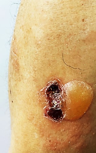
Some individuals have presented with bullosis diabeticorum at first presentation of a prediabetic state with impaired glucose tolerance.[12] Moreover, the literature has postulated disturbances in calcium, magnesium, and carbohydrate metabolism are also contributory.[2] Excessive exposure to ultraviolet light, especially in those with nephropathy, has also been suggested as a causative factor in a few cases. Individuals with end-stage renal failure may have mildly elevated plasma porphyrin levels, possibly contributing to the pathogenesis of blister formation (see Image. Bullosis Diabeticorum Blister).[3]
Histopathology
The histologic findings are heterogeneous. Bullosis diabeticorum shows bullae with inconsistent levels of skin layer separation. Initial reports described intraepidermal blisters (subcorneal or suprabasal) without acantholysis, situated in most cases in the superficial part of the prickle-cell layer; recent publications have usually described subepidermal bullae.[13] The combinations of subepidermal and suprabasal bullae are not infrequent.[2] There have been suggestions that the cleavage level depends on the blister’s age. Bullae are initially subepidermal, progressing to intraepidermal blisters, representing older lesions undergoing re-epithelialization. The blister cavity contains clear, sterile proteinaceous fluid with little or no inflammation. However, the sub-epidermal variant can be hemorrhagic. The surrounding epidermis does not exhibit any major alteration. However, there have been occasional cases of concomitant spongiosis and degenerative keratinocytic pallor.[14] The dermis displays minimal inflammation other than microvascular changes consistent with diabetes.[2][15] Caterpillar bodies, often found in patients with porphyria, can also be seen.
Direct immunofluorescence examination is usually negative, although one report described non-specific associated immunoglobulin M and complement C3 deposits within dermal blood vessel walls.[4] The absence or reduction of anchoring fibrils and hemidesmosomes characterize early blisters.[2] Electron microscopy of fresh blisters has revealed separation in a subepidermal location, residing in the lamina lucida or below the lamina densa.
History and Physical
Bullosis diabeticorum characteristically starts with a sudden appearance of 1 or more painless, tense vesicles or bullae within normal-appearing, non-inflamed skin, generally on the acral portions of the body.[16] The most frequent locations reported in the literature are the tips of the toes and the plantar surface of the feet, distal legs, hands, and forearms.[13] Bullae are rarely present on the trunk, and the lesions are usually asymptomatic; however, some patients may occasionally notice a mild burning sensation. Blisters often develop spontaneously and in the absence of known trauma. The vesicles and bullae range from a few millimeters to several centimeters in size. Lesions typically start as tense blisters, which become flaccid and asymmetrical in shape as they enlarge and sometimes possess an uneven shape, resembling a burn. Blisters contain a clear and sterile fluid, which may sometimes be hemorrhagic; they can be filled with pus if there has been a secondary bacterial infection.
Most patients with bullosis diabeticorum have longstanding diabetes. Bullosis diabeticorum has associations with both insulin- and noninsulin-dependent diabetes mellitus. Many patients have an associated polyneuropathy, retinopathy, or nephropathy. But occasionally, bullae can be a presenting sign of the disease.[13] The goals of the physical exam include finding the exact location and physical features of the lesions and coming up with a practical differential diagnosis. A biopsy may be required for an accurate diagnosis, and a culture may need to be collected to determine any secondary infections that require treatment.
Evaluation
Once the diagnosis of bullosis diabeticorum is considered, the patient should undergo evaluation for this metabolic condition, and their glycemic levels should be controlled. If diabetes mellitus is not a known diagnosis, perform a prompt screening. Patients with confirmed bullosis diabeticorum should be monitored for the development of secondary infections until lesions heal entirely.[17] Because of the higher prevalence of microangiopathic complications in patients with diabetes mellitus with bullosis diabeticorum, ophthalmological and neurological examinations are recommended. Evaluation of renal function, especially detecting microalbuminuria, is also necessary.
Treatment / Management
No firm consensus has emerged for managing bullosis diabeticorum.[18] The blisters have traditionally been deemed self-limiting, with bullae said to resolve if untreated within 2 to 6 weeks. Many authors have advised leaving the blistered skin intact since it constitutes an effective and sterile cover for the underlying wound.[19] Drug therapy, specifically antibiotics, should only be used if there is a secondary staphylococcal infection. Some authors advocate a small-bore needle aspiration or placement of a small window in the blister roof and the application of topical antiseptics or antibiotics to reduce discomfort and prevent secondary infections. In some cases, surgical management of bullosis diabeticorum is required when soft tissue infection is present, given the significant risk of osteomyelitis.[18] Immobilization can help keep the blister safe. Secondary tissue necrosis may necessitate debridement and possible tissue grafting. Aggressive wound management, as with diabetic ulcers, is necessary should the blister become unroofed. Recurrent bullosis diabeticorum has undergone successful treatment with autologous bone marrow mesenchymal cell transplantation therapy.[20](B2)
Differential Diagnosis
Several disorders should merit consideration in the differential diagnosis of bullosis diabeticorum. In porphyria cutanea tarda and pseudoporphyria, the blisters are usually infracentimetric and favor the hands rather than the feet and ankles, as in bullosis diabeticorum. Pseudoporphyria is not uncommon in patients with diabetes, as they may develop complications including both chronic renal failure and atherosclerotic cardiovascular disease; they may be receiving dialysis or diuretics, which can produce pseudoporphyria. A patient with significant involvement of the back of the hands requires an assessment of their porphyrin levels. Individuals with the bullous illness of diabetes exhibit normal levels. Increased levels suggest the presence of porphyria cutanea tarda or another type of blistering porphyria. Patients suffering from end-stage renal disease may have slightly increased amounts of porphyrins in their blood plasma, potentially contributing to blister formation.
The distal extremity is a common site for erythema multiforme and fixed drug eruptions, but their bullae usually develop on an inflammatory base. The differential diagnosis also includes friction bullae, bullae due to burns or edema, bullous fixed drug reaction, bullous pemphigoid, and epidermolysis bullosa acquisita.[21] Epidermolysis bullosa acquisita and localized bullous pemphigoid closely resemble bullosis diabeticorum clinically and histologically. However, they can be distinguished from bullosis diabeticorum by histologic examination and direct immunofluorescence, as well as by the accentuation of lesions at frictional sites in patients with epidermolysis bullosa acquisita. Immunofluorescence investigations may be necessary to exclude related medical disorders such as bullous pemphigoid, epidermolysis bullosa acquisita, and porphyrias. These conditions often exhibit the accumulation of C3 and immunoglobulin G along the basement membrane zone.[22] Bullous cellulitis must also be considered if there is surrounding inflammatory skin with erythema, warmth, and tenderness.
Prognosis
Bullosis diabeticorum is more frequently a self-limiting condition and usually resolves spontaneously within a few weeks. Lesions typically heal without post-inflammatory pigmentation or residual scarring. Nevertheless, repeated and recurrent episodes leading to ulceration and scarring [23] have been reported.[3] Bullous symptoms can reoccur over the years.
Complications
While lesions typically heal spontaneously within 2 to 6 weeks, they often recur in the same or different locations. Secondary infections characterized by cloudy blister fluid may develop and require a culture to verify and treat if indicated.[24] Additionally, there have been occurrences of osteomyelitis occurring at a location where the bullous disease of diabetes is present.[25] There are also reports of amputations due to infection.[13][26]
Deterrence and Patient Education
All patients with diabetes should receive comprehensive education on preventive foot self-care. A thorough foot examination should encompass the examination of the skin, the evaluation of foot deformities, the assessment of neurological function, and the evaluation of vascular fitness, including the assessment of pulses in the legs and feet. Antibiotic medication is unnecessary for wounds with no signs of soft tissue or bone infection. Empiric antibiotic therapy can be explicitly directed at gram-positive cocci in acute infections.
Enhancing Healthcare Team Outcomes
Recognizing bullosis diabeticorum is essential, as the differential diagnoses are extensive. The risk of consequent infection is the most pressing concern for the interprofessional team, including a primary care provider, nurse practitioner, internist, endocrinologist, and wound care nurse. The connection between bullosis diabeticorum and the degree of glycemic control in patients with diabetes mellitus remains unclear. Awareness of other potential vascular complications should always be in mind. Given that bullosis diabeticorum is generally self-limiting, treatment remains mainly supportive.
Media
(Click Image to Enlarge)
References
Cantwell AR Jr, Martz W. Idiopathic bullae in diabetics. Bullosis diabeticorum. Archives of dermatology. 1967 Jul:96(1):42-4 [PubMed PMID: 6067557]
Toonstra J. Bullosis diabeticorum. Report of a case with a review of the literature. Journal of the American Academy of Dermatology. 1985 Nov:13(5 Pt 1):799-805 [PubMed PMID: 3908510]
Level 3 (low-level) evidenceLarsen K, Jensen T, Karlsmark T, Holstein PE. Incidence of bullosis diabeticorum--a controversial cause of chronic foot ulceration. International wound journal. 2008 Oct:5(4):591-6. doi: 10.1111/j.1742-481X.2008.00476.x. Epub [PubMed PMID: 19006577]
Level 2 (mid-level) evidenceSonani H, Abdul Salim S, Garla VV, Wile A, Palabindala V. Bullosis Diabeticorum: A Rare Presentation with Immunoglobulin G (IgG) Deposition Related Vasculopathy. Case Report and Focused Review. The American journal of case reports. 2018 Jan 15:19():52-56 [PubMed PMID: 29332930]
Level 3 (low-level) evidenceArnold L, Vennard KC, Gilbert MP. Bullous Diabeticorum. The Journal of the American Osteopathic Association. 2018 Dec 1:118(12):832. doi: 10.7556/jaoa.2018.178. Epub [PubMed PMID: 30476996]
Chiriac A, Costache I, Podoleanu C, Naznean A, Stolnicu S. Bullosis Diabeticorum in a Young Child: Case Report of a Very Rare Entity and a Literature Review. Canadian journal of diabetes. 2017 Apr:41(2):129-131. doi: 10.1016/j.jcjd.2016.10.005. Epub 2016 Dec 21 [PubMed PMID: 28017292]
Level 3 (low-level) evidenceGupta V, Gulati N, Bahl J, Bajwa J, Dhawan N. Bullosis diabeticorum: rare presentation in a common disease. Case reports in endocrinology. 2014:2014():862912. doi: 10.1155/2014/862912. Epub 2014 Nov 18 [PubMed PMID: 25478251]
Level 3 (low-level) evidenceEl Fekih N, Zéglaoui F, Sioud A, Fazaa B, Kharfi M, Gaigi S, Kamoun R. [Bullosis diabeticorum: report of ten cases]. La Tunisie medicale. 2009 Nov:87(11):747-9 [PubMed PMID: 20209832]
Level 2 (mid-level) evidenceBernstein JE, Levine LE, Medenica MM, Yung CW, Soltani K. Reduced threshold to suction-induced blister formation in insulin-dependent diabetics. Journal of the American Academy of Dermatology. 1983 Jun:8(6):790-1 [PubMed PMID: 6863644]
Huntley AC. Threshold to suction-induced blister formation in insulin-dependent diabetics. Journal of the American Academy of Dermatology. 1984 Feb:10(2 Pt 1):305-6 [PubMed PMID: 6715607]
Level 3 (low-level) evidenceBraverman IM, Keh-Yen A. Ultrastructural abnormalities of the microvasculature and elastic fibers in the skin of juvenile diabetics. The Journal of investigative dermatology. 1984 Mar:82(3):270-4 [PubMed PMID: 6699427]
Lopez PR, Leicht S, Sigmon JR, Stigall L. Bullosis diabeticorum associated with a prediabetic state. Southern medical journal. 2009 Jun:102(6):643-4. doi: 10.1097/SMJ.0b013e3181a506d6. Epub [PubMed PMID: 19434030]
Level 3 (low-level) evidenceLipsky BA, Baker PD, Ahroni JH. Diabetic bullae: 12 cases of a purportedly rare cutaneous disorder. International journal of dermatology. 2000 Mar:39(3):196-200 [PubMed PMID: 10759959]
Level 3 (low-level) evidenceAlhadyani A, DeVore A, Gaster E, Elston D. Pediatric patient with linearly distributed bullae. JAAD case reports. 2023 Sep:39():67-69. doi: 10.1016/j.jdcr.2023.06.048. Epub 2023 Jul 17 [PubMed PMID: 37635858]
Level 3 (low-level) evidenceGrillo VTRDS, Botelho MS, Lange EP, Secanho MS, de Camargo PAB, Miot HA. Bullosis diabeticorum as a differential diagnosis for limb ulcers: case report. Jornal vascular brasileiro. 2022:21():e20210190. doi: 10.1590/1677-5449.202101901. Epub 2022 Sep 9 [PubMed PMID: 36187219]
Level 3 (low-level) evidenceWagner G, Meyer V, Sachse MM. [Bullosis diabeticorum : Two case studies]. Der Hautarzt; Zeitschrift fur Dermatologie, Venerologie, und verwandte Gebiete. 2018 Sep:69(9):751-755. doi: 10.1007/s00105-018-4132-7. Epub [PubMed PMID: 29468278]
Level 3 (low-level) evidenceBello F, Samaila OM, Lawal Y, Nkoro UK. 2 Cases of Bullosis Diabeticorum following Long-Distance Journeys by Road: A Report of 2 Cases. Case reports in endocrinology. 2012:2012():367218. doi: 10.1155/2012/367218. Epub 2012 Oct 16 [PubMed PMID: 23119191]
Level 3 (low-level) evidenceShahi N, Bradley S, Vowden K, Vowden P. Diabetic bullae: a case series and a new model of surgical management. Journal of wound care. 2014 Jun:23(6):326, 328-30 [PubMed PMID: 24920203]
Level 2 (mid-level) evidenceKansal NK, Anuragi RP. Bullous lesions in diabetes mellitus: bullous diabeticorum (diabetic bulla). BMJ case reports. 2020 Aug 27:13(8):. doi: 10.1136/bcr-2020-238617. Epub 2020 Aug 27 [PubMed PMID: 32859620]
Level 3 (low-level) evidenceChen Y, Ma Y, Li N, Wang H, Chen B, Liang Z, Ren R, Lu D, Boey J, Armstrong DG, Deng W. Efficacy and long-term longitudinal follow-up of bone marrow mesenchymal cell transplantation therapy in a diabetic patient with recurrent lower limb bullosis diabeticorum. Stem cell research & therapy. 2018 Apr 10:9(1):99. doi: 10.1186/s13287-018-0854-9. Epub 2018 Apr 10 [PubMed PMID: 29631615]
Taylor SP, Dunn K. Bullosis Diabeticorum. Journal of general internal medicine. 2017 Feb:32(2):220. doi: 10.1007/s11606-016-3802-3. Epub 2016 Jul 11 [PubMed PMID: 27400924]
Hines A, Alavi A, Davis MDP. Cutaneous Manifestations of Diabetes. The Medical clinics of North America. 2021 Jul:105(4):681-697. doi: 10.1016/j.mcna.2021.04.008. Epub [PubMed PMID: 34059245]
Vangipuram R, Hinojosa T, Lewis DJ, Downing C, Hixson C, Salas-Alanis JC, Tyring SK. Bullosis Diabeticorum: A Neglected Bullous Dermatosis. Skinmed. 2018:16(1):77-79 [PubMed PMID: 29551123]
Zhang AJ, Garret M, Miller S. Bullosis diabeticorum: case report and review. The New Zealand medical journal. 2013 Mar 15:126(1371):91-4 [PubMed PMID: 23793125]
Level 3 (low-level) evidenceTunuguntla A, Patel KN, Peiris AN, Zakaria WN. Bullosis diabeticorum associated with osteomyelitis. Tennessee medicine : journal of the Tennessee Medical Association. 2004 Nov:97(11):503-4 [PubMed PMID: 15620205]
Level 3 (low-level) evidenceWillcox MJ. Recurrent Blister Formation in Setting of Poorly Managed Diabetes Mellitus. Cureus. 2019 Jun 28:11(6):e5029. doi: 10.7759/cureus.5029. Epub 2019 Jun 28 [PubMed PMID: 31497455]
