Introduction
Chronic urticaria is a mast cell-mediated condition characterized by the recurrent occurrence of urticaria and/or angioedema.
The current definitions and classification stratify urticaria according to time course and etiology:
- Acute spontaneous urticaria: Spontaneous occurrence of wheals and/or angioedema for a total duration of fewer than six weeks.
- Chronic spontaneous urticaria (CSU): Spontaneous occurrence of wheals and/or angioedema for a total duration of six weeks or more. This is synonymous with "chronic urticaria" and "chronic idiopathic urticaria."
- Chronic inducible urticaria (CIndU): Occurrence of wheals for a total duration of six weeks or more, which is inducible by physical factors (e.g., touch, pressure extremes). This is synonymous with "physical urticaria."[1]
Etiology
Register For Free And Read The Full Article
Search engine and full access to all medical articles
10 free questions in your specialty
Free CME/CE Activities
Free daily question in your email
Save favorite articles to your dashboard
Emails offering discounts
Learn more about a Subscription to StatPearls Point-of-Care
Etiology
The etiology of CSU is yet to be fully established. The prevailing hypothesis is that it relates to autoimmune dysfunction involving autoantibodies targeting IgE and/or IgE receptors to activate histamine release from basophils and mast cells. Up to 40% of patients with CSU demonstrate a positive autologous serum skin test (ASST), whereby the patient’s serum injected into the dermis can induce urticaria. One-third of patients with CSU also has a positive basophil histamine release assay (BHRA), which tests for anti-FceRIa (an IgE receptor) or anti-IgE autoantibodies in the serum.[2][3]
Further support is lent by the increased prevalence of autoimmune disorders amongst CSU patients. Of these, autoimmune hypothyroidism is the most common, observed in up to 9.8%. Other associated conditions include rheumatoid arthritis, systemic lupus erythematosus, Sjogren syndrome, celiac disease, and type-1 diabetes mellitus.[4]
Infections by a variety of organisms have also been associated with CSU. These include bacteria (Helicobacter pylori, Streptococci, Staphylococci, Yersinia, Mycoplasma pneumoniae), virus (Hepatitis virus, Norovirus, Parvovirus B19), and parasites (Giardia lamblia, Entamoeba spp., Anisakis simplex). Causality remains unproven but may involve infection-mediated autoimmune response and molecular mimicry.[1][5]
Foods and additives, while implicated in acute IgE-mediated food allergy, are rarely the cause of CSU.[6]
Evidence for the association between malignancy and CSU is conflicting as different studies, including retrospective studies and systematic reviews, demonstrate varying results.[7][8]
Epidemiology
The prevalence of CSU is estimated at between 0.23% and 1.8% of the population in the U.S. and internationally. There is a strong female predilection, affecting women twice as often as men. Both children and adults are affected, although the prevalence is highest amongst those 40 to 60 years of age.[9][10]
Pathophysiology
Urticaria and angioedema result from the activation of mast cells and basophils. Mast cell degranulation leads to the release of immune mediators. The primary mediator is histamine, which binds to H-receptors located on endothelial cells and sensory nerves. However, prostaglandin, leukotrienes, and a variety of cytokines and chemokines are also involved. Ultimately, this induces vasodilatation - increased permeability of vessels - and in turn, dermal edema and recruitment of inflammatory cells.
Activation of mast cells can be divided into immunological and non-immunological processes.
- Immunological activation is mediated by effectors of adaptive immunity binding receptors on mast cells. In type 1 hypersensitivity, crosslinking of allergen with IgE on sensitized mast cells leads to acute urticaria and anaphylaxis. In CSU, however, immunological activation of mast cells is thought to occur independently of allergen-IgE complexes and may involve IgG (type 2 hypersensitivity), circulating immune complexes (type 3 hypersensitivity), and T-cells (type 4 hypersensitivity).
- Non-immunological activation can be mediated by physical factors, as in CIndU, as well as food and drug molecules independent of adaptive immunity. In CSU, it may be that the threshold for activation by external stimuli is reduced.[11]
Histopathology
The histopathological features of urticaria include interstitial edema involving the upper and mid-dermis, mixed inflammatory perivascular infiltrates, and dilated venules and lymphatic vessels that permit leakage of serum into the surrounding tissue.
Angioedema shares similar features, but they are observed deeper within the lower dermis and subcutaneous tissue.
A biopsy of involved skin for histology is not a routine aspect of the workup for CSU; however, it can be considered when the rash is atypical. Leukocytoclasis and vasculitis are not seen with CSU and must prompt consideration of alternate diagnoses.[12]
History and Physical
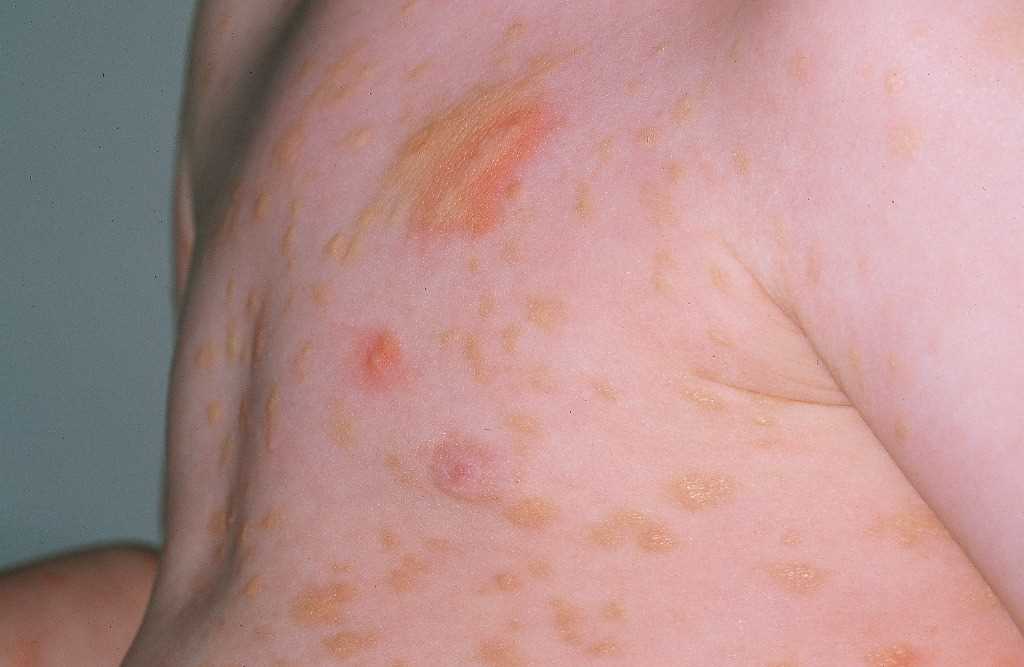
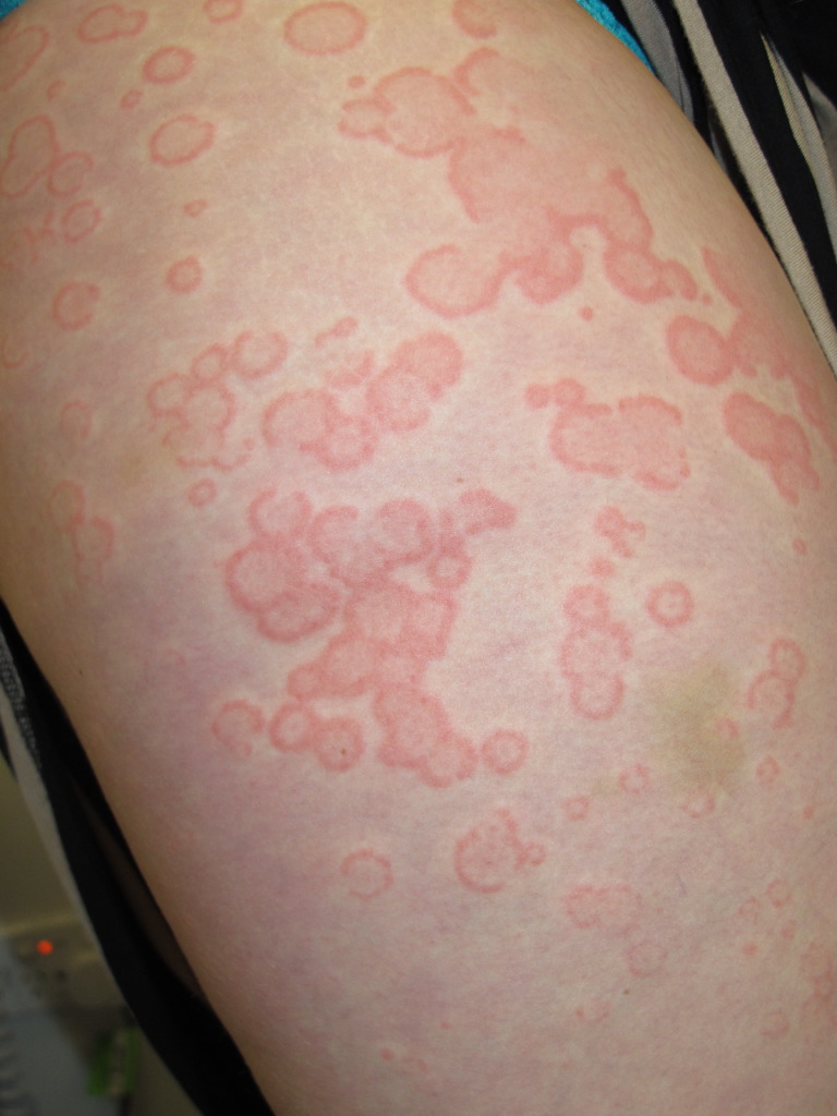
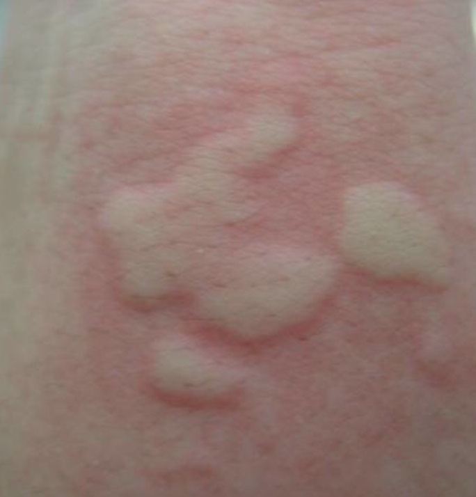
Urticaria is characterized by pruritic pink-to-red papules and plaques typically with central pallor. These can vary in size and shape as the plaques coalesce together. Urticaria is fleeting, and each wheal remains for fewer than 24 hours without any residual ecchymosis or pigmentation.[1] There may be a predilection for pressure-prone areas such as the waistline, axilla, and groin, but urticaria can generally affect any part of the skin.
Secondary changes from scratching may be evident on examination of the skin, including excoriations, erosions, and hemorrhagic crust.
Concurrent angioedema occurs in up to 40%, characterized by subcutaneous or submucosal edema affecting non-dependent areas, most commonly lips, peri-orbital, genitals, and extremities. Patients complain of discomfort or pain rather than pruritus. This may take longer to resolve - up to 72 hours - and is the primary manifestation of CSU in 10% of patients.[1][13]
Evaluation
A thorough history aims to identify potential triggers and exacerbating factors and to exclude differential diagnoses. This should explore time course and clinical features of urticarial rash, associated angioedema, associated systemic and infective symptoms, personal or family history of allergies and autoimmune diseases, social and occupational history, induction by physical factors, recent newly administered medications, relationship to foods, and any exacerbating factors.[1]
If the patient cannot recall the time course of wheals, drawing around an individual lesion with a skin marking pen is useful to document resolution within 24 hours. Where history is unrevealing, an external cause is highly unlikely to be identified for patients with CSU.[1]
Routine diagnostic work-up for CSU is limited to blood tests for complete blood count and inflammatory markers (C-reactive protein and/or erythrocyte sedimentation rate), mostly to rule out other potential diseases. Eosinophilia may be associated with atopy and parasitic infection. Elevated inflammatory markers should prompt consideration of associated systemic disease. Further investigations without clinical suspicion, as guided by history, are unlikely to yield any additional diagnoses.[14] Skin prick testing is not useful as CSU is rarely caused by type 1 allergy.[15] The utility and interpretation of autologous serum skin test as a screen for autoantibodies to IgE and IgE receptors is an area of active research.[16][17]
A number of tools have been developed to assess disease activity (e.g., urticaria activity score), disease control (e.g., urticaria control test), and impacts on quality of life (e.g., chronic urticaria quality of life index). Baseline assessments should be performed to help guide treatment decisions and monitor progress.[1]
Treatment / Management
The principles of management are to avoid exacerbating factors and to control symptoms as long as CSU persists. Pharmacological agents are directed at preventing mast cell degranulation and/or the effects of mast cell mediators released.
Second-generation H1-antihistamines (e.g., cetirizine, loratadine, fexofenadine), taken regularly, are the first-line pharmacological treatment. The dose can be up-titrated to 4 times standard dose if symptoms remain at 2 to 4-week intervals. Due to anticholinergic properties and the adverse effect profile on the central nervous system, the routine use of first-generation H1-antihistamines is no longer recommended.[1]
The primary difference in treatment between CSU and CIndU is that as needed, treatment may be used for the latter if the patient knows his trigger and is able to plan by taking an H1-antihistamine approximately two hours before being exposed to it.[1]
Omalizumab is a monoclonal antibody with a high affinity for free IgE. Given as a subcutaneous injection, the standard dosing regimen is 300 mg every 4 weeks. This has been shown to be efficacious as a second-line adjunct therapy for CSU that is unresponsive to H1-antihistamines.[18](A1)
Ciclosporin is recommended as a third-line agent for CSU that remains refractory to the combination of H1-antihistamine and omalizumab. CSU is an off-label indication, and the dose and duration of ciclosporin should be minimized to avoid adverse events, including nephrotoxicity and hypertension.[19](A1)
For acute flares of CSU, short courses only of systemic corticosteroids may help alleviate symptom severity and reduce the duration of the flare.[1] Topical corticosteroids do not have a role in CSU.[20](B2)
There is limited low-quality evidence for alternate therapeutic options, including leukotriene antagonists, dapsone, methotrexate, sulfasalazine, plasmapheresis, phototherapy, and intravenous immunoglobulin.[1]
Differential Diagnosis
- Urticarial vasculitis is a small vessel vasculitis characterized by wheals that persist for greater than 24 hours, associated with pain rather than an itch, and which resolve with residual ecchymosis and/or pigmentation.
- Papular urticaria is a persistent insect bite reaction characterized by clusters of pruritic papules, often with a central punctum, which persists for days to weeks.
- Mastocytosis encompasses a group of disorders resulting from the accumulation of clonal populations of mast cells in the skin and other organs. Darier sign, where a wheal can be induced by rubbing the affected skin, is pathognomonic.
- Auto-inflammatory syndromes such as cryopyrin-associated periodic syndromes (CAPS) and Schnitzler syndrome may present with urticarial eruptions in association with fever, arthralgia, and systemic symptoms.
- C1-esterase inhibitor deficiency, either hereditary or acquired, should be considered in patients who present with isolated angioedema in the absence of an urticarial rash.
- Bullous pemphigoid is an immunobullous disease affecting the elderly. The pre-bullous phase is characterized by pruritus and urticarial plaques, which evolve to tense bullae.
- Anaphylaxis is a potentially life-threatening type I hypersensitivity reaction that most often presents acutely with cutaneous symptoms of hives and angioedema.
Prognosis
CSU is typically self-limited, on average lasting 3 to 5 years.[21] The reported rate of remission in the first 12 months is as high as 80%.[22] However, up to 14% of patients may have persistent disease beyond 5 years.[23] Factors that portend a prolonged disease course include thyroid autoimmunity and concurrent angioedema.[21][24]
Complications
The pruritus associated with CSU can be of significant detriment to the patient’s quality of life and disrupt both activities of daily living and sleep.[25] Health status scores are also negatively affected by CSU.[1]
Deterrence and Patient Education
Patients should be educated on treatment goals. Regular use of H1-antihistamines confers better symptom control than as-needed administration.[1] Avoidance and understanding of factors that can further aggravate urticaria and pruritus also play an important role. These include:
- Drugs: Non-steroidal anti-inflammatory drugs (NSAIDs) and aspirin can cause non-allergic hypersensitivity reactions in a subset of patients.[26] Avoidance is recommended.
- Intercurrent illness: Urticaria may flare during bacterial and viral infections.
- Environmental factors: Tight clothing and heat (e.g., hot showers) can often aggravate symptoms.
Pearls and Other Issues
- Disease duration of less than and more than 6 weeks separates acute from chronic spontaneous urticaria.
- If the patient cannot recall the time course of urticarial lesions, drawing around an individual lesion with a skin marking pen is useful to document resolution or migration within 24 hours.
- Where history is unrevealing, an external cause is highly unlikely to be identified for patients with chronic spontaneous urticaria. Diagnostic workup is unrevealing.
- Skin prick test is not indicated in chronic spontaneous urticaria.
- Wheals that are painful rather than pruritic, that persist for greater than 24 hours, and/or resolve with ecchymosis or pigmentation should prompt consideration of urticarial vasculitis.
- The recommended stepwise approach to management begins with second-generation H1-antihistamines, followed by the addition of omalizumab, and subsequently ciclosporin.
- The rate of spontaneous remission in the first twelve months is as high as 80%.
Enhancing Healthcare Team Outcomes
Patients in whom symptoms remain refractory to H1-antihistamines should be referred to a dermatologist and/or clinical immunologist for review and initiation of second and third-line therapeutic agents. CSU follows a fluctuant disease course and may resolve spontaneously. Accordingly, efficacy and need for continuation of treatment should be evaluated periodically at 3- to- 6-month intervals.[1] The psychological burden of CSU should not be underestimated, and where appropriate, counseling and psychotherapy may enhance overall health outcomes.[27]
Media
(Click Image to Enlarge)
(Click Image to Enlarge)
(Click Image to Enlarge)
References
Zuberbier T, Aberer W, Asero R, Abdul Latiff AH, Baker D, Ballmer-Weber B, Bernstein JA, Bindslev-Jensen C, Brzoza Z, Buense Bedrikow R, Canonica GW, Church MK, Craig T, Danilycheva IV, Dressler C, Ensina LF, Giménez-Arnau A, Godse K, Gonçalo M, Grattan C, Hebert J, Hide M, Kaplan A, Kapp A, Katelaris CH, Kocatürk E, Kulthanan K, Larenas-Linnemann D, Leslie TA, Magerl M, Mathelier-Fusade P, Meshkova RY, Metz M, Nast A, Nettis E, Oude-Elberink H, Rosumeck S, Saini SS, Sánchez-Borges M, Schmid-Grendelmeier P, Staubach P, Sussman G, Toubi E, Vena GA, Vestergaard C, Wedi B, Werner RN, Zhao Z, Maurer M, Endorsed by the following societies: AAAAI, AAD, AAIITO, ACAAI, AEDV, APAAACI, ASBAI, ASCIA, BAD, BSACI, CDA, CMICA, CSACI, DDG, DDS, DGAKI, DSA, DST, EAACI, EIAS, EDF, EMBRN, ESCD, GA²LEN, IAACI, IADVL, JDA, NVvA, MSAI, ÖGDV, PSA, RAACI, SBD, SFD, SGAI, SGDV, SIAAIC, SIDeMaST, SPDV, TSD, UNBB, UNEV and WAO. The EAACI/GA²LEN/EDF/WAO guideline for the definition, classification, diagnosis and management of urticaria. Allergy. 2018 Jul:73(7):1393-1414. doi: 10.1111/all.13397. Epub [PubMed PMID: 29336054]
Konstantinou GN, Asero R, Ferrer M, Knol EF, Maurer M, Raap U, Schmid-Grendelmeier P, Skov PS, Grattan CE. EAACI taskforce position paper: evidence for autoimmune urticaria and proposal for defining diagnostic criteria. Allergy. 2013 Jan:68(1):27-36. doi: 10.1111/all.12056. Epub 2012 Nov 15 [PubMed PMID: 23157716]
Sabroe RA, Grattan CE, Francis DM, Barr RM, Kobza Black A, Greaves MW. The autologous serum skin test: a screening test for autoantibodies in chronic idiopathic urticaria. The British journal of dermatology. 1999 Mar:140(3):446-52 [PubMed PMID: 10233264]
Confino-Cohen R, Chodick G, Shalev V, Leshno M, Kimhi O, Goldberg A. Chronic urticaria and autoimmunity: associations found in a large population study. The Journal of allergy and clinical immunology. 2012 May:129(5):1307-13. doi: 10.1016/j.jaci.2012.01.043. Epub 2012 Feb 14 [PubMed PMID: 22336078]
Wedi B, Raap U, Kapp A. Chronic urticaria and infections. Current opinion in allergy and clinical immunology. 2004 Oct:4(5):387-96 [PubMed PMID: 15349038]
Level 3 (low-level) evidenceHsu ML, Li LF. Prevalence of food avoidance and food allergy in Chinese patients with chronic urticaria. The British journal of dermatology. 2012 Apr:166(4):747-52. doi: 10.1111/j.1365-2133.2011.10733.x. Epub 2012 Mar 2 [PubMed PMID: 22092280]
Lindelöf B, Sigurgeirsson B, Wahlgren CF, Eklund G. Chronic urticaria and cancer: an epidemiological study of 1155 patients. The British journal of dermatology. 1990 Oct:123(4):453-6 [PubMed PMID: 2095176]
Level 2 (mid-level) evidenceChen YJ, Wu CY, Shen JL, Chen TT, Chang YT. Cancer risk in patients with chronic urticaria: a population-based cohort study. Archives of dermatology. 2012 Jan:148(1):103-8. doi: 10.1001/archdermatol.2011.682. Epub [PubMed PMID: 22250240]
Level 2 (mid-level) evidenceWertenteil S, Strunk A, Garg A. Prevalence estimates for chronic urticaria in the United States: A sex- and age-adjusted population analysis. Journal of the American Academy of Dermatology. 2019 Jul:81(1):152-156. doi: 10.1016/j.jaad.2019.02.064. Epub 2019 Mar 11 [PubMed PMID: 30872154]
Lee SJ, Ha EK, Jee HM, Lee KS, Lee SW, Kim MA, Kim DH, Jung YH, Sheen YH, Sung MS, Han MY. Prevalence and Risk Factors of Urticaria With a Focus on Chronic Urticaria in Children. Allergy, asthma & immunology research. 2017 May:9(3):212-219. doi: 10.4168/aair.2017.9.3.212. Epub [PubMed PMID: 28293927]
Hennino A, Bérard F, Guillot I, Saad N, Rozières A, Nicolas JF. Pathophysiology of urticaria. Clinical reviews in allergy & immunology. 2006 Feb:30(1):3-11 [PubMed PMID: 16461989]
Antia C, Baquerizo K, Korman A, Bernstein JA, Alikhan A. Urticaria: A comprehensive review: Epidemiology, diagnosis, and work-up. Journal of the American Academy of Dermatology. 2018 Oct:79(4):599-614. doi: 10.1016/j.jaad.2018.01.020. Epub [PubMed PMID: 30241623]
Saini SS, Kaplan AP. Chronic Spontaneous Urticaria: The Devil's Itch. The journal of allergy and clinical immunology. In practice. 2018 Jul-Aug:6(4):1097-1106. doi: 10.1016/j.jaip.2018.04.013. Epub [PubMed PMID: 30033911]
Kozel MM, Bossuyt PM, Mekkes JR, Bos JD. Laboratory tests and identified diagnoses in patients with physical and chronic urticaria and angioedema: A systematic review. Journal of the American Academy of Dermatology. 2003 Mar:48(3):409-16 [PubMed PMID: 12637921]
Level 1 (high-level) evidenceAugey F, Gunera-Saad N, Bensaid B, Nosbaum A, Berard F, Nicolas JF. Chronic spontaneous urticaria is not an allergic disease. European journal of dermatology : EJD. 2011 May-Jun:21(3):349-53. doi: 10.1684/ejd.2011.1285. Epub [PubMed PMID: 21616748]
Konstantinou GN, Asero R, Maurer M, Sabroe RA, Schmid-Grendelmeier P, Grattan CE. EAACI/GA(2)LEN task force consensus report: the autologous serum skin test in urticaria. Allergy. 2009 Sep:64(9):1256-68. doi: 10.1111/j.1398-9995.2009.02132.x. Epub 2009 Jul 24 [PubMed PMID: 19650847]
Level 3 (low-level) evidenceEl-Sharkawy REE, Abd-Elmaged WM, Ahmed DA, Abdel-Wahed SAE. Pattern of chronic urticaria and value of autologous serum skin test in Sohag Province, Upper Egypt. Electronic physician. 2018 May:10(5):6781-6788. doi: 10.19082/6781. Epub 2018 May 5 [PubMed PMID: 29997762]
Kaplan A, Ledford D, Ashby M, Canvin J, Zazzali JL, Conner E, Veith J, Kamath N, Staubach P, Jakob T, Stirling RG, Kuna P, Berger W, Maurer M, Rosén K. Omalizumab in patients with symptomatic chronic idiopathic/spontaneous urticaria despite standard combination therapy. The Journal of allergy and clinical immunology. 2013 Jul:132(1):101-9. doi: 10.1016/j.jaci.2013.05.013. Epub [PubMed PMID: 23810097]
Level 1 (high-level) evidenceKulthanan K, Chaweekulrat P, Komoltri C, Hunnangkul S, Tuchinda P, Chularojanamontri L, Maurer M. Cyclosporine for Chronic Spontaneous Urticaria: A Meta-Analysis and Systematic Review. The journal of allergy and clinical immunology. In practice. 2018 Mar-Apr:6(2):586-599. doi: 10.1016/j.jaip.2017.07.017. Epub 2017 Sep 12 [PubMed PMID: 28916431]
Level 1 (high-level) evidenceEllingsen AR, Thestrup-Pedersen K. Treatment of chronic idiopathic urticaria with topical steroids. An open trial. Acta dermato-venereologica. 1996 Jan:76(1):43-4 [PubMed PMID: 8721491]
Level 2 (mid-level) evidenceChampion RH, Roberts SO, Carpenter RG, Roger JH. Urticaria and angio-oedema. A review of 554 patients. The British journal of dermatology. 1969 Aug:81(8):588-97 [PubMed PMID: 5801331]
Gaig P, Olona M, Muñoz Lejarazu D, Caballero MT, Domínguez FJ, Echechipia S, García Abujeta JL, Gonzalo MA, Lleonart R, Martínez Cócera C, Rodríguez A, Ferrer M. Epidemiology of urticaria in Spain. Journal of investigational allergology & clinical immunology. 2004:14(3):214-20 [PubMed PMID: 15552715]
Level 2 (mid-level) evidenceToubi E, Kessel A, Avshovich N, Bamberger E, Sabo E, Nusem D, Panasoff J. Clinical and laboratory parameters in predicting chronic urticaria duration: a prospective study of 139 patients. Allergy. 2004 Aug:59(8):869-73 [PubMed PMID: 15230821]
Kulthanan K, Jiamton S, Thumpimukvatana N, Pinkaew S. Chronic idiopathic urticaria: prevalence and clinical course. The Journal of dermatology. 2007 May:34(5):294-301 [PubMed PMID: 17408437]
Level 2 (mid-level) evidenceO'Donnell BF, Lawlor F, Simpson J, Morgan M, Greaves MW. The impact of chronic urticaria on the quality of life. The British journal of dermatology. 1997 Feb:136(2):197-201 [PubMed PMID: 9068731]
Level 2 (mid-level) evidenceGrattan CE. Aspirin sensitivity and urticaria. Clinical and experimental dermatology. 2003 Mar:28(2):123-7 [PubMed PMID: 12653694]
Berrino AM, Voltolini S, Fiaschi D, Pellegrini S, Bignardi D, Minale P, Troise C, Maura E. Chronic urticaria: importance of a medical-psychological approach. European annals of allergy and clinical immunology. 2006 May:38(5):149-52 [PubMed PMID: 17058846]


