Introduction
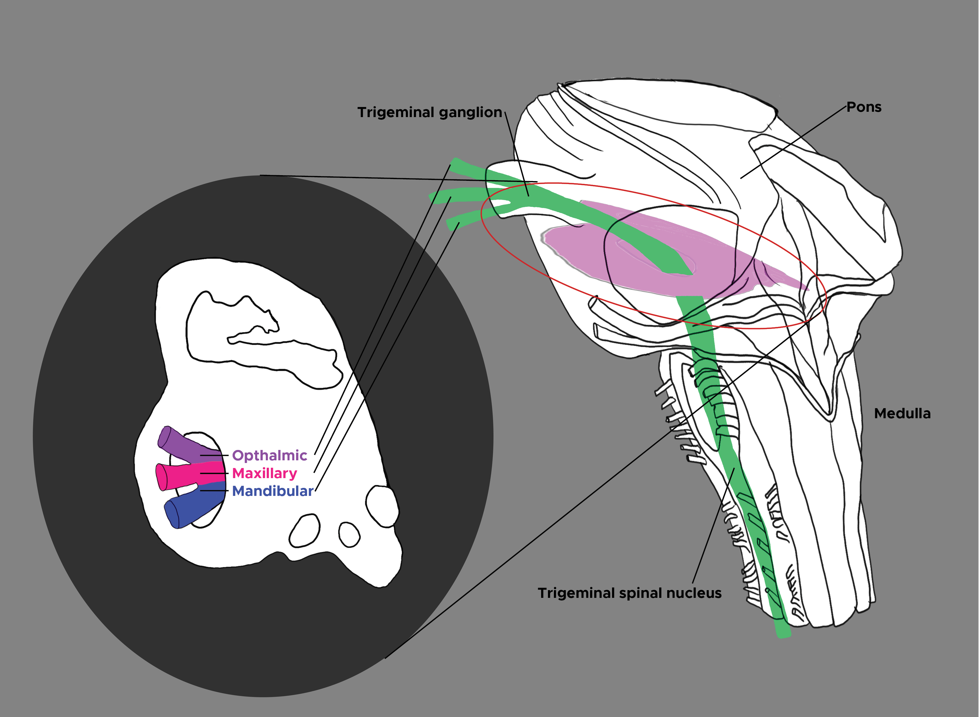
The spinal trigeminal nucleus (SN) is a sensory tract located in the lateral medulla of the brain stem whose principal function is relaying pain and temperature sensations from the oral cavity and the surface of the face (see Image. Trigeminal Nucleus). This is subdivided into 3 segments representing topographical regions of the face in an inverted fashion; the forehead is represented ventrally (distally), and the oral region is represented dorsally (proximally). Lesions of the SN cause important clinical syndromes owing to the crucial utility in the daily life of the orofacial region.
Structure and Function
Register For Free And Read The Full Article
Search engine and full access to all medical articles
10 free questions in your specialty
Free CME/CE Activities
Free daily question in your email
Save favorite articles to your dashboard
Emails offering discounts
Learn more about a Subscription to StatPearls Point-of-Care
Structure and Function
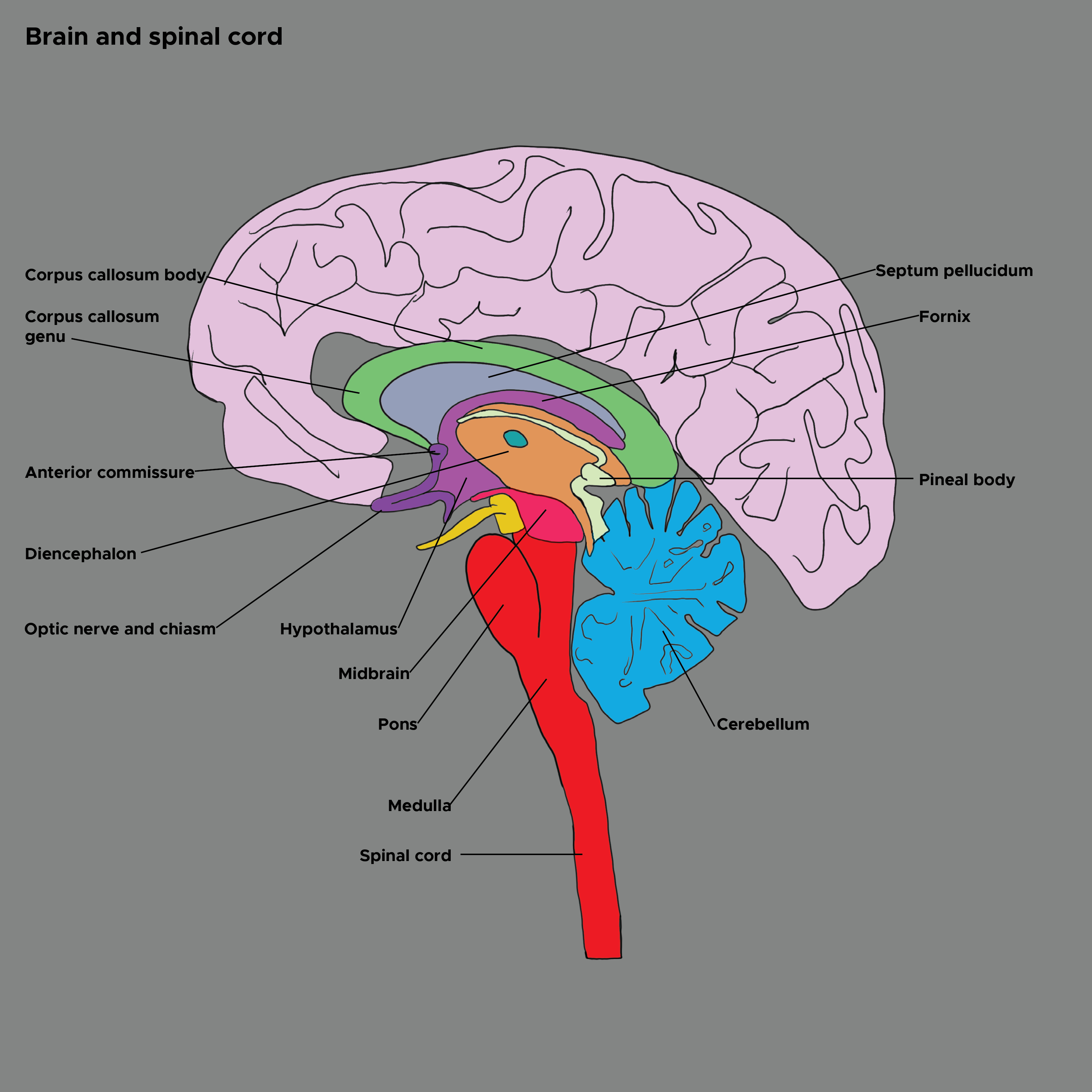
The spinal trigeminal nucleus (SN) is located in the lateral medulla of the brainstem (see Image. Brain and Spinal Cord and Image. Brain Stem, Labels). The SN extends down to the level of the C3 vertebra, at which point it becomes continuous with the dorsal spinal horn.[1] The SN is responsible for relaying various sensory modalities, including temperature, touch, and pain from the ipsilateral portion of the face, as well as nociceptive inputs from the supratentorial dura mater.[2]
The SN incorporates sensory information from different cranial nerves, but its inputs and physiological importance come principally from the three extracranial divisions of the trigeminal nerve (V1, ophthalmic; V2, maxillary; V3, mandibular). These extracranial nerves initially fuse at the trigeminal ganglion, entering the cranial vault through the superior orbital fissure, foramen rotundum, and foramen ovale.[3] After the trigeminal ganglion, the fibers divide into sensory and motor rootlets, which are then distributed to the trigeminal nuclei (mesencephalic nucleus, motor nucleus, principal sensory nucleus, and spinal nucleus) before projecting onwards to the cortex.[4]
The spinal nucleus of the trigeminal nerve is a longitudinally-shaped nucleus situated in the caudal pons and medulla, which receives sensory afferents relating to pain and temperature from the orofacial region supplied by the trigeminal nerve and, together with the principal sensory nucleus, is sometimes termed the trigeminal sensory nuclear complex (TSNC).[1] The SN also receives afferent fibers from other cranial nerves, including the facial nerve (CN VII) via the geniculate ganglion, the glossopharyngeal nerve (CN IX) via the petrosal ganglion, and the vagus nerve (CN X) via the nodose ganglion.[5]
The spinal trigeminal nucleus is further subdivided anatomically into the pars oralis, pars interporalis, and pars caudalis.[6] The pars oralis is the termination of afferent connections from the oral and nasal regions and contains circuits involved in brain stem reflexes. The pars interpolaris receives inputs from the ipsilateral face and forms the ascending anterior trigeminothalamic tract with the pars oralis. The pars caudalis also receives input from the face, forehead, cheek, and jaw, as well as being involved in transmitting temperature sensation from this area. The topographical representation of the inputs to the spinal trigeminal nucleus is inverted, such that the area encoding the forehead is represented ventrally (distally), and the area encoding the oral region is represented dorsally (proximally). The SN projects both contralaterally and ipsilaterally to the ventral posteromedial nucleus of the thalamus via the ventral and dorsal trigeminothalamic tracts.[1] This sensory information is relayed from the thalamus to the primary motor cortex via the primary sensory cortex, allowing response to stimuli of the face. This ability to react to noxious stimuli of the face and remove ourselves from harm makes the spinal trigeminal nucleus a pivotal component of the sensory pathway.
Embryology
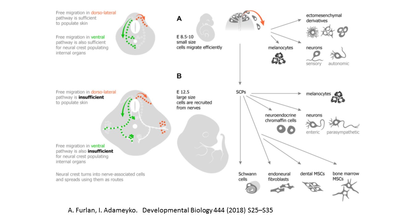
During embryological development, the cranial nerve nuclei are some of the earliest structures in the brain to emerge and are distinguishable by day 28. The trigeminal sensory nuclei develop from neural crest cells, a highly migratory population of multipotent cells that originates in the dorsal neural tube (see Diagram. Neural Crest Derivatives.)[7] The trigeminal nuclei develop initially in the metencephalon and myelencephalon before being partly displaced into the mesencephalon.[8]
Blood Supply and Lymphatics
The area of the brainstem where the spinal trigeminal nucleus is situated is supplied by the posterior inferior cerebellar artery (PICA).[9] The PICA is the four branches from the paired vertebral arteries and arises approximately 16 mm before the vertebral arteries join to form the basilar artery.[10] This is further divided into 5 anatomical segments: the anterior medullary segment, lateral medullary segment, tonsillomedullary segment, telovelotonsillar segment, and cortical segment.[11] Specifically related to the spinal nucleus of the trigeminal nerve are the first and second of these segments, which pass over the ventrolateral part of the brainstem. Any disruption of blood flow through the vertebral artery or posterior inferior cerebellar artery would interrupt the processing of sensory information from the trigeminal nerve. Such pathologies include vertebral artery dissection, thrombotic occlusion, and stenosis from extrinsic compression, such as from a tumor or fibrous band.[12]
Nerves
The spinal trigeminal nucleus incorporates sensory information from all three branches of the trigeminal nerve (CN V). CN V1, also known as the ophthalmic nerve, relays sensory information from the face in regions located above the orbit of the eyes. CN V2, also known as the maxillary nerve, is primarily responsible for sensory information between the orbit and the mouth. The final branch is CN V3, also known as the mandibular nerve. CN V3 has both a sensory component as well as a motor component; however, only the sensory component projects to the SN. The spinal trigeminal nucleus also consolidates sensory input from the facial, glossopharyngeal, and vagus nerves; this is a minor role of SN in comparison to its integration of sensory information from the three branches of the trigeminal nerve.
Surgical Considerations
Because the spinal trigeminal tract extends inferiorly down to the level of C3, this tract can be involved clinically in patients with craniovertebral junction pathologies. For the same reason, surgical intervention at this level, for example, posterior fossa decompression, could affect this area and related brainstem structures. The caudal end of the spinal trigeminal nucleus terminates at the obex of the fourth ventricle in a raised tubercle, termed the trigeminal tubercle. This is a valuable landmark for neurosurgeons operating in this region.
Clinical Significance
Lateral Medullary Syndrome
Lateral medullary syndrome, also known as medullary or posterior inferior cerebellar artery syndrome, occurs due to the occlusion of the posterior inferior cerebellar artery or the vertebral artery. An occlusion to either of these arteries can result in a lack of blood flow to the lateral medulla, the location of the spinal trigeminal nucleus. The primary ischemic events that trigger lateral medullary syndrome can result from stenotic blood vessels, as seen in adults, often with other comorbid conditions. In children, however, ischemia can be due to injury to the vertebral artery following a hyperextension neck injury.[13]
The lateral medullary syndrome presents with many symptoms, including contralateral loss of pain and temperature sensation in the body with ipsilateral Horner syndrome, dysphagia, and loss of sensation in the face.[14] This ipsilateral loss of facial sensation is due to a lack of blood supply via the posterior inferior cerebellar artery to the spinal trigeminal nucleus. This nucleus is responsible for relaying sensory information from the trigeminal nerve to the primary sensory cortex, making it a key player in the presentation of lateral medullary syndrome.
Trigeminal Neuralgia
Trigeminal neuralgia is a condition that most often presents with unilateral, stabbing, and paroxysmal painful sensations from the face along the trigeminal nerve distribution.[15] There are myriad theories regarding the underlying mechanism of this condition. The etiologies have been broken down into idiopathic, primary, and secondary trigeminal neuralgia. Suggested peripheral mechanisms involve defects along the trigeminal nerve pathway before entering the brainstem. A recent hypothesis attributes the pain of trigeminal neuralgia to a central mechanism involving the pars oralis of the spinal trigeminal nucleus.[16]
This theory has as its basis the characterization of trigeminal neuralgia as focal epileptic and neuronal hyperactivity. An increase in activity at the spinal trigeminal nucleus has been shown to precipitate the pain seen in trigeminal neuralgia in both cats and monkeys. Furthermore, the administration of anti-epileptic medications in cats and monkeys was able to decrease the intensity and duration of the attacks. The difficulty of treatment approaches makes the understanding of trigeminal neuralgia imperative. Though there is no consensus on the underlying mechanism of trigeminal neuralgia, recent evidence suggests that the spinal trigeminal nucleus, particularly the pars oralis, plays a vital role in this process.[17]
Chronic Orofacial Pain
Understanding chronic orofacial pain is important as it has been determined as a source of significant psychological distress in patients. This distress is likely because procedures and medications often cannot relieve symptoms.[18] A better understanding of the underlying mechanism may help create new solutions to this lack of treatment efficacy. In processing orofacial pain, the spinal trigeminal nucleus, specifically the pars caudalis, projects to the ventral posteromedial thalamic nucleus and the parabrachial nucleus.[19]
A recent study was conducted in humans to demonstrate changes in synapses of patients with chronic orofacial neuropathic pain. Utilizing T1-weighted MRI imaging, the study showed that alterations in the anatomy of primary synapses of the trigeminal nerve, particularly in the pars oralis, are critical for both the generation and maintenance of chronic pain in the distribution of the trigeminal nerve. These changes included significant regional gray matter volume reduction, a decrease in mean diffusivity, and a fractional anisotropy increase.[20] Imaging of the peripheral pathways of the trigeminal nerve showed no significant change in the anatomy in the setting of chronic orofacial pain, thus decreasing the likelihood of its involvement in the pathophysiology of this condition. Anatomical changes to the pars oralis of the spinal trigeminal nerve are responsible for the presentation of chronic orofacial pain.
Craniocervical Dystonia
Craniocervical dystonia is a poorly understood neurological disorder, part of the group of primary focal dystonias that present with involuntary muscle contractions, sustained or intermittent.[21] No cause or candidate pathological gene has been found, and treatment for this condition, which has significant deleterious effects on quality of life, remains limited.[22] Trigeminal reflexes, which indicate the functioning pathway of the trigeminal nerve and nuclei (and therefore the SN), include head retraction, corneal reflex, and jaw jerk reflexes.[23]
The observation that these reflexes associated with the trigeminal ganglia are aberrant in patients with craniocervical dystonia has led to the hypothesis that aberrant plasticity in the basal ganglia leading to tonic inhibition of the trigeminal sensory complex may be involved in the pathological process of this condition.[24] Furthermore, anatomical and electrophysiological experimental evidence has demonstrated the need for the normal function of the trigeminal sensory complex for normal motor control of head and neck muscles.[6] For these reasons, it is hypothesized that neuromodulation of this area, including the trigeminal spinal nucleus, either by invasive or non-invasive means, may provide therapeutic benefits in patients with this condition.[6]
Other Issues
There is evidence in rats suggesting the presence of oxytocin receptors in both the medulla oblongata and pons.[25] These oxytocin receptors are present in the spinal trigeminal nucleus; however, as is seen with oxytocin receptors in other regions of the medulla and pons, they disappear by postnatal day 10. At the time of birth, there is an increase in the maternal release of oxytocin, likely acting on these oxytocin receptors as well; this indicates an early and transient role of oxytocin in the neuronal development of the neonatal period. The full extent of the effect of oxytocin on neuronal development postnatally is yet to be determined.
Media
(Click Image to Enlarge)

Neural Crest Derivatives. Migratory neural crest and nerve-associated Schwann cell precursors represent a long-lasting source of multipotent progenitors available in any body location due to diversification of dissemination strategies. Outline of dissemination routes in the mouse embryo: E8.5-E10 NCCs (A) and E12.5-onwards nerve-dependent SCPs (B). NCCs and SCPs form most of the peripheral nervous system elements and other non-neuronal cell types (shown on the right). NCCs: neural crest cells; SCPs: Schwann cell precursors; MSC: mesenchymal stem cells.
Furlan A, Adameyko I. Schwann cell precursor: a neural crest cell in disguise? Developmental Biology. 2018;444 (suppl 1):S25–S35 doi: 10.1016/j.ydbio.2018.02.008.
(Click Image to Enlarge)
(Click Image to Enlarge)
(Click Image to Enlarge)
References
Henssen DJ, Kurt E, Kozicz T, van Dongen R, Bartels RH, van Cappellen van Walsum AM. New Insights in Trigeminal Anatomy: A Double Orofacial Tract for Nociceptive Input. Frontiers in neuroanatomy. 2016:10():53. doi: 10.3389/fnana.2016.00053. Epub 2016 May 10 [PubMed PMID: 27242449]
Bartsch T, Goadsby PJ. Increased responses in trigeminocervical nociceptive neurons to cervical input after stimulation of the dura mater. Brain : a journal of neurology. 2003 Aug:126(Pt 8):1801-13 [PubMed PMID: 12821523]
Level 3 (low-level) evidenceMarur T, Tuna Y, Demirci S. Facial anatomy. Clinics in dermatology. 2014 Jan-Feb:32(1):14-23. doi: 10.1016/j.clindermatol.2013.05.022. Epub [PubMed PMID: 24314374]
Erzurumlu RS,Murakami Y,Rijli FM, Mapping the face in the somatosensory brainstem. Nature reviews. Neuroscience. 2010 Apr [PubMed PMID: 20179712]
Level 3 (low-level) evidenceHenssen DJHA, Derks B, van Doorn M, Verhoogt NC, Staats P, Vissers K, Van Cappellen van Walsum AM. Visualizing the trigeminovagal complex in the human medulla by combining ex-vivo ultra-high resolution structural MRI and polarized light imaging microscopy. Scientific reports. 2019 Aug 5:9(1):11305. doi: 10.1038/s41598-019-47855-5. Epub 2019 Aug 5 [PubMed PMID: 31383932]
Bradnam L, Barry C. The role of the trigeminal sensory nuclear complex in the pathophysiology of craniocervical dystonia. The Journal of neuroscience : the official journal of the Society for Neuroscience. 2013 Nov 20:33(47):18358-67. doi: 10.1523/JNEUROSCI.3544-13.2013. Epub [PubMed PMID: 24259561]
Méndez-Maldonado K, Vega-López GA, Aybar MJ, Velasco I. Neurogenesis From Neural Crest Cells: Molecular Mechanisms in the Formation of Cranial Nerves and Ganglia. Frontiers in cell and developmental biology. 2020:8():635. doi: 10.3389/fcell.2020.00635. Epub 2020 Aug 7 [PubMed PMID: 32850790]
Wang CZ,Shi M,Yang LL,Yang RQ,Luo ZG,Jacquin MF,Chen ZF,Ding YQ, Development of the mesencephalic trigeminal nucleus requires a paired homeodomain transcription factor, Drg11. Molecular and cellular neurosciences. 2007 Jun [PubMed PMID: 17482477]
Level 3 (low-level) evidenceNovy J. Spinal cord syndromes. Frontiers of neurology and neuroscience. 2012:30():195-8. doi: 10.1159/000333682. Epub 2012 Feb 14 [PubMed PMID: 22377894]
Saylam C, Ucerler H, Orhan M, Cagli S, Zileli M. The relationship of the posterior inferior cerebellar artery to cranial nerves VII-XII. Clinical anatomy (New York, N.Y.). 2007 Nov:20(8):886-91 [PubMed PMID: 17907205]
Miao HL, Zhang DY, Wang T, Jiao XT, Jiao LQ. Clinical Importance of the Posterior Inferior Cerebellar Artery: A Review of the Literature. International journal of medical sciences. 2020:17(18):3005-3019. doi: 10.7150/ijms.49137. Epub 2020 Oct 18 [PubMed PMID: 33173421]
Cloud GC,Markus HS, Diagnosis and management of vertebral artery stenosis. QJM : monthly journal of the Association of Physicians. 2003 Jan [PubMed PMID: 12509646]
Ruedrich ED, Chikkannaiah M, Kumar G. Wallenberg's lateral medullary syndrome in an adolescent. The American journal of emergency medicine. 2016 Nov:34(11):2254.e1-2254.e2. doi: 10.1016/j.ajem.2016.05.022. Epub 2016 May 12 [PubMed PMID: 27246963]
Day GS, Swartz RH, Chenkin J, Shamji AI, Frost DW. Lateral medullary syndrome: a diagnostic approach illustrated through case presentation and literature review. CJEM. 2014 Mar:16(2):164-70 [PubMed PMID: 24626124]
Level 3 (low-level) evidenceCruccu G. Trigeminal Neuralgia. Continuum (Minneapolis, Minn.). 2017 Apr:23(2, Selected Topics in Outpatient Neurology):396-420. doi: 10.1212/CON.0000000000000451. Epub [PubMed PMID: 28375911]
Peker S, Sirin A. Primary trigeminal neuralgia and the role of pars oralis of the spinal trigeminal nucleus. Medical hypotheses. 2017 Mar:100():15-18. doi: 10.1016/j.mehy.2017.01.008. Epub 2017 Jan 16 [PubMed PMID: 28236840]
Lee KH. Facial pain: trigeminal neuralgia. Annals of the Academy of Medicine, Singapore. 1993 Mar:22(2):193-6 [PubMed PMID: 8363331]
Merrill RL, Goodman D. Chronic Orofacial Pain and Behavioral Medicine. Oral and maxillofacial surgery clinics of North America. 2016 Aug:28(3):247-60. doi: 10.1016/j.coms.2016.03.007. Epub [PubMed PMID: 27475505]
Okada S, Katagiri A, Saito H, Lee J, Ohara K, Iinuma T, Bereiter DA, Iwata K. Differential activation of ascending noxious pathways associated with trigeminal nerve injury. Pain. 2019 Jun:160(6):1342-1360. doi: 10.1097/j.pain.0000000000001521. Epub [PubMed PMID: 30747907]
Wilcox SL, Gustin SM, Macey PM, Peck CC, Murray GM, Henderson LA. Anatomical changes at the level of the primary synapse in neuropathic pain: evidence from the spinal trigeminal nucleus. The Journal of neuroscience : the official journal of the Society for Neuroscience. 2015 Feb 11:35(6):2508-15. doi: 10.1523/JNEUROSCI.3756-14.2015. Epub [PubMed PMID: 25673845]
Albanese A, Bhatia K, Bressman SB, Delong MR, Fahn S, Fung VS, Hallett M, Jankovic J, Jinnah HA, Klein C, Lang AE, Mink JW, Teller JK. Phenomenology and classification of dystonia: a consensus update. Movement disorders : official journal of the Movement Disorder Society. 2013 Jun 15:28(7):863-73. doi: 10.1002/mds.25475. Epub 2013 May 6 [PubMed PMID: 23649720]
Level 3 (low-level) evidenceLohmann K, Klein C. Genetics of dystonia: what's known? What's new? What's next? Movement disorders : official journal of the Movement Disorder Society. 2013 Jun 15:28(7):899-905. doi: 10.1002/mds.25536. Epub [PubMed PMID: 23893446]
Serrao M, Rossi P, Parisi L, Perrotta A, Bartolo M, Cardinali P, Amabile G, Pierelli F. Trigemino-cervical-spinal reflexes in humans. Clinical neurophysiology : official journal of the International Federation of Clinical Neurophysiology. 2003 Sep:114(9):1697-703 [PubMed PMID: 12948799]
Blood AJ, Kuster JK, Woodman SC, Kirlic N, Makhlouf ML, Multhaupt-Buell TJ, Makris N, Parent M, Sudarsky LR, Sjalander G, Breiter H, Breiter HC, Sharma N. Evidence for altered basal ganglia-brainstem connections in cervical dystonia. PloS one. 2012:7(2):e31654. doi: 10.1371/journal.pone.0031654. Epub 2012 Feb 22 [PubMed PMID: 22384048]
Murata Y, Li MZ, Masuko S. Developmental expression of oxytocin receptors in the neonatal medulla oblongata and pons. Neuroscience letters. 2011 Sep 20:502(3):157-61. doi: 10.1016/j.neulet.2011.07.032. Epub 2011 Jul 29 [PubMed PMID: 21820489]
Level 3 (low-level) evidence

