Introduction
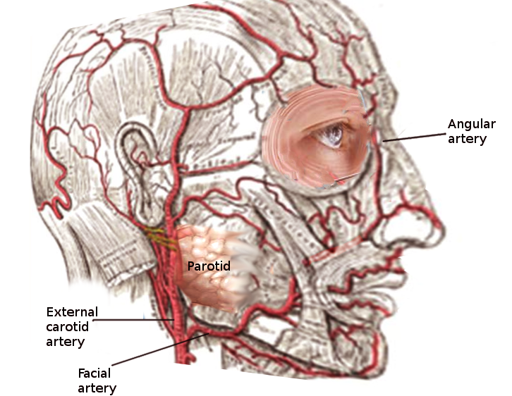
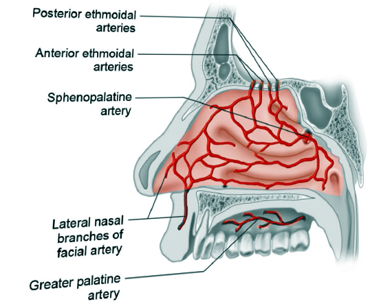
The blood supply of the external nose and the nasal cavity is derived from anastomosing terminal branches of the internal and external carotid artery systems (see Image. Nasal Blood Supply). The blood supply of the external nose is provided primarily by the facial artery, particularly along the lateral aspect and in the lower one-third, with the anterior lateral nasal artery supplying the majority of blood flow to the nasal tip (see Image. Facial Artery).[1] Branches of the internal carotid artery perfuse the middle and upper third of the external nose with a contribution from the maxillary artery.[2] The blood supply of the nasal cavity is similarly convoluted. Still, its flow likewise ultimately stems from the carotid system, with the anterior lateral nasal artery traversing the nasal alar soft tissue and perfusing the anterior aspect of the lateral nasal wall while the posterior lateral nasal artery, a branch of the sphenopalatine artery, supplies the posterior aspect of the lateral nasal wall.[3]
Structure and Function
Register For Free And Read The Full Article
Search engine and full access to all medical articles
10 free questions in your specialty
Free CME/CE Activities
Free daily question in your email
Save favorite articles to your dashboard
Emails offering discounts
Learn more about a Subscription to StatPearls Point-of-Care
Structure and Function
Both the anterior and posterior lateral nasal arteries are distal branches of the external carotid arteries, each measuring approximately 2 mm in diameter, with the anterior artery slightly larger than the posterior.[4][5] The anterior lateral nasal artery is the remaining branch of the facial artery after it gives off the superior labial artery near the oral commissure. As the anterior lateral nasal artery turns anteriorly along the nasal ala, it gives off the angular artery, which courses superiorly along the lateral aspect of the external nose. The anterior lateral nasal artery supplies the superficial and deep tissues of the tip of the nose. It contributes to the perfusion of the entire external nose via anastomoses with the columellar artery (a branch of the superior labial branch of the facial artery), the infraorbital artery (a branch of the internal maxillary artery off the external carotid artery), the external nasal artery, which is a branch of the anterior ethmoidal artery (from the ophthalmic branch of the internal carotid artery) that emerges into the external nose via a gap between the upper lateral cartilage and the nasal bone, and the dorsal nasal artery (a terminal branch of the ophthalmic artery), which emerges from the orbit just inferior to the trochlea of the superior oblique muscle. Internally, the anterior lateral nasal artery perfuses the anterior aspect of the inferior turbinate and the lateral nasal wall, superiorly anastomosing with the anterior ethmoidal artery.[3]
The posterior lateral nasal artery is a branch of the sphenopalatine artery, itself a branch of the internal maxillary division of the external carotid artery, that arises along with the posterior septal artery at a bifurcation that occurs just before the arteries' entrance into the nasal cavity via the sphenopalatine foramen.[6] The posterior lateral nasal artery perfuses the posterior aspect of the lateral nasal wall. It gives off branches to the middle and inferior turbinates, while the posterior septal artery feeds the superior turbinate. The posterior lateral nasal artery then anastomoses with the posterior ethmoidal artery superiorly and the anterior lateral nasal artery via the inferior turbinate.[3]
Embryology
During the fourth and fifth weeks of embryological development, the aortic arches form parallel with the branchial arches and ultimately develop into the arteries of the face, neck, and chest. The third aortic arch develops into the common carotid artery, which branches into the external and internal carotid arteries.[7] The external carotid artery gives off the facial artery, from which the anterior lateral nasal artery branches. The posterior lateral nasal artery is a branch of the sphenopalatine artery, itself a branch of the internal maxillary artery, which derives from the first aortic arch and is the primary artery of the first branchial arch; it ultimately joins with the external carotid artery to perfuse the deep structures of the face.[8]
Nerves
The anterior and posterior-lateral nasal arteries anastomose along the lateral walls of the nasal cavity with the anterior and posterior ethmoidal arteries, helping to perfuse the anterior and posterior ethmoidal nerves. These nerves are terminal branches of the nasociliary nerve, itself a branch of the ophthalmic division of the trigeminal nerve (cranial nerve V).
Muscles
The anterior lateral nasal artery perfuses the intrinsic muscles of the nose, the transverse and alar portions of the nasalis muscle, also known as the compressor naris and the dilator naris muscles. It also feeds the nearby muscles of the medial midface, specifically the levator labii superioris alaeque nasi muscle and, potentially, the levator labii superioris muscle.[9]
Physiologic Variants
The branching pattern of the facial artery in the area of the nasofacial junction is variable. In most individuals, the lateral nasal artery arises at the facial artery's bifurcation, giving off the superior labial artery, just superior to the oral commissure. In some cases, however, the lateral nasal artery may branch directly from the superior labial artery instead of the facial artery. If the lateral nasal artery is a branch of the superior labial artery, it ascends the face until it reaches the ala. At the ala, the lateral nasal artery ascends the nose, turns medially towards the tip, and gives off the angular artery to continue its ascent along the nasofacial junction.[10]
Surgical Considerations
As mentioned, the blood supply to the external nose is complicated and redundant, which permits reliable healing and infection resistance even after extensive trauma or surgical intervention. The anterior lateral nasal arteries represent the primary perfusion to the tip of the nose. Without their contribution to blood flow, dividing the columellar artery during open rhinoplasty would likely lead to more healing complications.[1] Likewise, dermal fillers may compress small arteries and cause soft tissue ischemia when injected into the nose. Still, the collateral circulation afforded by the lateral nasal arteries and other branches often prevents severe complications.[11] The anterior lateral nasal artery also passes underneath the levator labii superioris alaeque nasi muscle and nourishes it; this muscle can be used as a regional flap to reconstruct full-thickness nasal defects.[9] The commonly used nasolabial flap has also been described based on the anterior lateral nasal artery in addition to its often employed random blood supply; the nasolabial flap has myriad applications within the midface but is most commonly used for alar reconstruction.[12] Numerous other local and regional soft tissue flaps that derive their blood supply from the anterior lateral nasal artery or its branches have also been described; they are particularly useful for lateral nasal wall, columellar, and alar reconstruction.[13][4][14]
Internally, the posterior and anterior lateral nasal arteries comprise the major blood supply to the inferior turbinate. In addition to serving a major role in warmth, humidification, and regulation of nasal airflow, the inferior turbinate also provides a significant amount of soft tissue available for use in regional flap reconstruction. The inferior turbinate can be raised as an anteriorly-based flap, perfused by the anterior lateral nasal artery, and inset to repair nasal septal perforations, nasal cavity mucosal defects, and skull base defects.[15][16] The inferior turbinate may also be raised as a posteriorly-based flap, pedicled on the posterior lateral nasal artery, and employed to reconstruct select skull base defects.[17] The robust blood supply at the posterior aspect of the inferior turbinate does come with a drawback, however, which is the risk of posterior epistaxis that may result from instrumentation of the nasopharynx, particularly when the posterolateral nasal cavity's venous drainage, Woodruff's plexus, is disrupted.
Clinical Significance
The anterior and posterior-lateral nasal arteries are branches of the external carotid system, and between them, significant portions of the external nose and the nasal cavity are perfused. The excellent collateral circulation of the nose permits rapid healing and infection resistance, as well as the flexibility to manipulate tissue extensively for reconstructive purposes.[18] This collateral circulation, particularly in the case of the posterior lateral nasal artery, also results in the potential for significant epistaxis due to trauma or iatrogenic injury. Still, the benefits of robust perfusion outweigh the drawbacks.
Media
(Click Image to Enlarge)
References
Rohrich RJ, Gunter JP, Friedman RM. Nasal tip blood supply: an anatomic study validating the safety of the transcolumellar incision in rhinoplasty. Plastic and reconstructive surgery. 1995 Apr:95(5):795-9; discussion 800-1 [PubMed PMID: 7708862]
Pilsl U, Anderhuber F. The External Nose: The Nasal Arteries and Their Course in Relation to the Nasolabial Fold and Groove. Plastic and reconstructive surgery. 2016 Nov:138(5):830e-835e. doi: 10.1097/PRS.0000000000002626. Epub [PubMed PMID: 27782991]
MacArthur FJ, McGarry GW. The arterial supply of the nasal cavity. European archives of oto-rhino-laryngology : official journal of the European Federation of Oto-Rhino-Laryngological Societies (EUFOS) : affiliated with the German Society for Oto-Rhino-Laryngology - Head and Neck Surgery. 2017 Feb:274(2):809-815. doi: 10.1007/s00405-016-4281-1. Epub 2016 Aug 27 [PubMed PMID: 27568352]
Lombardo GA, Tamburino S, Tracia L, Tarico MS, Perrotta RE. Lateral Nasal Artery Perforator Flaps: Anatomic Study and Clinical Applications. Archives of plastic surgery. 2016 Jan:43(1):77-83. doi: 10.5999/aps.2016.43.1.77. Epub 2016 Jan 15 [PubMed PMID: 26848450]
Prades JM, Asanau A, Timoshenko AP, Faye MB, Martin Ch. Surgical anatomy of the sphenopalatine foramen and its arterial content. Surgical and radiologic anatomy : SRA. 2008 Oct:30(7):583-7. doi: 10.1007/s00276-008-0390-x. Epub 2008 Jul 23 [PubMed PMID: 18648719]
El-Shaarawy EAA, Hassan SS. The sphenopalatine foramen in man: anatomical, radiological and endoscopic study. Folia morphologica. 2018:77(2):345-355. doi: 10.5603/FM.a2017.0104. Epub 2017 Nov 13 [PubMed PMID: 29131280]
Rosen RD, Bordoni B. Embryology, Aortic Arch. StatPearls. 2025 Jan:(): [PubMed PMID: 31985966]
Ansari A, Bordoni B. Embryology, Face. StatPearls. 2024 Jan:(): [PubMed PMID: 31424786]
Moore K 2nd, Thompson R, Lian T. The pedicled levator labii superioris alaeque nasi flap: A durable single-stage option for reconstruction of full-thickness nasal defects. American journal of otolaryngology. 2019 Mar-Apr:40(2):279-281. doi: 10.1016/j.amjoto.2018.09.015. Epub 2018 Oct 2 [PubMed PMID: 30473167]
Jung DH, Kim HJ, Koh KS, Oh CS, Kim KS, Yoon JH, Chung IH. Arterial supply of the nasal tip in Asians. The Laryngoscope. 2000 Feb:110(2 Pt 1):308-11 [PubMed PMID: 10680935]
Lee W, Kim JS, Oh W, Koh IS, Yang EJ. Nasal dorsum augmentation using soft tissue filler injection. Journal of cosmetic dermatology. 2019 Oct:18(5):1254-1260. doi: 10.1111/jocd.13018. Epub 2019 Jun 3 [PubMed PMID: 31157508]
Zimman OA, Felice F. The lateral nasal artery pedicle nasolabial island flap. Plastic and reconstructive surgery. 2008 Jun:121(6):2177. doi: 10.1097/PRS.0b013e31817072fe. Epub [PubMed PMID: 18520919]
Level 3 (low-level) evidenceJang SY, Kim WS, Kim HK, Bae TH. Nasal Columellar Reconstruction With Reverse Lateral Nasal Artery Pedicled Nasolabial Island Flap. The Journal of craniofacial surgery. 2018 May:29(3):e250-e251. doi: 10.1097/SCS.0000000000004268. Epub [PubMed PMID: 29381622]
Aynehchi BB, Westreich RW. Lateral nasal artery pedicled island flap for repair of nasal alar defects. Otolaryngology--head and neck surgery : official journal of American Academy of Otolaryngology-Head and Neck Surgery. 2012 Mar:146(3):382-4. doi: 10.1177/0194599811428036. Epub 2011 Nov 7 [PubMed PMID: 22063733]
Murakami CS, Kriet JD, Ierokomos AP. Nasal reconstruction using the inferior turbinate mucosal flap. Archives of facial plastic surgery. 1999 Apr-Jun:1(2):97-100 [PubMed PMID: 10937085]
Level 2 (mid-level) evidenceGil Z, Margalit N. Anteriorly based inferior turbinate flap for endoscopic skull base reconstruction. Otolaryngology--head and neck surgery : official journal of American Academy of Otolaryngology-Head and Neck Surgery. 2012 May:146(5):842-7. doi: 10.1177/0194599811434516. Epub 2012 Jan 18 [PubMed PMID: 22261494]
Fortes FS, Carrau RL, Snyderman CH, Prevedello D, Vescan A, Mintz A, Gardner P, Kassam AB. The posterior pedicle inferior turbinate flap: a new vascularized flap for skull base reconstruction. The Laryngoscope. 2007 Aug:117(8):1329-32 [PubMed PMID: 17597634]
Level 2 (mid-level) evidenceKoziej M, Trybus M, Hołda M, Polak J, Wnuk J, Brzegowy P, Popiela T, Walocha J, Chrapusta A. Anatomical Map of the Facial Artery for Facial Reconstruction and Aesthetic Procedures. Aesthetic surgery journal. 2019 Oct 15:39(11):1151-1162. doi: 10.1093/asj/sjz028. Epub [PubMed PMID: 30721996]

