Introduction
Osteoporotic compression fractures are the most common fragility fractures today. Individuals with osteoporosis may experience a vertebral compression fracture from seemingly insignificant trauma, such as sitting down abruptly. A higher-energy axial loading force is required to compress the vertebral body in younger individuals. Non-osteoporotic lumbar compression fractures are typically observed in motor vehicle accidents or falls from heights. Infectious and malignant processes that weaken vertebrae can also lead to an eventual compression fracture.[1]
These fractures can lead to significant physical limitations, including back pain and functional disability.[2] Compression fractures are prone to progression over time and may facilitate the compression of adjacent vertebrae due to compensatory increases in axial load. Due to the high prevalence of this injury, there is a considerable socioeconomic burden associated with the disease, and there is significant controversy regarding optimal treatment.
Vertebroplasty and kyphoplasty are percutaneous interventional procedures used to treat symptomatic, nonhealing fragility fractures of the spine by injecting polymethyl methacrylate into the vertebral body, providing structural support (see Image. Fluoroscopic Image of Cement Injection During Percutaneous Kyphoplasty). These procedures have faced scrutiny due to the lack of significant clinical improvement noted in 2 early randomized controlled trials, which had significant methodological limitations. Some of these limitations included the inclusion of patients with chronic fractures and those with less severe pain or disability, as well as the performance of an alternative intervention rather than a sham procedure.[3] Although the effectiveness of vertebroplasty and kyphoplasty has been debated, substantial evidence currently demonstrates its advantages over nonoperative treatment.[4] Vertebroplasty is associated with a higher incidence of polymethyl methacrylate extravasation than kyphoplasty; consequently, kyphoplasty has become the preferred surgical intervention for osteoporotic compression fractures.
Anatomy and Physiology
Register For Free And Read The Full Article
 Search engine and full access to all medical articles
- 10 free questions in your specialty
- Free CME/CE Activities
- Free daily question in your email
- Save favorite articles to your dashboard
- Emails offering discounts
Learn more about a Subscription to StatPearls Point-of-Care
Anatomy and Physiology
Bone mineral density decreases with age following peak bone mass, leading to osteoporotic bone. Although osteoporotic bone retains normal quality, its quantity is reduced. Cancellous bone exhibits decreased trabecular quantity, whereas cortical bone becomes thinner, leading to a decreased load-to-failure capacity and a higher likelihood of fracture. Vertebral compression fractures occur when axial forces exceed the structural integrity of the vertebral bone, resulting in a loss of height in the anterior, middle, or posterior vertebral height.[5] The thoracolumbar spine (T12-L2) is the most frequently affected site (60%-75%), followed by the lower lumbar region (L2-L5).[2]
Compression fractures primarily affect the anterior column of the spine, which includes the anterior two-thirds of the vertebral body, the anterior two-thirds of the intervertebral disc, and the anterior longitudinal ligament. Unlike vertebral burst or Chance fractures, compression fractures do not compromise the posterior tension band.[6] The posterior tension band is characterized by muscles, ligaments, processes, and pedicles that maintain spinal stability.[6] Therefore, compression fractures are deemed stable fractures.
Posterior tension band components include the following:
- Posterior ligamentous complex: Supraspinous and interspinous ligaments, ligamentum flavum
- Musculature: Longissimus, iliocostalis, spinalis, semispinalis, rotatores, intertransversarii, multifidus
- Bone: Transverse and spinous processes, pedicles, facets
Indications
Understanding the clinical indications for vertebroplasty and kyphoplasty helps guide appropriate treatment decisions, optimize patient outcomes, and reduce complications associated with vertebral compression fractures. Patients considered for vertebroplasty and kyphoplasty typically meet all of the following criteria.
Failure of Nonoperative Treatment
Standard treatment options for a vertebral compression fracture include conservative therapies such as analgesics, external orthosis, physical therapy, and bed rest. Most patients can be treated with observation and a gradual return to activity. Symptoms typically resolve within 4 to 6 weeks.[1]
Although conservative management is commonly perceived as safe, it is neither entirely benign nor without risks. Bed rest and immobilization have well-established risks. In the attempt at early mobilization, the fractures may rapidly progress, resulting in vertebral height loss, increased kyphosis, and potentially osseous retropulsion into the spinal canal.
Early intervention in osteoporotic vertebral compression fractures aligns closely with established orthopedic principles for prompt stabilization in extremity fractures. Prompt stabilization alleviates pain and yields superior biomechanical and functional outcomes.[4] Major professional medical societies report that vertebroplasty and kyphoplasty are more effective compared to prolonged nonoperative management in patients experiencing painful vertebral compression fractures who have not achieved adequate pain relief or functional improvement with nonoperative therapy.[7] However, the optimal timing to transition from conservative management to vertebroplasty and kyphoplasty remains uncertain.[8]
Active Vertebral Body Fracture
Active vertebral body fractures may result from osteoporosis or neoplastic processes. Such fractures are identified by hyperintense signals on short tau inversion recovery or fat-suppressed T2-weighted magnetic resonance imaging (MRI) sequences. Although hyperintensity typically indicates acute fractures, similar signal characteristics may also appear in certain chronic fractures, suggesting ongoing fracture activity. If MRI is not feasible, single-photon emission computed tomography can be used to confirm the active status of the fracture.[9]
Correlation of Pain with the Fracture Site
The pain location and characteristics must correlate with the site of the pain. Typically, pain is located at the level of the fracture, worsens with spinal movement, and may radiate.
High Pain Intensity
Pain intensity must be high enough to justify the invasive intervention. Studies have used variable thresholds. Some studies have used the Numeric Rating Scale pain score of 7 or greater (out of 10), whereas others use 5 or greater.[9][10] In clinical practice, this decision is individualized, based on the impact of pain on the patient's functional status.
Contraindications
Contraindications to vertebroplasty and kyphoplasty include absolute and relative conditions that may adversely affect patient safety or procedural efficacy. Absolute contraindications include burst fractures, retropulsed bone fragments, and significant breaches of the posterior vertebral wall that jeopardize neural structures. In addition, spinal instability and symptomatic myelopathy or radiculopathy typically preclude safe and effective treatment with vertebroplasty and kyphoplasty.
Vertebra plana describes near-total collapse of the vertebral body, typically contraindicating vertebroplasty and kyphoplasty due to a minimal expected benefit. In this condition, insufficient residual bone volume prevents meaningful restoration of vertebral height. Furthermore, anterior column support in vertebra plana cases is partly maintained by the proximity of adjacent intervertebral discs, reducing the necessity for vertebroplasty and kyphoplasty.
Uncorrectable coagulopathies; systemic infections, such as bacteremia; and spinal infections, such as osteomyelitis or discitis, must be treated before performing vertebroplasty and kyphoplasty. Injecting cement under these conditions risks contaminating the cement itself, complicating infection management due to the persistence of an infected foreign body.
Allergies to bone cement or contrast agents and pregnancy are additional absolute contraindications. Severe cardiopulmonary comorbidities represent a critical concern, given the heightened risk of a potential cement or marrow embolism. Medical conditions precluding an emergent open surgical decompression in the event of a complication must be considered during decision-making.
Relative contraindications include significant vertebral collapse exceeding 75% of the original height. Tumors extending into the spinal canal and moderate coagulopathies that can be corrected also require cautious consideration. Active fractures in patients without osteoporosis, multiple compression fractures, and asymptomatic compression fractures can pose clinical dilemmas. Deficiencies in the posterior vertebral cortex raise concerns about cement leakage and potential neural element compromise. The decision to treat a nonfractured vertebra, especially when adjacent fractures are present, remains debated among experts. Chronic fractures generally represent a contraindication unless advanced imaging reveals a visible fracture cleft or increased signal intensity. Pedicle fractures also raise concerns about proper cement containment and can represent a relative contraindication if stability is compromised.
Equipment
Vertebroplasty and kyphoplasty are best performed using high-quality fluoroscopy to confirm the needle placement. C-arm fluoroscopy is the preferred imaging technique for this procedure. This procedure can be performed with 1 C-arm transitioning between anteroposterior and lateral images, or with 2 C-arms—1 positioned for anteroposterior, and the other for lateral images (biplanar fluoroscopy). The 2 C-arm technique alleviates the need to constantly change the C-arm position. A list of types of equipment is as follows:
- Fluoroscopy
- Bone needle, typically a Jamshidi needle
- Bone needle stylets, with diamond-shaped multi-bevel and single-bevel
- Bone cement, typically: Polymethyl methacrylate
- Kyphoplasty balloon catheter (optional) [1]
Personnel
Vertebroplasty and kyphoplasty are performed primarily by interventional radiologists, interventional pain management physicians, and neurosurgical/orthopedic spine surgeons. Intraoperative personnel may also include a fluoroscopy technician, a nurse, and a company representative. An anesthesiologist is also present if general anesthesia or monitored anesthesia care is implemented.
Preparation
According to the Society of Interventional Radiology Standards of Practice Consensus Guidelines, vertebroplasty and kyphoplasty are considered high-bleeding-risk procedures. International normalized ratio (INR) and complete blood count should be obtained before the procedure.[11] Recommendations are as follows:
- INR corrected to be within the range of ≤1.5 to 1.8
- Transfuse for platelets <50,000
- Aspirin: Hold 3 to 5 days pre-procedure
- Clopidogrel: Hold 5 days pre-procedure
- Low-molecular-weight heparin (LMWH): Hold 1 dose pre-procedure if a prophylactic dose is used. Hold 2 doses pre-procedure if a therapeutic dose.
Antibiotic prophylaxis is achieved by administering 1 g of intravenous (IV) cefazolin, 1 h pre-procedure (first-line). Once intubated, the patient is placed in the prone position on a well-padded radiolucent table. Proper positioning of the fluoroscopic C-arm is crucial. The affected vertebral level is isolated, proper views are obtained before the start of the procedure, and the C-arm position is noted so it can be replicated after surgical preparation. On the anteroposterior image, the vertebral body should be positioned in a direct anteroposterior view with the endplates parallel to the x-ray beam and the spinous process centered between the pedicles.[12] The pedicles are then marked with a marking pen, and the surgical area is prepped and draped in a sterile fashion.
Technique or Treatment
The setup and techniques of vertebroplasty and kyphoplasty procedures are highly similar, the only difference being that kyphoplasty involves creating a cavity in the vertebral body through balloon expansion (see Image. Typical Procedural Steps for Percutaneous Vertebroplasty and Kyphoplasty). Vertebroplasty is performed in the thoracic and lumbar spine using a transpedicular or parapedicular approach.[12] There may be variations in technique based on surgeon preference, equipment used, and patient-specific anatomy or pathology.
Typical Procedural Steps for Percutaneous Vertebroplasty and Kyphoplasty
Step 1: The bone needle (yellow) is advanced until bone contact is established. The goal is to dock the needle onto the lateral aspect of the pedicle on the anteroposterior view for a transpedicular approach. A more lateral entry point allows for a more oblique trajectory known as parapedicular or extrapedicular, as the more dorsal part of the needle trajectory is lateral to the pedicle. The skin incision (small stab incision) is typically placed 1 to 2 cm lateral to the pedicle on the anteroposterior view; however, this distance increases with greater tissue thickness between the skin surface and bone entry site. A parapedicular approach requires a more laterally placed skin entry point. Some surgeons prefer the starting point along the superior lateral edge of the pedicle with variable angulation depending on the pedicle level being accessed.[13] The medial and inferior walls of the pedicle must be clearly visualized to decrease injury to nerve roots and the spinal cord.[14] If the trajectory is appropriate on the anteroposterior view, the C-arm is then transitioned to the lateral view to confirm proper pedicle placement and superior or inferior trajectory in the vertebral body.
Step 2: The bone needle is advanced by gently tapping with a mallet for approximately 2 cm. Typically, at this point, the needle tip reaches the posterior vertebral body wall (magenta line). At this stage, confirming that the needle tip remains lateral to the medial border of the pedicle (red line) on the anteroposterior view is crucial. A needle tip positioned medial to the pedicle indicates a spinal canal breach, requiring immediate repositioning. Serial fluoroscopic images are typically obtained during the first 2 cm of needle advancement to ensure a safe needle trajectory and avoid violating the pedicle's medial boundary.
Step 3: The needle is advanced further into the vertebral body.
Step 4: A twist drill or bone curette (green) may be used to deepen the access tract. The tract should reach the anterior to middle third of the vertebral body as close to the midline as possible.[1]
Step 5 (Kyphoplasty only): A balloon tamp (blue) is inserted into the prepared tract for kyphoplasty.
Step 6 (Kyphoplasty only): The balloon (light blue) is inflated with contrast fluid, allowing fluoroscopic visualization. Inflation continues until the manufacturer's recommended pressure or volume is reached, vertebral height is restored, or the balloon approaches the anatomical limits of the vertebral body, whichever occurs first. In this illustration, the balloon inflation has partially restored the vertebral body height by a partial reduction of the superior endplate fracture. The balloon is then deflated, and the balloon tamp is removed. Recently developed vertebral augmentation devices, resembling a mechanical jack or a vascular stent, may also be used instead of a balloon. Steps 1 to 6 are repeated contralaterally if a bilateral procedure is indicated. The unipedicular approach is commonly favored due to reduced operative time and radiation exposure, mainly if a steerable bone curette and a directional cement injection cannula are used. However, the bipedicular approach may still allow for better fracture reduction and more extensive cement filling of the vertebral body.
Step 7: The cement injection cannula (purple) is inserted. Cement is mixed and allowed to reach a paste-like consistency before injection, minimizing the risk of leakage in severely osteoporotic bone.[15]
Step 8: Cement injection is typically performed in aliquots of up to 0.5 cc, under live fluoroscopic visualization or serial imaging, enabling precise monitoring for potential cement leakage. If the cement approaches the anatomical boundaries of the vertebral body, injection must be halted immediately. Subsequently, the clinician must decide whether to terminate the injection or reposition the needle for further cement delivery. The volume of cement injected closely matches the volume of contrast used during balloon inflation. Cement injection typically begins anteriorly within the vertebral body and progresses posteriorly as the cement applicator is gradually withdrawn. After completing the injection, the cement injection cannula remains briefly in place until cement curing (turning into a solid, hardened material) occurs, after which the cement applicator and the bone needle are removed.
Complications
Complications occur in approximately 50% of patients undergoing vertebroplasty, but about 95% are clinically asymptomatic.[16] Common complications include the following:
- Infection and bleeding: Universally recognized risks of any interventional procedure.
- Radiculopathy or spinal cord injury: If the spinal needle violates the inferior or medial wall of the pedicle during entry, there is a significant risk of damage to a nerve root or the spinal cord.
- Cement leakage into the surrounding tissue, intradiscal space, or spinal canal (see Image. Bone Cement Leakage in Vertebroplasty and Kyphoplasty). This event is more common for vertebroplasty because no cavity is created as in kyphoplasty, and increased pressure is needed to inject the cement.[17] Cement leakage can lead to further serious complications, such as:
- Pulmonary embolization: Cement particles introduced into a vein can potentially embolize to the lungs; however, this is a rare complication.
- Spinal stenosis: Cement leakage into the epidural space can essentially cause iatrogenic spinal stenosis; however, this is also a rare complication.
The types of cement leakage include the following:
- Cement leakage superiorly or inferiorly into the disc space: This type of leakage is usually well-tolerated by patients and can be visible on both anteroposterior and lateral views.
- Cement leakage laterally: This type is visible only on anteroposterior views.
- Cement leakage anteriorly: This type is visible only on lateral views.
- Cement leakage posteriorly: This type is visible only on lateral views (see Image. Cement Leakage Complications in Vertebroplasty and Kyphoplasty).
Cement leakage types 2 and 3 can lead to pulmonary embolism. Cement leakage type 4 can lead to nerve or spinal cord compression, requiring emergent open surgery for decompression.
Cement leakage represents the most frequent complication following vertebroplasty and kyphoplasty procedures. Recognizing risk factors can significantly reduce complication rates. A meta-analysis identified intravertebral clefts and cortical disruption as structural factors significantly increasing leakage risk. Cement characteristics also affect leakage risk, particularly low cement viscosity and larger injection volumes. Conversely, age, sex, fracture type, vertebral level, and surgical approach do not significantly impact leakage likelihood.[15]
Developing vertebral compression fractures at adjacent vertebrae following percutaneous kyphoplasty is another potential complication. Risk factors include advanced age, reduced bone mineral density, elevated postoperative visual analogue scale pain scores (>2.5), excessive vertebral height restoration, and significant vertebral kyphosis angle correction. Substantial restoration of vertebral height or aggressive correction of vertebral kyphosis may increase biomechanical stress on adjacent vertebrae, elevating the risk of subsequent fractures.[18]
Clinical Significance
Osteoporotic Vertebral Compression Fractures
Percutaneous vertebroplasty and kyphoplasty significantly impact clinical outcomes in patients with osteoporotic vertebral compression fractures, offering notable benefits in pain relief, vertebral body height restoration, and quality of life enhancement. Recent randomized controlled trials have provided robust evidence supporting their clinical benefits in different fracture contexts.
The VAPOUR trial specifically assessed vertebroplasty for acute osteoporotic vertebral compression fractures (fracture duration less than 6 weeks) and demonstrated substantial pain relief compared to placebo, with benefits persisting up to 6 months post-intervention. Importantly, this study emphasized early intervention with vertebroplasty, showing improved functional outcomes and quality of life, especially for fractures at the thoracolumbar junction.[9]
The VERTOS IV trial compared vertebroplasty to sham intervention in acute osteoporotic vertebral compression fractures, concluding that vertebroplasty significantly protected against progressive vertebral height loss. This trial found no increase in the risk of new adjacent or distant vertebral fractures following vertebroplasty, addressing earlier concerns about vertebral stiffness and altered spinal biomechanics potentially contributing to new fractures.[19]
The VERTOS V randomized controlled trial expanded on these findings by evaluating chronic painful osteoporotic vertebral compression fractures (symptoms persisting for more than 3 months). Results indicated superior pain relief and improved health-related quality of life for patients receiving vertebroplasty compared to active control (anesthetic infiltration) over 12 months. Notably, the VERTOS V trial highlighted a sustained benefit in pain management and quality of life, reinforcing vertebroplasty's use for selected patients with chronic, unresolved vertebral fractures.[10] These robust randomized trials (VAPOUR, VERTOS IV, and VERTOS V) consistently support the clinical efficacy of vertebroplasty in acute and chronic osteoporotic vertebral fractures.
A recent systematic review and meta-analysis investigated the timing of kyphoplasty in treating osteoporotic vertebral compression fractures.[8] Early intervention was associated with superior restoration of vertebral height and better correction of kyphotic deformities compared to delayed treatment. Furthermore, the incidence of adjacent vertebral fractures was significantly lower with early kyphoplasty intervention. Importantly, this study demonstrated that early intervention did not increase the risk of cement leakage compared to delayed procedures, supporting early surgical timing to achieve optimal clinical outcomes. The included studies varied in their definitions of early intervention (ranging from less than 24 hours to less than 10 weeks) and late intervention (ranging from more than 2 weeks to more than 16 weeks). Therefore, a specific time point representing an early kyphoplasty cannot be defined.
Another important meta-analysis evaluated overlapping data comparing vertebroplasty with kyphoplasty.[20] The analysis provided robust evidence indicating that both procedures effectively alleviate pain and improve functional outcomes in patients with osteoporotic vertebral compression fractures. However, kyphoplasty demonstrated a significantly reduced incidence of cement leakage compared to vertebroplasty, highlighting a significant safety advantage. This advantage may be attributed to the lower injection pressure and higher cement viscosity achievable with kyphoplasty due to cavity creation by balloon expansion, reducing leakage risk and related complications. The data highlight that, with careful patient selection and timely intervention, vertebroplasty and kyphoplasty significantly improves pain management, restores vertebral stability, and enhances overall quality of life in patients with osteoporotic vertebral compression fractures.
Multiple Myeloma-Related Vertebral Compression Fractures
Vertebral compression fractures are a common and debilitating complication in patients with multiple myeloma, significantly impacting their quality of life due to pain, spinal deformity, and mobility limitations. Multiple myeloma-related vertebral compression fractures (MMVCF) are frequently severe and predominantly occur in the thoracic spine. Key clinical predictors of increased fracture burden and severity include elevated β2-microglobulin, serum creatinine, and higher disease burden markers such as myeloma protein levels and serum light chains.[21] Further highlighting the aggressive characteristics of multiple myeloma-related vertebral compression fractures, these fractures may exhibit rapid radiographic progression, especially in the thoracic spine.[22] Patients with initially less severe fractures progress the fastest, emphasizing the importance of early spine specialist referral.[23]
Vertebroplasty and kyphoplasty provide sustained and effective pain relief in managing multiple myeloma-related vertebral compression fractures. Kyphoplasty offers superior restoration of vertebral body height and significant correction of kyphotic deformity compared to vertebroplasty or non-surgical management. In addition, kyphoplasty demonstrates a lower incidence of cement leakage, enhancing procedural safety. Importantly, kyphoplasty has proven safe and effective even with posterior vertebral wall defects, previously viewed as contraindications. Early intervention (within 6–8 weeks post-fracture) further optimizes functional outcomes and reduces analgesic needs, emphasizing timely referral and management within multidisciplinary care teams.[24]
Enhancing Healthcare Team Outcomes
Improved outcomes following vertebroplasty and kyphoplasty rely on coordinated interprofessional collaboration and clear, structured communication. Clinicians and advanced practice providers must rigorously assess indications and contraindications, optimizing patient selection for procedural safety and effectiveness. Only patients with acute compression fractures should be considered for vertebroplasty and kyphoplasty. The primary care physician and interventionalist are responsible for verifying the fracture's acuity using diagnostic imaging. Careful consideration of bleeding risks helps prevent a potentially life-threatening spinal hematoma or hemorrhage. Ethical practice requires transparent communication regarding procedural risks, benefits, and alternative treatments, empowering patients to make informed decisions about their care.
Nursing professionals play a critical role in perioperative patient care, pain management, and early detection of complications. Nurses assist in careful post-procedure follow-up, ensuring that the desired analgesic effect has been achieved. Interventionalists, rehabilitation specialists, and physical therapists collaborate to help patients return to their daily activities and restore their quality of life.
Interprofessional communication ensures the timely sharing of vital patient information among team members, enhancing procedural safety and continuity of care. Regular interdisciplinary meetings facilitate the alignment of patient-centered goals, risk assessment, and coordinated post-procedural care plans. Ongoing education and training for all healthcare team members ensure procedural competency, enhance patient outcomes, and support professional accountability.
Media
(Click Image to Enlarge)
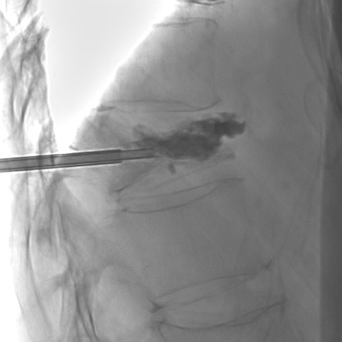
Fluoroscopic Image of Cement Injection During Percutaneous Kyphoplasty. The image shows a needle inserted into the vertebral body, with polymethyl methacrylate cement being injected into the vertebra.
Contributed by M Jaber, MD
(Click Image to Enlarge)

Typical Procedural Steps for Percutaneous Vertebroplasty and Kyphoplasty. The left-sided image for each step represents the anteroposterior fluoroscopic view, and the right-sided image shows the lateral fluoroscopic view.
Contributed by K Margetis, MD, PhD
(Click Image to Enlarge)

Bone Cement Leakage in Vertebroplasty and Kyphoplasty. Left image: Anteroposterior view showing bone cement leakage lateral to the vertebral body (red arrow). Right image: Lateral view showing bone cement leakage inferiorly into the disc space (purple arrow).
Contributed by K Margetis MD, PhD
(Click Image to Enlarge)

Cement Leakage Complications in Vertebroplasty and Kyphoplasty. The left-sided image for each type of cement leakage represents the anteroposterior fluoroscopic view, and the right-sided image shows the lateral fluoroscopic view. (1) Cement leakage superiorly (green) or inferiorly (not shown) into the disc space. This type of leakage is typically tolerated and is visible on both anteroposterior and lateral views. (2) Cement leakage laterally (dangerous-red). This type of leakage is visible only on anteroposterior views. (3) Cement leakage anteriorly (dangerous-red). This type of leakage is visible only on lateral views. (4) Cement leakage posteriorly (dangerous-red). This type of leakage is visible only on lateral views.
Contributed by K Margetis, MD, PhD
References
Jay B, Ahn SH. Vertebroplasty. Seminars in interventional radiology. 2013 Sep:30(3):297-306. doi: 10.1055/s-0033-1353483. Epub [PubMed PMID: 24436552]
Hoyt D, Urits I, Orhurhu V, Orhurhu MS, Callan J, Powell J, Manchikanti L, Kaye AD, Kaye RJ, Viswanath O. Current Concepts in the Management of Vertebral Compression Fractures. Current pain and headache reports. 2020 Mar 20:24(5):16. doi: 10.1007/s11916-020-00849-9. Epub 2020 Mar 20 [PubMed PMID: 32198571]
Bono CM, Heggeness M, Mick C, Resnick D, Watters WC 3rd. North American Spine Society: Newly released vertebroplasty randomized controlled trials: a tale of two trials. The spine journal : official journal of the North American Spine Society. 2010 Mar:10(3):238-40. doi: 10.1016/j.spinee.2009.09.007. Epub 2009 Oct 12 [PubMed PMID: 19822459]
Level 1 (high-level) evidenceGozel T, Ortiz AO. Vertebral Augmentation for Osteoporotic Vertebral Compression Fractures: What is the Current Evidence Pro and Con? Radiologic clinics of North America. 2024 Nov:62(6):979-991. doi: 10.1016/j.rcl.2024.03.004. Epub 2024 Apr 23 [PubMed PMID: 39393856]
Dewar C. Diagnosis and treatment of vertebral compression fractures. Radiologic technology. 2015 Jan-Feb:86(3):301-20; quiz 321-3 [PubMed PMID: 25739109]
Pizones J, Zúñiga L, Sánchez-Mariscal F, Alvarez P, Gómez-Rice A, Izquierdo E. MRI study of post-traumatic incompetence of posterior ligamentous complex: importance of the supraspinous ligament. Prospective study of 74 traumatic fractures. European spine journal : official publication of the European Spine Society, the European Spinal Deformity Society, and the European Section of the Cervical Spine Research Society. 2012 Nov:21(11):2222-31. doi: 10.1007/s00586-012-2403-z. Epub 2012 Jun 22 [PubMed PMID: 22722921]
Barr JD, Jensen ME, Hirsch JA, McGraw JK, Barr RM, Brook AL, Meyers PM, Munk PL, Murphy KJ, O'Toole JE, Rasmussen PA, Ryken TC, Sanelli PC, Schwartzberg MS, Seidenwurm D, Tutton SM, Zoarski GH, Kuo MD, Rose SC, Cardella JF, Society of Interventional Radiology, American Association of Neurological Surgeons, Congress of Neurological Surgeons, American College of Radiology, American Society of Neuroradiology, American Society of Spine Radiology, Canadian Interventional Radiology Association, Society of Neurointerventional Surgery. Position statement on percutaneous vertebral augmentation: a consensus statement developed by the Society of Interventional Radiology (SIR), American Association of Neurological Surgeons (AANS) and the Congress of Neurological Surgeons (CNS), American College of Radiology (ACR), American Society of Neuroradiology (ASNR), American Society of Spine Radiology (ASSR), Canadian Interventional Radiology Association (CIRA), and the Society of NeuroInterventional Surgery (SNIS). Journal of vascular and interventional radiology : JVIR. 2014 Feb:25(2):171-81. doi: 10.1016/j.jvir.2013.10.001. Epub 2013 Dec 8 [PubMed PMID: 24325929]
Level 3 (low-level) evidenceLiu D, Xu J, Wang Q, Zhang L, Yin S, Qian B, Li X, Wen T, Jia Z. Timing of Percutaneous Balloon Kyphoplasty for Osteoporotic Vertebral Compression Fractures. Pain physician. 2023 May:26(3):231-243 [PubMed PMID: 37192225]
Level 2 (mid-level) evidenceClark W, Bird P, Gonski P, Diamond TH, Smerdely P, McNeil HP, Schlaphoff G, Bryant C, Barnes E, Gebski V. Safety and efficacy of vertebroplasty for acute painful osteoporotic fractures (VAPOUR): a multicentre, randomised, double-blind, placebo-controlled trial. Lancet (London, England). 2016 Oct 1:388(10052):1408-1416. doi: 10.1016/S0140-6736(16)31341-1. Epub 2016 Aug 17 [PubMed PMID: 27544377]
Level 1 (high-level) evidenceCarli D, Venmans A, Lodder P, Donga E, van Oudheusden T, Boukrab I, Schoemaker K, Smeets A, Schonenberg C, Hirsch J, de Vries J, Lohle P. Vertebroplasty versus Active Control Intervention for Chronic Osteoporotic Vertebral Compression Fractures: The VERTOS V Randomized Controlled Trial. Radiology. 2023 Jul:308(1):e222535. doi: 10.1148/radiol.222535. Epub [PubMed PMID: 37462495]
Level 1 (high-level) evidencePatel IJ, Rahim S, Davidson JC, Hanks SE, Tam AL, Walker TG, Wilkins LR, Sarode R, Weinberg I. Society of Interventional Radiology Consensus Guidelines for the Periprocedural Management of Thrombotic and Bleeding Risk in Patients Undergoing Percutaneous Image-Guided Interventions-Part II: Recommendations: Endorsed by the Canadian Association for Interventional Radiology and the Cardiovascular and Interventional Radiological Society of Europe. Journal of vascular and interventional radiology : JVIR. 2019 Aug:30(8):1168-1184.e1. doi: 10.1016/j.jvir.2019.04.017. Epub 2019 Jun 20 [PubMed PMID: 31229333]
Level 3 (low-level) evidenceBeall DP, Braswell JJ, Martin HD, Stapp AM, Puckett TA, Stechison MT. Technical strategies and anatomic considerations for parapedicular access to thoracic and lumbar vertebral bodies. Skeletal radiology. 2007 Jan:36(1):47-52 [PubMed PMID: 17013657]
Level 2 (mid-level) evidenceStockton R, Albano J, Lentz J, Ganz M, Grewal K, Katsigiorgis G. A comparison of lumbar transverse pedicle angles between ethnic groups: a retrospective review. BMC musculoskeletal disorders. 2019 Mar 18:20(1):114. doi: 10.1186/s12891-019-2507-2. Epub 2019 Mar 18 [PubMed PMID: 30885189]
Level 2 (mid-level) evidencePark SY, Modi HN, Suh SW, Hong JY, Noh W, Yang JH. Epidural cement leakage through pedicle violation after balloon kyphoplasty causing paraparesis in osteoporotic vertebral compression fractures - a report of two cases. Journal of orthopaedic surgery and research. 2010 Aug 6:5():54. doi: 10.1186/1749-799X-5-54. Epub 2010 Aug 6 [PubMed PMID: 20691094]
Level 3 (low-level) evidenceZhan Y, Jiang J, Liao H, Tan H, Yang K. Risk Factors for Cement Leakage After Vertebroplasty or Kyphoplasty: A Meta-Analysis of Published Evidence. World neurosurgery. 2017 May:101():633-642. doi: 10.1016/j.wneu.2017.01.124. Epub 2017 Feb 10 [PubMed PMID: 28192270]
Level 1 (high-level) evidenceSaracen A, Kotwica Z. Complications of percutaneous vertebroplasty: An analysis of 1100 procedures performed in 616 patients. Medicine. 2016 Jun:95(24):e3850. doi: 10.1097/MD.0000000000003850. Epub [PubMed PMID: 27310966]
Xiao H, Yang J, Feng X, Chen P, Li Y, Huang C, Liang Y, Chen H. Comparing complications of vertebroplasty and kyphoplasty for treating osteoporotic vertebral compression fractures: a meta-analysis of the randomized and non-randomized controlled studies. European journal of orthopaedic surgery & traumatology : orthopedie traumatologie. 2015 Jul:25 Suppl 1():S77-85. doi: 10.1007/s00590-014-1502-4. Epub 2014 Jul 3 [PubMed PMID: 24989933]
Level 2 (mid-level) evidenceWu F, Chen X, Jiang R, Li L, Qin L, Qi W, Hao C, Tang J. Risk factor analysis of adjacent vertebral compression fracture following the surgery of percutaneous kyphoplasty in postmenopausal women. Scientific reports. 2025 Feb 17:15(1):5772. doi: 10.1038/s41598-025-85381-9. Epub 2025 Feb 17 [PubMed PMID: 39962092]
Level 2 (mid-level) evidenceFiranescu CE, de Vries J, Lodder P, Schoemaker MC, Smeets AJ, Donga E, Juttmann JR, Klazen CAH, Elgersma OEH, Jansen FH, van der Horst I, Blonk M, Venmans A, Lohle PNM. Percutaneous Vertebroplasty is no Risk Factor for New Vertebral Fractures and Protects Against Further Height Loss (VERTOS IV). Cardiovascular and interventional radiology. 2019 Jul:42(7):991-1000. doi: 10.1007/s00270-019-02205-w. Epub 2019 Apr 2 [PubMed PMID: 30941490]
Liu D, Wen T, Li X, Xie Z, Wei M, Wang Y, Tang H, Jia Z. Percutaneous Vertebroplasty Versus Balloon Kyphoplasty in the Treatment of Osteoporotic Vertebral Compression Fractures: Evaluating the Overlapping Meta-analyses. Pain physician. 2024 May:27(4):E383-E394 [PubMed PMID: 38805534]
Miller JA, Bowen A, Morisada MV, Margetis K, Lubelski D, Lieberman IH, Benzel EC, Mroz TE. Radiologic and clinical characteristics of vertebral fractures in multiple myeloma. The spine journal : official journal of the North American Spine Society. 2015 Oct 1:15(10):2149-56. doi: 10.1016/j.spinee.2015.05.026. Epub 2015 May 22 [PubMed PMID: 26008684]
Xiao R, Miller JA, Margetis K, Lubelski D, Lieberman IH, Benzel EC, Mroz TE. Predicting the progression of vertebral fractures in patients with multiple myeloma. The spine journal : official journal of the North American Spine Society. 2016 Apr:16(4):510-5. doi: 10.1016/j.spinee.2015.12.014. Epub 2015 Dec 15 [PubMed PMID: 26704858]
Xiao R, Miller JA, Margetis K, Lubelski D, Lieberman IH, Benzel EC, Mroz TE. Radiographic progression of vertebral fractures in patients with multiple myeloma. The spine journal : official journal of the North American Spine Society. 2016 Jul:16(7):822-32. doi: 10.1016/j.spinee.2015.10.033. Epub 2015 Oct 26 [PubMed PMID: 26515398]
Eseonu KC, Panchmatia JR, Streetly MJ, Grauer JN, Fakouri B. The role of Vertebral Augmentation Procedures in the management of vertebral compression fractures secondary to multiple myeloma. Hematological oncology. 2023 Aug:41(3):323-334. doi: 10.1002/hon.3102. Epub 2022 Dec 7 [PubMed PMID: 36440820]