Introduction
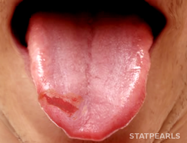
The tongue is a muscular organ essential to a person’s ability to communicate and to experience food via taste, chewing and swallowing. Injuries to the tongue, therefore, have a considerable impact on quality of life. Tongue lacerations can result from a variety of means including seizures, self-harm, blunt force facial trauma, oral trauma while intubated, and child abuse.[1][2][3] There have also been reported cases during electroconvulsive therapy.[4] The use of electronic cigarettes has also led to explosions causing intraoral trauma.[5] The question of which tongue lacerations require repair and which can be allowed to heal by secondary intention is still a topic of debate.[6]
Anatomy and Physiology
Register For Free And Read The Full Article
Search engine and full access to all medical articles
10 free questions in your specialty
Free CME/CE Activities
Free daily question in your email
Save favorite articles to your dashboard
Emails offering discounts
Learn more about a Subscription to StatPearls Point-of-Care
Anatomy and Physiology
Most tongue lacerations are on the anterior dorsum of the tongue followed by the mid-dorsum and anterior ventrum. Posterior tongue lacerations are less common. Complete loss of tissue from the tip or lateral edge of the tongue leaves nothing to be repaired and is unlikely to cause a permanent deficit. After these types of injuries, the tongue will hypertrophy and obscure the deficit over time. An injury to the base of the tongue is potentially more problematic than other locations due to the location of the hypoglossal nerve.[7]
Indications
The decision to suture a tongue laceration depends on the size of the laceration or the gaping nature of the wound. Performing tongue assessment is best when the tongue is at rest inside the mouth as this is the most common position, as opposed to protruding outside the mouth. Complex lacerations are those that involve large flaps, active bleeding or through-and-through injuries of the tongue and are more likely to require repair. Lacerations or avulsions that are small can potentially be left to heal without intervention due to the tongue’s ability to bulk up via hypertrophy. The ability to hypertrophy can often fill in the gaps of the wound so that no deficit is identifiable.[8]
Contraindications
Contraindication to repair in the emergency department would include a significant delay in presentation after the injury in which case the wound can be allowed to heal secondarily. Tongue amputation requires specialist repair and is not suitable for emergency medicine or primary care independent management.
Equipment
Required equipment could include the following:
- Suction
- Gauze
- Laceration repair kit/Instruments: needle driver, scissors
- Anesthetic (topical, local, block): Lidocaine with or without epinephrine, LET, bupivacaine with or without epinephrine
- Normal saline
- Needles
- Syringes
- Absorbable suture: 3-0, 4-0 chromic gut or vicryl
- Tongue depressor
- Bite block
*Repair in children may also require procedural sedation, or intranasal pain medications to facilitate the procedure.[8]
Personnel
Personnel could include an emergency medicine physician, advanced practice provider, oral maxillofacial surgeon, ear nose and throat specialist, other specialist or nursing staff depending on the complexity of the injury, patient population, and resources available.
Preparation
Once wound assessment is complete and a decision is made to repair the wound, it should undergo inspection for retained foreign bodies; specifically, broken pieces of teeth are a concern. The wound should be irrigated thoroughly and hemostasis achieved before repair.[7] The patient should receive appropriate pain medication, local anesthetic, or sedation as needed. Extensive lacerations, avulsions, or amputation may require general anesthesia for repair by a specialist in the OR. An inferior alveolar nerve block or lingual nerve block can be used to anesthetize the anterior two-thirds of the tongue.[9] A piece of gauze can be used to hold the tongue outside the mouth, or a silk suture can be placed through the tongue and the strands used to manipulate it during repair.
Technique or Treatment
Large lacerations may require deep sutures or layered repair to relieve muscle tension and allow approximation.[7] Layered repair can also help prevent a hematoma from forming. The suture used requires tensile strength given the mobility and tension it will experience as part of normal tongue function. A literature review showed the use of vicryl and chromic gut as most common. Sutures were placed to approximate edges, but tied loose enough to allow for swelling and prevent tissue necrosis. In addition to sutures, one case report detailed the use of dermabond for wound approximation in a seven-year-old. While dermabond does not have approval for use on oral mucosa, it was used in this case as the child’s mother was resistant to procedural sedation or local anesthetic. A piece of gauze was used to hold and dry the child’s tongue. Additional pressurized air was used to dry the tongue immediately prior to dermabond use. At next day and two-week wound checks, the dermabond had held and achieved a well approximated and healed wound. The laceration in the case report was located to the dorsum of the tongue and did not involve an edge.[10]
Complications
Sutures in tongue lacerations with a non-absorbable suture can develop a granuloma because of a foreign body reaction. As with all laceration repairs, there is a possibility of a noticeable scar, but this is also possible in patients that heal by secondary intention.[8] Literature review shows a discussion of the risk of infection in tongue lacerations. The use of antibiotics is controversial with the ultimate decision being left to the physician as no clear guidelines exist at present.[11][12] Criteria that may favor prophylactic antibiotics include immunocompromised state, heavily contaminated wounds, and delayed closure. Antibiotics of choice should cover gram-positive and anaerobic organisms.[13] Other complications include swelling, impaired speech, impaired swallowing or even difficulty breathing if swelling or hemorrhage is significant.
Clinical Significance
Tongue lacerations can occur in any age group with a variety of injuries making the development of a standard of care difficult. Initial assessment for severity of injury, contamination, and barriers to healing can be considered similar to lacerations on other parts of the body. The decision to repair a tongue laceration or allow it to heal by secondary intention has been a hotly debated topic. A large population study comparing the two management methods does not exist, so the decision has been left up to individual clinicians. General descriptions of suture repair and the use of dermabond in repair exist, as well as examples of well-healed wounds that received no intervention. The Zurich Tongue Scheme aims to give the practitioner an idea of factors that favor repair, ways to repair and common complications to consider when they need to care for a patient with a tongue laceration.[8]
Enhancing Healthcare Team Outcomes
From minor tongue lacerations to tongue amputation, the repair of a tongue injury requires a range of abilities and specialties to achieve a good result. More complex lacerations will need the primary physician, emergency department physician, and nurse practitioner to adequately communicate the nature of the wound to advanced care specialists such as otolaryngologist and oral maxillofacial surgeons. Lacerations in children might require the teamwork of physician, nursing and respiratory therapist to sedate and repair the wound. The pharmacy may have involvement depending on the decision to use antibiotics prophylactically. The lack of large trial or comparison studies on the repair of tongue lacerations means that communication between providers on past experiences will be an integral part of each new patient's plan of care as part of an interprofessional team approach. [Level V]
Media
References
George CLS, Theesfeld SSN, Wang Q, Hudson MJ, Harper NS. Identification and Characterization of Oral Injury in Suspected Child Abuse Cases: One Health System's Experience. Pediatric emergency care. 2021 Oct 1:37(10):494-497. doi: 10.1097/PEC.0000000000001715. Epub [PubMed PMID: 30601344]
Level 3 (low-level) evidenceBeena JP. Management of tongue and lip laceration due to dystonia in a 1-year-old infant. Journal of the Indian Society of Pedodontics and Preventive Dentistry. 2017 Jan-Mar:35(1):90-93. doi: 10.4103/0970-4388.199223. Epub [PubMed PMID: 28139490]
Costacurta M, Benavoli D, Arcudi G, Docimo R. Oral and dental signs of child abuse and neglect. ORAL & implantology. 2015 Apr-Sep:8(2-3):68-73. doi: 10.11138/orl/2015.8.2.068. Epub 2016 Jul 25 [PubMed PMID: 27555907]
Woo SW, Do SH. Tongue laceration during electroconvulsive therapy. Korean journal of anesthesiology. 2012 Jan:62(1):101-2. doi: 10.4097/kjae.2012.62.1.101. Epub 2012 Jan 25 [PubMed PMID: 22323965]
Vaught B, Spellman J, Shah A, Stewart A, Mullin D. Facial trauma caused by electronic cigarette explosion. Ear, nose, & throat journal. 2017 Mar:96(3):139-142 [PubMed PMID: 28346645]
Lamell CW, Fraone G, Casamassimo PS, Wilson S. Presenting characteristics and treatment outcomes for tongue lacerations in children. Pediatric dentistry. 1999 Jan-Feb:21(1):34-8 [PubMed PMID: 10029965]
Das UM, Gadicherla P. Lacerated tongue injury in children. International journal of clinical pediatric dentistry. 2008 Sep:1(1):39-41. doi: 10.5005/jp-journals-10005-1007. Epub 2008 Dec 26 [PubMed PMID: 25206087]
Level 3 (low-level) evidenceSeiler M, Massaro SL, Staubli G, Schiestl C. Tongue lacerations in children: to suture or not? Swiss medical weekly. 2018 Oct 22:148():w14683. doi: 10.4414/smw.2018.14683. Epub 2018 Oct 28 [PubMed PMID: 30378089]
Balasubramanian S, Paneerselvam E, Guruprasad T, Pathumai M, Abraham S, Krishnakumar Raja VB. Efficacy of Exclusive Lingual Nerve Block versus Conventional Inferior Alveolar Nerve Block in Achieving Lingual Soft-tissue Anesthesia. Annals of maxillofacial surgery. 2017 Jul-Dec:7(2):250-255. doi: 10.4103/ams.ams_65_17. Epub [PubMed PMID: 29264294]
Kazzi MG, Silverberg M. Pediatric tongue laceration repair using 2-octyl cyanoacrylate (dermabond(®)). The Journal of emergency medicine. 2013 Dec:45(6):846-8. doi: 10.1016/j.jemermed.2013.05.004. Epub 2013 Jul 1 [PubMed PMID: 23827167]
Level 3 (low-level) evidenceSteele MT, Sainsbury CR, Robinson WA, Salomone JA 3rd, Elenbaas RM. Prophylactic penicillin for intraoral wounds. Annals of emergency medicine. 1989 Aug:18(8):847-52 [PubMed PMID: 2502938]
Level 1 (high-level) evidenceMark DG, Granquist EJ. Are prophylactic oral antibiotics indicated for the treatment of intraoral wounds? Annals of emergency medicine. 2008 Oct:52(4):368-72 [PubMed PMID: 18819178]
Armstrong BD. Lacerations of the mouth. Emergency medicine clinics of North America. 2000 Aug:18(3):471-80, vi [PubMed PMID: 10967735]
