 Anatomy, Bony Pelvis and Lower Limb: Thigh Semitendinosus Muscle
Introduction
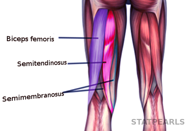
The semitendinosus muscle is a member of the posterior component of the thigh, which also includes the biceps femoris and the semimembranosus muscles. These three muscles together are collectively referred to as the hamstring muscle complex, which serves as hip extensors and knee flexors that are integral to gait and running.[1][2] Injuries to the hamstring muscle complex are commonly seen in athletes and are usually brought on through explosive or high-speed running.
Structure and Function
Register For Free And Read The Full Article
Search engine and full access to all medical articles
10 free questions in your specialty
Free CME/CE Activities
Free daily question in your email
Save favorite articles to your dashboard
Emails offering discounts
Learn more about a Subscription to StatPearls Point-of-Care
Structure and Function
The semitendinosus, just medial to the biceps femoris, arises from three locations. The medial border of the ischial tuberosity, the medial border of the proximal tendon of the long head of the biceps femoris, and the proximal aponeurosis.[3] Distal from the ischial tuberosity, the semitendinosus becomes fusiform until it reaches below the middle of the thigh. Here it transitions into a long tendon that also serves as the medial border of the popliteal fossa.[4] The tendon then curves over the pes anserine bursa, which is superficial to the medial collateral ligament, and inserts into the superior anteromedial surface of the tibia. The sartorius and gracilis muscles also terminate at this common insertion point to make the pes anserinus. The attachment of the semitendinosus is most posterior behind the tendinous insertions of the sartorius and gracilis muscles.[5][6][7]
The semitendinosus muscle, collectively with the other two muscles of the posterior compartment of the thigh, works to extend at the hip and flex at the knee. The semitendinosus muscle, in particular, has the added functionality of assisting the popliteus muscle in rotating the leg internally.
Embryology
The gastrulation phase of the embryo gives rise to three germ layers: the ectoderm, endoderm, and mesoderm. The paraxial mesoderm specifically develops into striated skeletal muscle and the skeleton except for the skull.[8] Therefore, the bony insertions of the semitendinosus muscle and the muscle itself derive from the mesoderm. As a whole, the lower limb begins to form towards the end of the fourth week of the embryonic period. The limb continuously develops and becomes well-differentiated towards the eighth week as the embryo transitions to the fetal period.[9]
Blood Supply and Lymphatics
The semitendinosus muscle predominantly receives vascular supply from the profunda femoris artery, also known as the deep artery of the thigh, which is the largest branch of the femoral artery. Specifically, the perforating branches of the profunda femoris artery are responsible for the vascular supply of the muscle.[10][11] Similarly, vascular drainage of the semitendinosus muscle is by the perforating veins of the profunda femoris vein, which drains into the femoral vein.[12]
Nerves
The tibial nerve, an offshoot of the sciatic nerve, innervates the semitendinosus muscle. Proximally, the sacral plexus branches into the sciatic nerve, which involve nerve roots from L4 to S2. The sciatic nerve then travels through the posterior compartment, which branches into the common fibular nerve and the tibial nerve. The tibial nerve, either before or after its separation from the common fibular nerve, innervates the semitendinosus and all other muscles of the posterior compartment of the thigh except for the short head of the biceps femoris.[13]
Muscles
The semitendinosus muscle accompanies the biceps femoris muscle and the semimembranosus muscle in the posterior compartment of the thigh. These three muscles compose the hamstring muscle complex. The semimembranosus muscle lies deep to the semitendinosus muscle and is the most medial muscle of the posterior compartment of the thigh.
The semimembranosus muscle originates from the superolateral aspect of the ischial tuberosity and inserts into the medial tibial condyle, posterior oblique ligament, the posterior joint capsule, and arcuate ligament.
The biceps femoris muscle is the most lateral muscle in the posterior compartment, with the long head originating from the ischial tuberosity and the short head rising from the lateral lip of the linea aspera of the femur. The biceps femoris then inserts into the head of the fibula and lateral condyle of the tibia.
Physiologic Variants
Case studies have reported anatomical variations of the semitendinosus muscle. One such case was during an anterior cruciate ligament reconstructive surgery where during graft harvesting, the semitendinosus muscle had given off an additional tendinous attachment. Along with the original insertion of the semitendinosus muscle, the muscle gave off an extra tendinous slip that was attached to the gracilis tendon at its insertion point.[14] The gracilis also followed suit with an additional tendinous insertion into the semitendinosus tendon. Therefore, there was a “double” pes anserinus. There have also been reports of the semitendinosus tendon inserting into the crural fascia of the leg rather than the superior anteromedial tibia.[15]
Surgical Considerations
The semitendinosus and gracilis muscle tendons are commonly used as an alternative to the bone-patellar-tendon-bone graft (BPTB) for ACL reconstruction. When grafting for ACL reconstruction, the sartorius fascia is incised to expose the underlying semitendinosus and gracilis tendon. These two tendons are not adherent to the bone outside of the attachment sites, which allows differentiation of the tendons from the medial collateral ligament, which lies deep to the semitendinosus and gracilis muscles. Compared to the BPTB graft, the diameter of the hamstring graft is predetermined by the original diameter of the semitendinosus and gracilis tendons. In the BPTB graft, the diameter of the graft can be determined by the surgeon during the harvesting of the graft. Unlike the hamstring autograft, the BPTB graft includes bone which is believed to improve graft incorporation.[16][17]
The BPTB graft is the most common autograft used in the United States and has originally been considered the gold-standard graft for ACL reconstruction. However, there is a debate about which autograft is superior. Hamstring autografts have the benefits of decreased anterior knee pain, osteoarthritis, and donor site morbidity. The benefits of the BPTB autograft include faster graft incorporation, reduced risk of rupture, and a larger proportion of patients returning to preinjury activity levels. Despite BTBP having a lower failure rate, both graft choices have low failure rates. Therefore, utilizing the semitendinosus and gracilis tendons as an autograft remains a reasonable option for reconstruction, and its use should be evaluated on a case-by-case basis.[18]
MRI studies have also shown that the semitendinosus tendon has also been found to regenerate in 75% of patients after harvesting, with the remaining 25% of patients having an increase in the size of the semimembranosus muscle as compensation. Outside the common use of the semitendinosus and gracilis tendon grafts in ACL reconstruction, these tendons have also found use in repairing massive rotator cuff tears that have already undergone retraction and degeneration and muscle atrophy.[19]
Clinical Significance
Hamstring injuries are among the most frequently encountered muscle injuries in athletes and correlate with significant time away from the sport. Injuries typically occur during rapid acceleration or high-speed running. The biceps femoris generally is the most commonly injured muscle of the posterior compartment; however, the semitendinosus muscle has also been implicated in hamstring strains.[20]
Media
References
Koulouris G, Connell D. Hamstring muscle complex: an imaging review. Radiographics : a review publication of the Radiological Society of North America, Inc. 2005 May-Jun:25(3):571-86 [PubMed PMID: 15888610]
Pérez-Bellmunt A, Miguel-Pérez M, Brugué MB, Cabús JB, Casals M, Martinoli C, Kuisma R. An anatomical and histological study of the structures surrounding the proximal attachment of the hamstring muscles. Manual therapy. 2015 Jun:20(3):445-50. doi: 10.1016/j.math.2014.11.005. Epub 2014 Nov 21 [PubMed PMID: 25515332]
Woodley SJ, Mercer SR. Hamstring muscles: architecture and innervation. Cells, tissues, organs. 2005:179(3):125-41 [PubMed PMID: 15947463]
Beltran L, Ghazikhanian V, Padron M, Beltran J. The proximal hamstring muscle-tendon-bone unit: a review of the normal anatomy, biomechanics, and pathophysiology. European journal of radiology. 2012 Dec:81(12):3772-9. doi: 10.1016/j.ejrad.2011.03.099. Epub 2011 Apr 27 [PubMed PMID: 21524864]
Lee JH, Kim KJ, Jeong YG, Lee NS, Han SY, Lee CG, Kim KY, Han SH. Pes anserinus and anserine bursa: anatomical study. Anatomy & cell biology. 2014 Jun:47(2):127-31. doi: 10.5115/acb.2014.47.2.127. Epub 2014 Jun 20 [PubMed PMID: 24987549]
Ridley WE, Xiang H, Han J, Ridley LJ. Pes anserinus: Normal anatomy. Journal of medical imaging and radiation oncology. 2018 Oct:62 Suppl 1():148. doi: 10.1111/1754-9485.23_12786. Epub [PubMed PMID: 30309084]
Zhong S, Wu B, Wang M, Wang X, Yan Q, Fan X, Hu Y, Han Y, Li Y. The anatomical and imaging study of pes anserinus and its clinical application. Medicine. 2018 Apr:97(15):e0352. doi: 10.1097/MD.0000000000010352. Epub [PubMed PMID: 29642176]
Tickle C. How the embryo makes a limb: determination, polarity and identity. Journal of anatomy. 2015 Oct:227(4):418-30. doi: 10.1111/joa.12361. Epub 2015 Aug 7 [PubMed PMID: 26249743]
Farfán E, Rojas S, Olivé-Vilás R, Rodríguez-Baeza A. Morphological study on the origin of the semitendinosus muscle in the long head of biceps femoris. Scandinavian journal of medicine & science in sports. 2021 Dec:31(12):2282-2290. doi: 10.1111/sms.14045. Epub 2021 Sep 12 [PubMed PMID: 34472147]
Davis DD, Ginglen JG, Kwon YH, Kahwaji CI. EMS Traction Splint. StatPearls. 2023 Jan:(): [PubMed PMID: 29939619]
de Athayde Soares R, Matielo MF, Brochado Neto FC, Martins Cury MV, Matoso Chacon AC, Nakamura ET, Sacilotto R. The importance of the superficial and profunda femoris arteries in limb salvage following endovascular treatment of chronic aortoiliac occlusive disease. Journal of vascular surgery. 2018 Nov:68(5):1422-1429. doi: 10.1016/j.jvs.2018.02.052. Epub 2018 May 24 [PubMed PMID: 29804745]
Repella TL, Lopez O, Abraham CZ, Azarbal AF, Liem TK, Mitchell EL, Landry GJ, Moneta GL, Jung E. Characterization of profunda femoris vein thrombosis. Journal of vascular surgery. Venous and lymphatic disorders. 2018 Sep:6(5):585-591. doi: 10.1016/j.jvsv.2018.01.012. Epub 2018 Apr 19 [PubMed PMID: 29681458]
Kouzaki K, Nakazato K, Mizuno M, Yonechi T, Higo Y, Kubo Y, Kono T, Hiranuma K. Sciatic Nerve Conductivity is Impaired by Hamstring Strain Injuries. International journal of sports medicine. 2017 Oct:38(11):803-808. doi: 10.1055/s-0043-115735. Epub 2017 Sep 11 [PubMed PMID: 28895622]
Patel S, Trehan RK, Railton GT. Successful ACL reconstruction with a variant of the pes anserinus. Journal of orthopaedics and traumatology : official journal of the Italian Society of Orthopaedics and Traumatology. 2009 Dec:10(4):203-5. doi: 10.1007/s10195-009-0075-1. Epub 2009 Nov 18 [PubMed PMID: 19921482]
Level 3 (low-level) evidenceRizvi A, Iwanaga J, Oskouian RJ, Loukas M, Tubbs RS. Additional Attachment of the Semitendinosus and Gracilis Muscles to the Crural Fascia: A Review and Case Illustration. Cureus. 2018 Aug 7:10(8):e3116. doi: 10.7759/cureus.3116. Epub 2018 Aug 7 [PubMed PMID: 30338191]
Level 3 (low-level) evidenceCharalambous CP, Kwaees TA. Anatomical considerations in hamstring tendon harvesting for anterior cruciate ligament reconstruction. Muscles, ligaments and tendons journal. 2012 Oct:2(4):253-7 [PubMed PMID: 23738306]
Eriksson K, Hamberg P, Jansson E, Larsson H, Shalabi A, Wredmark T. Semitendinosus muscle in anterior cruciate ligament surgery: Morphology and function. Arthroscopy : the journal of arthroscopic & related surgery : official publication of the Arthroscopy Association of North America and the International Arthroscopy Association. 2001 Oct:17(8):808-17 [PubMed PMID: 11600977]
Samuelsen BT, Webster KE, Johnson NR, Hewett TE, Krych AJ. Hamstring Autograft versus Patellar Tendon Autograft for ACL Reconstruction: Is There a Difference in Graft Failure Rate? A Meta-analysis of 47,613 Patients. Clinical orthopaedics and related research. 2017 Oct:475(10):2459-2468. doi: 10.1007/s11999-017-5278-9. Epub [PubMed PMID: 28205075]
Level 1 (high-level) evidenceGigante A, Bottegoni C, Milano G, Riccio M, Dei Giudici L. Semitendinosus and gracilis free muscle-tendon graft for repair of massive rotator cuff tears: surgical technique. Joints. 2016 Jul-Sep:4(3):189-192 [PubMed PMID: 27900313]
Cooper DE, Conway JE. Distal semitendinosus ruptures in elite-level athletes: low success rates of nonoperative treatment. The American journal of sports medicine. 2010 Jun:38(6):1174-8. doi: 10.1177/0363546509361016. Epub 2010 Mar 29 [PubMed PMID: 20351198]
Level 2 (mid-level) evidence