Introduction
Riehl melanosis is an acquired pigmentary disorder that predominantly affects darker-skinned individuals, particularly older women. This condition is characterized by brown-gray reticulated-to-diffuse hyperpigmented macules or patches on the face, neck, and upper chest (see Image. Clinical Photographs of Riehl Melanosis).[1] The etiology is presumed to be a type IV hypersensitivity reaction triggered by contact allergens such as personal fragrances, textiles, and cosmetics. However, genetic factors, autoimmune conditions, and UV light exposure may also contribute to the condition's development.[2]
Despite the distinctive clinical presentation of Riehl melanosis, a thorough evaluation is necessary for its differential diagnosis, which may involve various techniques such as skin biopsy, patch testing, dermoscopy, reflectance confocal microscopy, or newer diagnostic tools following an initial clinical examination of the patients. These tools are crucial for diagnosing and distinguishing Riehl melanosis from other differential diagnoses, including conditions such as lichen planus pigmentosus and erythema dyschromicum perstans, all falling under the spectrum of acquired dermal macular hyperpigmentation. Histopathological examination of a skin biopsy will reveal basal layer vacuolar degeneration accompanied by colloid bodies and pigment incontinence.[3]
Apart from allergen avoidance, management of this condition involves sun protection and various tailored regular skincare measures. Treatment options include topical therapies (such as hydroquinone, azelaic acid, and retinoids), oral therapies (such as tranexamic acid, glycyrrhizin, and mycophenolate mofetil), chemical peels (such as salicylic acid and glycolic acid), and lasers and light therapy (such as 1064-nm quality-switched neodymium-doped yttrium aluminum garnet laser and intense pulsed light) to meet individual patient needs.[4] Regular monitoring using the dermal pigmentation area and severity index (DPASI) is necessary to gauge treatment efficacy and mitigate potential psychosocial impacts. This involves ensuring regular observation for improvement of hyperpigmentation, which can have deleterious effects if left untreated, particularly in patients with Riehl melanosis.[5]
Riehl melanosis was first described by Gustav Riehl in 1917, who observed striking rough, indurated brown-gray pigmentation on the forehead, temporal, and zygomatic regions on patients' faces (see Image. Riehl Melanosis on the Left Cheek).[6] Initially, it was believed to be related to nutritional substitutions resulting from wartime food measures, such as poor-quality flour. However, it later became evident that it was also present on the faces of women who used cosmetic products.[6][7] Consequently, the condition transitioned from being termed war dermatosis to melanosis faciei feminae, eventually becoming known simply as Riehl melanosis.[1]
Riehl melanosis belongs to a group of conditions collectively referred to as acquired dermal macular hyperpigmentation (previously known as macular pigmentation of uncertain etiology). This group may encompass Riehl melanosis, erythema dyschromicum perstans, lichen planus pigmentosus, melasma, exogenous ochronosis, idiopathic macular eruptive pigmentation, and pigmented contact dermatitis.[2][8] The acquired dermal macular pigmentation disorders have similar clinical and histopathological findings.[9]
Pigmented contact dermatitis is considered a variant of Riehl melanosis, with an identified contact allergen, such as cosmetics like kumkum (see Image. Kumkum-Induced Pigmented Contact Dermatitis).[9] Although pigmented contact dermatitis shares similarities with Riehl melanosis, the extent of inflammation observed creates distinctions between them. Some experts consider pigmented contact dermatitis and Riehl melanosis identical; however, this view may be controversial, as pigmented contact dermatitis typically exhibits minimal to no inflammation compared to Riehl melanosis. Thus, this group of conditions may be classified as acquired dermal macular pigmentation with or without contact sensitization.[1] Please see StatPearls' companion resource, "Contact Dermatitis," for further information.
Etiology
Register For Free And Read The Full Article
Search engine and full access to all medical articles
10 free questions in your specialty
Free CME/CE Activities
Free daily question in your email
Save favorite articles to your dashboard
Emails offering discounts
Learn more about a Subscription to StatPearls Point-of-Care
Etiology
The etiology of Riehl melanosis is unknown, but many intrinsic and extrinsic factors have been proposed.[1] These factors include genetic predisposition to Riehl melanosis, predilection for autoimmune conditions, type IV hypersensitivity reactions, and UV light exposure.[10][11][12][13][14]
Etiopathogenesis of Riehl Melanosis
Although not all features must be present for Riehl melanosis, the below-mentioned factors may contribute to its etiopathogenesis.
Type IV hypersensitivity reaction: Riehl melanosis typically arises in areas of fragrance or cosmetic product application, suggesting allergic contact dermatitis as its primary cause.[1] The presence of basal cell liquefaction and degeneration on reflectance confocal microscopy implies a chronic inflammatory state that may surpass the more commonly encountered contact dermatitis.[15] Inflammatory changes are believed to disturb melanin synthesis, distribution, and degradation, resulting in dispersed melanin granules within the superficial dermis, which are subsequently phagocytosed by melanophages, leading to pigment incontinence on histopathology.[3] This reaction is associated with a stratum basale cytolytic reaction rather than an eczematous stratum spinosum reaction.[7]
Genetic factors: Elevated guanine deaminase function is observed in patients with Riehl melanosis, correlating with upregulation of stem cell factor, endothelin-1, c-kit gene products, and estrogen and progesterone receptors.[11] Next-generation sequencing has identified increased guanine deaminase function in Riehl melanosis, potentially predisposing individuals to basement membrane or keratinocyte damage.[16]
Autoimmune association: The significant correlation between Riehl melanosis and concurrent autoimmune conditions suggests a potential autoimmune association with Riehl melanosis. Riehl melanosis has been associated with Sjögren syndrome, AIDS, systemic lupus erythematosus, and thyroid disease.[10][1] Riehl melanosis has also been associated with anti-SSA (anti-Ro) antibodies. Notably, it has been postulated that these antibodies on keratinocytes destabilize the extracellular matrix and predispose patients to immune dysregulation.[17][1][18] Additionally, Riehl melanosis may exhibit immune dysregulation through alterations in human leukocyte antigen (HLA), particularly in HLA-A2, HLA-DPA1, and HLA-DPB1. Other proposed pathways include the activation of T-cells with CD8 and CD45RO markers, adrenocorticotropin-related hormone, or melanocortin-1 receptor (MC1R).[1][18]
UV light exposure: The photodistributed presentation of Riehl melanosis has led to speculation about its relationship with the etiopathogenesis of the condition. It is suggested that it may be associated with photosensitization of allergens, as photopatch testing has been observed to replicate pigmented reactions similar to Riehl melanosis.[10][19] UV light exposure may activate dermal fibroblast CCN1 protein, which in turn induces melanogenesis by binding to integrin α6β1, p38 mitogen-activated protein kinase, and extracellular signal-regulated kinases (ERK) 1 and 2. These signaling cascades activate microphthalmia-associated transcription factor (MITF), tyrosinase, and tyrosinase-related protein-1 (TRP-1).[1][12]
Several contact allergens, including airborne allergens, have been associated with Riehl melanosis and are listed in the table below (see Table).[20][21][7][22] Additional allergens, including medications (such as minoxidil 5%) and endogenous factors (such as digestive disorders), have also been implicated.[22][6][23]
Table. Commons Allergens in Riehl Melanosis
| Sources of Allergens | Common Allergens |
| Cosmetics |
|
| Fragrance |
|
| Textile |
|
| Occupation |
|
Epidemiology
A typical patient with Riehl melanosis is an older woman with a darker skin type who applies cosmetic products to her face or neck. Riehl melanosis is predominantly observed in women than men and has been noted to correspond with the reproductive cycle, suggesting hormonal mediation.[25] The condition is particularly observed to be prevalent in individuals with darker skin tones, notably those of Asian or Hispanic origin.[26] Riehl melanosis is more frequently encountered in older individuals, often associated with practices such as hair coloring with henna, and is rare among younger patients.[13] Although precise data on the incidence and prevalence of Riehl melanosis remain unknown, evidence suggests an increasing trend.[27]
Histopathology
A skin biopsy is considered the gold standard for diagnosing Riehl melanosis, although in some cases, a thorough clinical history may be adequate without the need for a biopsy.[1] When a skin biopsy is performed, histopathological features characteristic of Riehl melanosis may include the following findings:[28][29][3]
- Basal vacuolar degeneration with colloid bodies
- Pigment incontinence with pilosebaceous involvement
- Increased dermal melanophages and increased epidermal melanocytes
- Perivascular, epidermal, or dermal inflammatory infiltrates (such as mononuclear cells, eosinophils, and lymphocytes)
- Dilated blood vessels
- Interface changes
- Hypergranulosis (although more common in lichen planus pigmentosus)
- Epidermal spongiosis
Perilesional skin may show similar features but to a lesser extent. Generally, the histopathology of Riehl melanosis may be nonspecific. The use of melanin stains (such as Melan-A and Fontana-Masson) may reveal increased melanin deposition.[29]
History and Physical
Patients with Riehl melanosis may present with erythema, edema, and pruritus preceding pigmentation or with pigmentation alone. This initial inflammatory phase putatively underlies the onset of allergic contact dermatitis.[20][30] Upon physical examination, patients may exhibit patchy-to-diffuse hyperpigmentation, which may appear scaly and accompanied by nearby satellite perifollicular pigmented macules. These macules are typically a few millimeters in size, in contrast to the larger patches commonly observed in conditions such as lichen planus pigmentosus and erythema dyschromicum perstans (also known as ashy dermatosis).[1] A reticulated pattern is often evident at the center of the pigmented areas.[20][7] The shades of pigmentation, which may vary from brown to blue or gray, depend on factors such as severity, skin type, and causal agents (see Image. Riehl Melanosis on Bilateral Cheeks).[20][7]
The usual presentation of Riehl melanosis occurs on the face, with the forehead, temple, or zygomatic areas being the most frequently affected regions. In cases associated with textiles, Riehl melanosis manifests on the anterior thighs or axillae while sparing the vault.[20][22] When the implicated allergens are photosensitizers, pigmentation tends to be more pronounced in sun-exposed areas such as the chest, neck, scalp, or hands.[31]
Evaluation
Diagnosing and evaluating Riehl melanosis can pose challenges. Diagnostic tools include skin biopsy, dermoscopy, reflectance confocal microscopy, and patch testing. While a skin biopsy may aid in diagnosing Riehl melanosis, patients may decline due to concerns about scarring or undesirable cosmetic outcomes, especially in cosmetically sensitive areas affected by the condition. Skin biopsies are often unnecessary or inappropriate if a contact allergen can be identified.[1] Ongoing research explores newer diagnostic tools, including serum biomarkers, such as guanine deaminase, and optical coherence tomography. Cellular resolution optical coherence tomography imaging can reveal melanocyte capping, basement membrane degeneration, telangiectasias, and dermal melanophages.[32]
DermoscopyDermoscopy reveals pseudo networks characterized by brown or gray dots and globules arranged in an arcuate, semiarcuate, or hexagonal pattern.[33] Additionally, telangiectasias, scale, perifollicular hypopigmented halos, and follicular keratotic plugs have been observed.[3] A grading scale based on dermoscopy categorizes the dermoscopic features of Riehl melanosis into 4 grades, which include:[5]
- Grade 1: Randomly distributed sparse dots with light brown macules and patches.
- Grade 2: Dots with occasional globules, appearing as broken or semiarcuate formations.
- Grade 3: Reticulate dots and globules with occasional blotches.
- Grade 4: Thick spots, globules, and blotches with pigment network destruction, clinically presenting as widespread brown-black discoloration
Reflectance Confocal MicroscopyUpon correlation with conventional histopathologic examination, findings consistent with disorders of increased pigmentation are observed.[28] These findings include multiple bright, refractile oval-to-stellate melanophages and basal layer liquefactive changes, which are characteristic of acquired dermal macular hyperpigmentation.[1][34] Reflectance confocal microscopy mirrors the following features seen in histopathology:[28][15]
- Epidermal or superficial dermal inflammatory cell infiltrates, such as monocytes.
- Basal layer vacuolar degeneration with obscured papillary rims and obliteration of the ring-like dermal papillae.
- Pigment incontinence.
- Dilated vessels with prominent surrounding linear or round canalicular structures.
- Dilated, hyperkeratotic infundibula with highly refractive material
Patch TestingPatch testing is recommended to identify a potential causative agent that patients may continue to use, promoting low-level chronic inflammation consistent with the condition.[3] Closed patch testing should be performed with standard series, cosmetic series, fragrance series, and other patient-provided products.[1] For patients with suspected photoallergy, photopatch testing should be pursued. Patch testing has been shown to identify the cause of Riehl melanosis in approximately 80% of cases.[3] Standard closed-patch testing may prove inadequate for diagnosis due to the low allergen concentration. In cases where patients exhibit a negative test result but maintain a high clinical suspicion for Riehl melanosis or when the result is equivocal, a repeated open application test (ROAT) is recommended.[1] If the ROAT yields equivocal results on the forearm, further evaluation on the face or other affected areas should be conducted.[22]
MonitoringClinical improvement can be measured with the DPASI, an objective scoring system of 6 facial segments: forehead, left cheek, right cheek, central face (from the glabella to the chin extending laterally from the medial canthus of the eye to the lateral edges of the oral commissure).[35] In each segment, dermoscopy grading (which is correlated with histopathologic findings) on a 1 to 4 grading scale is multiplied by a factor based on the surface area of the segment. The DPASI was validated in a study of 55 patients as a reliable indicator of acquired dermal macular hyperpigmentation severity.[9]
The following calculation results in a score ranging from 0 to 40:
DPASI = Forehead + Left cheek + Right cheek + Central face + Left neck + Right neck, where
- Forehead = % forehead involvement × dermoscopy grade × 2
- Left cheek = % left cheek involvement × dermoscopy grade × 2
- Right cheek = % right cheek involvement × dermoscopy grade × 2
- Central face = % central face involvement × dermoscopy grade × 1
- Left neck = % left neck involvement × dermoscopy grade × 1.5
- Right neck = % right neck involvement × dermoscopy grade × 1.5 [36][35]
Treatment / Management
All individuals with Riehl melanosis should avoid using products containing identified allergens and adhere to regular sun protection measures.[13] Although several therapeutic approaches have been proposed with variable results, a definitive treatment or cure for Riehl melanosis is yet to be established, and treatment outcomes may vary depending on the chosen modality.[20][23] Studied treatment modalities include both medical and procedural treatments.(B3)
Medical Therapies
Topical therapies encompass a range of options, including hydroquinone (2% or 4% cream), retinoids, topical corticosteroids, kojic acid, glycolic acid, azelaic acid, and lignin peroxidase.[4][1][23][1] Lignin peroxidase is an enzyme derived from Phanerochaete chrysosporium, a fungus in woody plants, which exhibits melanin decolorization properties.[37] In a split-face study involving 60 women with facial dyspigmentation, lignin peroxidase demonstrated notable improvements in skin texture, roughness, and overall appearance by week 2, with a reduction in spot size observed by week 12 compared to untreated areas. Compared to hydroquinone, lignin peroxidase exhibited similar efficacy but showed superior outcomes in skin texture and roughness.[38] Lignin peroxidase lotion has demonstrated efficacy in treating melasma, with reported improvements in luminance.[39] In a randomized, double-blind, placebo-controlled, split-face single-center study involving 51 patients, the use of lignin peroxidase cream twice daily significantly reduced the melanin index compared to both placebo and 2% hydroquinone cream.[40](A1)
A few oral therapies used to treat Riehl melanosis are listed below.
Tranexamic acid: Tranexamic acid is a lysine derivative utilized in the treatment of Riehl melanosis functions by obstructing melanocyte-keratinocyte interactions through the plasmin-plasminogen system. This acid putatively inhibits melanin synthesis in melanocytes and inhibits neovascularization.[4] A regimen of oral tranexamic acid (500 mg) combined with glycyrrhizin compound (150 mg) daily over a period of 3 weeks has shown efficacy in treating Riehl melanosis. In a study involving 10 patients, 7 exhibited marked improvement in both the Melanin Index and the Erythema Index.[41] (B3)
Glycyrrhizin: Glycyrrhizic acid (glycyrrhizin), the active component of licorice root, exhibits steroid-like properties. In oral form, it demonstrates anti-inflammatory and immunomodulatory effects, making it useful in various pigmentation disorders, including vitiligo.[42][43] In patients with Riehl melanosis, glycyrrhizin decreases inflammation, pruritus, erythema, and edema.[4] However, caution is warranted due to potential adverse effects stemming from its impact on the renin-angiotensin-aldosterone system.[44](B3)
Mycophenolate mofetil: Mycophenolate mofetil, an immunomodulator and immunosuppressive agent, acts by targeting purine production to decrease melanin content in affected areas.[4] In an open-label pilot study involving 43 patients with acquired dermal macular hyperpigmentation, the use of mycophenolate mofetil resulted in significant improvement in DPASI scores.[45][19](B3)
Procedural TherapiesProcedural treatments for Riehl melanosis include chemical peels and laser or light therapy. Chemical peels induce controlled epidermal and dermal destruction, promoting skin remodeling and rejuvenation, thus improving Riehl melanosis.[46] Salicylic acid, azelaic acid, and glycolic acid peels have demonstrated efficacy in Riehl melanosis treatment.[47][23][20] Additionally, pulsed-type microneedling radiofrequency has shown promise in 44 patients with melasma or Riehl melanosis. Patients received 5 treatments at 2-week intervals, resulting in clinical improvement evidenced by decreases in the Erythema Index, Melanin Index, transepidermal water loss, and histopathologic findings indicating reduced melanin, melanophages, and dermal vascular proliferation, along with decreased CD44 and basic-fibroblast growth factor expression.[48](B2)
Various laser modalities have shown promise in treating Riehl melanosis. The 1064-nm quality-switched (Q-switched) neodymium-doped yttrium aluminum garnet (Nd:YAG) laser used at lower fluence (to reduce adverse effects like postinflammatory hyperpigmentation) is the most studied laser for Riehl melanosis, and it has even been used for cases refractory to other laser treatments like intense pulsed light therapy.[49][50][51][52] Other laser and light treatments for Riehl melanosis include the 755-nm ps alexandrite laser, nonablative 1927-nm fractional thulium fiber laser, and intense pulsed light therapy.[53][54][55] Combining fractional picosecond laser with pulsed dye laser to target dermal melanin and vessels has shown greater efficacy than using a single laser to target pigmentation alone, as measured by DPASI.[56] However, adverse effects such as postinflammatory hyperpigmentation, hypopigmentation, rebound aggravation, and guttate hypopigmentation may occur with laser treatments.[1] (B3)
Combination TherapiesCombining multiple therapies may be necessary for recalcitrant forms and could even be considered an initial treatment.[47][41][57] Combinations of treatment modalities for Riehl melanosis include:(B3)
- Combining low-fluence 1064-nm quality-switched (Q-switched) neodymium-doped yttrium aluminum garnet (Nd:YAG) laser, hydroquinone cream, and oral tranexamic acid was found to improve Riehl melanosis in 8 patients significantly.[57] (B3)
- In a retrospective study of 10 patients in Korea, combining 1064-nm Q-switched Nd:YAG laser with hydroquinone resulted in near-total improvement for most patients, with a mean of 12.1 and 14.6 low-fluence Nd:YAG treatments for marked and near-total improvement, respectively.[58] (B2)
- Glycyrrhizin compound at a dosage of 150 mg/d and vitamin C at 100 mg/d, along with salicylic acid peels at a concentration of 30% once every 2 weeks, proved to be a safe and effective treatment for Riehl melanosis in 3 patients.[47]
- Oral tranexamic acid at a dosage of 250 mg/d combined with 4% hydroquinone application and Nd:YAG laser treatment administered every 3 weeks showed improvement in mixed melasma.[59]
Differential Diagnosis
Riehl melanosis must be differentiated from other conditions categorized under acquired dermal macular pigmentation (previously termed macular pigmentation of uncertain etiology). This spectrum includes Riehl melanosis, erythema dyschromia perstans (also known as ashy dermatosis), lichen planus pigmentosus, melasma, exogenous ochronosis, idiopathic macular eruptive pigmentation, and pigmented contact dermatitis.[2][8] The acquired dermal macular pigmentation disorders have similar clinical and histopathological findings.[9]
Some differential diagnoses related to Riehl melanosis include the following conditions:
- Lichen planus pigmentosus presents as a diffuse slate-gray or black pigmentation affecting overexposed areas and flexures. This condition is generally associated with classic lichen planus.
- Poikiloderma of Civatte manifests clinically as dark pigmentation with a reticulate distribution, telangiectasia, and atrophy on the sides of the neck. Photodynamic substances in cosmetics may have a significant role in its development.
- Erythromelanosis follicularis of the face and neck is characterized by follicular papules surrounded by red-brown pigmentation and causes hair loss. The etiology of this condition is unknown.
- Erythrosis pigmentosa mediofacialis (also known as erythrose peribuccale pigmentaire or Brocq) typically appears as a brown-red pigmentation around the mouth, sparing the vermillion border. Photodynamic molecules in cosmetics are believed to contribute to its development.
- Linea fusca is characterized by an arciform brown-yellow pigmentation near the hair implantation, primarily affecting the forehead and temporal areas. Other dark patches may appear across the face. This condition is often associated with the use of cosmetics and hat ribbons, although it could also indicate underlying conditions such as encephalitis, central nervous tumors, or neurosyphilis.
- Erythema dyschromicum perstans (also known as ashy dermatosis) manifests as multiple asymptomatic, gradually enlarging hyperpigmented macules with a raised erythematous border. These lesions may merge and cover extensive areas of the face, trunk, and limbs.[20][61][23]
Histologically, these diseases exhibit a lichenoid reaction along with other characteristic findings. Other conditions to consider in the differential diagnosis include drug-induced melanoderma (causative drugs include tetracyclines, salicylamides, sulfonamides, chlorpromazine, chlorothiazides, some oral antidiabetics, phenothiazines, minocycline, antimalarials, psychotropic drugs, chemotherapeutic agents, and psoralens), melasma, Nevus of Ota, Hori nevus, Addison disease, exogenous ochronosis, postinflammatory hyperpigmentation, idiopathic eruptive macular pigmentation, allergic contact dermatitis, and irritant dermatitis.
Prognosis
Riehl melanosis is not considered life-threatening; however, it poses an increased risk of psychosocial effects as the pigmentation may cause distress to patients.[33] Although the pigmentation tends to lighten over time, it may persist for months or even years. Resistant cases or relapses of Riehl melanosis are not uncommon, particularly if a causative allergen is identified but not avoided.
Complications
Complications of Riehl melanosis may include poor cosmesis, anxiety, depression, or poor quality of life, usually related to the physical appearance associated with the condition.
Deterrence and Patient Education
Patients should diligently adhere to instructions aimed at minimizing the appearance of Riehl melanosis, which may include:
- Avoiding exposure to the causative allergen.
- Using skincare products free of allergens when applicable.
- Implementing sun protection measures such as applying broad-spectrum sunscreen (with protection against visible light) and wearing appropriate clothing.
- Steering clear of peak sunlight exposure in affected and other susceptible areas.
- Maintaining continued treatment, as noticeable improvement may take several weeks or months to manifest.
Enhancing Healthcare Team Outcomes
Patients with Riehl melanosis may require prolonged treatment courses along with regular education to improve this condition. Early identification and intervention for individuals with Riehl melanosis are crucial in mitigating morbidity. The care for patients with Riehl melanosis requires a collaborative approach among healthcare professionals to ensure patient-centered care and enhance overall outcomes. Dermatologists, primary care physicians, cosmetic surgeons, plastic surgeons, allergists, advanced practitioners, nurses, pharmacists, and other healthcare providers involved in the treatment process should possess the requisite clinical skills and knowledge to accurately diagnose and manage Riehl melanosis. This includes proficiency in recognizing diverse clinical presentations and comprehending the intricacies of diagnostic techniques such as dermoscopy and reflectance confocal microscopy. By working together, these teams can enhance the detection and treatment of Riehl melanosis. Educating patients and caregivers about triggering factors, medication adherence, and optimal practices is crucial for preventing morbidity associated with Riehl melanosis.
A strategic approach is equally crucial, entailing evidence-based strategies to optimize treatment plans while mitigating adverse effects. Ethical considerations must guide decision-making, ensuring informed consent and respecting patient autonomy in treatment decisions. Each healthcare professional must understand their responsibilities and contribute their unique expertise to the patient's care plan, fostering a multidisciplinary approach. Effective interprofessional communication is paramount, enabling seamless information exchange and collaborative decision-making among team members. Care coordination plays a pivotal role in ensuring that the patient's journey from diagnosis to treatment and follow-up is well-managed, minimizing errors and enhancing patient safety. By prioritizing skill, strategy, ethics, responsibilities, interprofessional communication, and care coordination, healthcare professionals can deliver patient-centered care, ultimately improving patient outcomes and enhancing team performance in the management of Riehl melanosis.
Media
(Click Image to Enlarge)
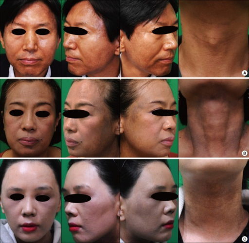
Clinical Photographs of Riehl Melanosis. The images show brown mottled patches on the face and neck (A), slate-gray patches on the lateral face and neck (B), and gray patches on the face and neck (C) of patients with Riehl melanosis.
Lee YJ, Park JH, Lee DY, Lee JH. Acquired bilateral dyspigmentation on face and neck: clinically appropriate approaches. J Korean Med Sci. 2016;31(12):2042-2050. doi: 10.3346/ jkms.2016.31.12.2042.
(Click Image to Enlarge)
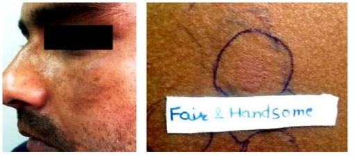
Riehl Melanosis on the Left Cheek. The left image shows a brown-gray patch on the left cheek, while the right image depicts positive patch testing for Fair & Handsome cream in a patient with Riehl melanosis.
Prabha N, Mahajan VK, Mehta KS, Chauhan PS, Gupta M. Cosmetic contact sensitivity in patients with melasma: results of a pilot study. Dermatol Res Pract. 2014;2014:316219. doi: 10.1155/2014/316219.
(Click Image to Enlarge)
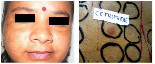
Riehl Melanosis on Bilateral Cheeks. The left image displays brown-gray reticulated, mottled patches on the bilateral cheeks, while the right image shows a positive patch test for cetrimide in a patient with Riehl melanosis.
Prabha N, Mahajan VK, Mehta KS, Chauhan PS, Gupta M. Cosmetic contact sensitivity in patients with melasma: results of a pilot study. Dermatol Res Pract. 2014;2014:316219. doi: 10.1155/2014/316219.
(Click Image to Enlarge)
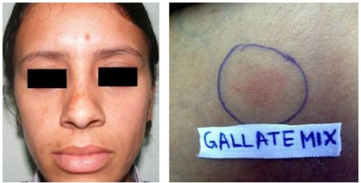
Riehl Melanosis due to Gallate. The left image shows mottled brown-gray face discoloration, while the right image displays a positive patch test result for gallate mix in a patient with Riehl melanosis.
Prabha N, Mahajan VK, Mehta KS, Chauhan PS, Gupta M. Cosmetic contact sensitivity in patients with melasma: results of a pilot study. Dermatol Res Pract. 2014;2014:316219. doi: 10.1155/2014/316219.
(Click Image to Enlarge)
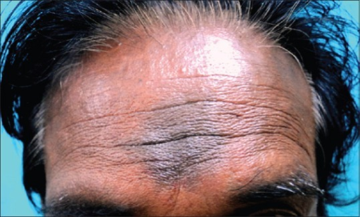
Kumkum-Induced Pigmented Contact Dermatitis. The image depicts a brown-gray hyperpigmented plaque on the glabella resulting from kumkum-induced pigmented contact dermatitis.
Gupta D, Thappa DM. Dermatoses due to Indian cultural practices. Indian J Dermatol. 2015;60(1):3-12. doi:10.4103/0019-5154.147778.
References
Ding Y, Xu Z, Xiang LF, Zhang C. Unveiling the mystery of Riehl's melanosis: An update from pathogenesis, diagnosis to treatment. Pigment cell & melanoma research. 2023 Nov:36(6):455-467. doi: 10.1111/pcmr.13108. Epub 2023 Jul 4 [PubMed PMID: 37401632]
Shah S, Baskaran N, Vinay K, Bishnoi A, Parsad D, Kumaran MS. Acquired dermal macular hyperpigmentation: an overview of the recent updates. International journal of dermatology. 2023 Dec:62(12):1447-1457. doi: 10.1111/ijd.16859. Epub 2023 Sep 28 [PubMed PMID: 37767951]
Level 3 (low-level) evidenceSitohang IBS, Prayogo RL, Rihatmadja R, Sirait SP. The diagnostic conundrum of Riehl melanosis and other facial pigmentary disorders: a case report with overlapping clinical, dermoscopic, and histopathological features. Acta dermatovenerologica Alpina, Pannonica, et Adriatica. 2020 Jun:29(2):81-83 [PubMed PMID: 32566956]
Level 3 (low-level) evidenceXu L, Huang Q, Wu T, Mu Y. Research Advances in the Treatment of Riehl's Melanosis. Clinical, cosmetic and investigational dermatology. 2023:16():1181-1189. doi: 10.2147/CCID.S403090. Epub 2023 May 4 [PubMed PMID: 37168093]
Level 3 (low-level) evidenceVinay K, Bishnoi A, Parsad D, Saikia UN, Sendhil Kumaran M. Dermatoscopic evaluation and histopathological correlation of acquired dermal macular hyperpigmentation. International journal of dermatology. 2017 Dec:56(12):1395-1399. doi: 10.1111/ijd.13782. Epub 2017 Oct 3 [PubMed PMID: 28971471]
rorsman H. Riehl's melanosis. International journal of dermatology. 1982 Mar:21(2):75-8 [PubMed PMID: 7068304]
Nakayama H, Matsuo S, Hayakawa K, Takhashi K, Shigematsu T, Ota S. Pigmented cosmetic dermatitis. International journal of dermatology. 1984 Jun:23(5):299-305 [PubMed PMID: 6746179]
Wang RF, Ko D, Friedman BJ, Lim HW, Mohammad TF. Disorders of hyperpigmentation. Part I. Pathogenesis and clinical features of common pigmentary disorders. Journal of the American Academy of Dermatology. 2023 Feb:88(2):271-288. doi: 10.1016/j.jaad.2022.01.051. Epub 2022 Feb 11 [PubMed PMID: 35151757]
Sarkar R, Vinay K, Bishnoi A, Poojary S, Gupta M, Kumaran MS, Jain A, Gurumurthy C, Arora P, Kandhari R, Rathi S, Zawar V, Gupta V, Ravivarma VN, Rodrigues M, Parsad D. A Delphi consensus on the nomenclature and diagnosis of lichen planus pigmentosus and related entities. Indian journal of dermatology, venereology and leprology. 2023 Jan-Frebuary:89(1):41-46. doi: 10.25259/IJDVL_804_2021. Epub [PubMed PMID: 35593293]
Level 3 (low-level) evidenceLai K, Zheng X, Wei S, Zhou H, Zeng X, Liang G, Zhang Z, Zhang W. Coexistence of Riehl's Melanosis, Lupus Erythematosus and Thyroiditis in a Patient. Clinical, cosmetic and investigational dermatology. 2022:15():1809-1813. doi: 10.2147/CCID.S376614. Epub 2022 Sep 8 [PubMed PMID: 36105748]
Jung JM, Noh TK, Jo SY, Kim SY, Song Y, Kim YH, Chang SE. Guanine Deaminase in Human Epidermal Keratinocytes Contributes to Skin Pigmentation. Molecules (Basel, Switzerland). 2020 Jun 5:25(11):. doi: 10.3390/molecules25112637. Epub 2020 Jun 5 [PubMed PMID: 32517074]
Xu Z, Chen L, Jiang M, Wang Q, Zhang C, Xiang LF. CCN1/Cyr61 Stimulates Melanogenesis through Integrin α6β1, p38 MAPK, and ERK1/2 Signaling Pathways in Human Epidermal Melanocytes. The Journal of investigative dermatology. 2018 Aug:138(8):1825-1833. doi: 10.1016/j.jid.2018.02.029. Epub 2018 Mar 3 [PubMed PMID: 29510193]
Woo YR, Kim JS, Lim JH, Choi JY, Kim M, Yu DS, Park YM, Park HJ. Acquired diffuse slate-grey facial dyspigmentation due to henna: an unrecognized cause of pigment contact dermatitis in Korean patients. European journal of dermatology : EJD. 2018 Oct 1:28(5):644-648. doi: 10.1684/ejd.2018.3404. Epub [PubMed PMID: 30530434]
Sugai T, Takahashi Y, Takagi T. Pigmented cosmetic dermatitis and coal tar dyes. Contact dermatitis. 1977 Oct:3(5):249-56 [PubMed PMID: 589997]
Lu Q, Jiang G. Progress in the application of reflectance confocal microscopy in dermatology. Postepy dermatologii i alergologii. 2021 Oct:38(5):709-715. doi: 10.5114/ada.2021.110077. Epub 2021 Nov 5 [PubMed PMID: 34849113]
Kim NH, Lee AY. Growth Factors Upregulated by Uric Acid Affect Guanine Deaminase-Induced Melanogenesis. Biomolecules & therapeutics. 2023 Jan 1:31(1):89-96. doi: 10.4062/biomolther.2022.137. Epub 2022 Dec 22 [PubMed PMID: 36549672]
Miyoshi K, Kodama H. Riehl's melanosis-like eruption associated with Sjögren's syndrome. The Journal of dermatology. 1997 Dec:24(12):784-6 [PubMed PMID: 9492444]
Level 3 (low-level) evidenceTakeo N, Sakai T, Saito-Shono T, Ishikawa K, Hatano Y, Katagiri K, Takahashi Y, Kawano K, Kimoto K, Kubota T, Eshima N, Kojima H, Fujiwara S. Three cases of pigmented cosmetic dermatitis-like eruptions associated with primary Sjögren's syndrome or anti-SSA antibody. The Journal of dermatology. 2016 Aug:43(8):947-50. doi: 10.1111/1346-8138.13300. Epub 2016 Feb 19 [PubMed PMID: 26892480]
Level 3 (low-level) evidenceVinay K, Bishnoi A, Kamat D, Chatterjee D, Kumaran MS, Parsad D. Acquired Dermal Macular Hyperpigmentation: An Update. Indian dermatology online journal. 2021 Sep-Oct:12(5):663-673. doi: 10.4103/idoj.IDOJ_881_20. Epub 2021 Aug 2 [PubMed PMID: 34667751]
Khanna N, Rasool S. Facial melanoses: Indian perspective. Indian journal of dermatology, venereology and leprology. 2011 Sep-Oct:77(5):552-63; quiz 564. doi: 10.4103/0378-6323.84046. Epub [PubMed PMID: 21860153]
Level 3 (low-level) evidenceSerrano G, Pujol C, Cuadra J, Gallo S, Aliaga A. Riehl's melanosis: pigmented contact dermatitis caused by fragrances. Journal of the American Academy of Dermatology. 1989 Nov:21(5 Pt 2):1057-60 [PubMed PMID: 2808836]
Level 3 (low-level) evidenceShenoi SD, Rao R. Pigmented contact dermatitis. Indian journal of dermatology, venereology and leprology. 2007 Sep-Oct:73(5):285-7 [PubMed PMID: 17921604]
Pérez-Bernal A, Muñoz-Pérez MA, Camacho F. Management of facial hyperpigmentation. American journal of clinical dermatology. 2000 Sep-Oct:1(5):261-8 [PubMed PMID: 11702317]
Kozuka T, Tashiro M, Sano S, Fujimoto K, Nakamura Y, Hashimoto S, Nakaminami G. Brilliant Lake Red R as a cause of pigmented contact dermatitis. Contact dermatitis. 1979 Sep:5(5):297-304 [PubMed PMID: 509931]
Level 3 (low-level) evidenceWoo YR, Jung Y, Jeong SW, Park HJ. Paracrine roles of hormone receptors in Riehl's melanosis: A quantitative analysis of oestrogen and progesterone receptor expression patterns. Experimental dermatology. 2021 Mar:30(3):396-401. doi: 10.1111/exd.14233. Epub 2020 Dec 10 [PubMed PMID: 33141431]
Kang HY. Melasma and aspects of pigmentary disorders in Asians. Annales de dermatologie et de venereologie. 2012 Dec:139 Suppl 4():S144-7. doi: 10.1016/S0151-9638(12)70126-6. Epub [PubMed PMID: 23522629]
Woo YR, Jung Y, Kim M, Park HJ. Impact of Riehl's melanosis on quality of life in Korean patients: A cross-sectional comparative study. The Journal of dermatology. 2020 Aug:47(8):893-897. doi: 10.1111/1346-8138.15382. Epub 2020 Jun 26 [PubMed PMID: 32592174]
Farabi B, Khan S, Jamgochian M, Atak MF, Jain M, Rao BK. The role of reflectance confocal microscopy in the diagnosis and management of pigmentary disorders: A review. Journal of cosmetic dermatology. 2023 Dec:22(12):3213-3222. doi: 10.1111/jocd.15827. Epub 2023 Sep 27 [PubMed PMID: 37759421]
Kim SM, Lee ES, Sohn S, Kim YC. Histopathological Features of Riehl Melanosis. The American Journal of dermatopathology. 2020 Feb:42(2):117-121. doi: 10.1097/DAD.0000000000001515. Epub [PubMed PMID: 31990700]
Ebihara T, Nakayama H. Pigmented contact dermatitis. Clinics in dermatology. 1997 Jul-Aug:15(4):593-9 [PubMed PMID: 9255469]
Lautenschlager S, Itin PH. Reticulate, patchy and mottled pigmentation of the neck. Acquired forms. Dermatology (Basel, Switzerland). 1998:197(3):291-6 [PubMed PMID: 9812039]
Shen PC, Chan YP, Huang CH, Ng CY. Riehl's Melanosis: A Multimodality, In Vivo, Real-Time Skin Imaging Study with Cellular Resolution Optical Coherence Tomography and Advanced Skin Diagnosis System in a Tertiary Medical Center. Bioengineering (Basel, Switzerland). 2022 Aug 26:9(9):. doi: 10.3390/bioengineering9090419. Epub 2022 Aug 26 [PubMed PMID: 36134965]
Krueger L, Saizan A, Stein JA, Elbuluk N. Dermoscopy of acquired pigmentary disorders: a comprehensive review. International journal of dermatology. 2022 Jan:61(1):7-19. doi: 10.1111/ijd.15741. Epub 2021 Jul 7 [PubMed PMID: 34235719]
Wang L, Xu AE. Four views of Riehl's melanosis: clinical appearance, dermoscopy, confocal microscopy and histopathology. Journal of the European Academy of Dermatology and Venereology : JEADV. 2014 Sep:28(9):1199-206. doi: 10.1111/jdv.12264. Epub 2013 Sep 7 [PubMed PMID: 24010902]
Kumaran MS, Dabas G, Vinay K, Parsad D. Reliability assessment and validation of the dermal pigmentation area and severity index: a new scoring method for acquired dermal macular hyperpigmentation. Journal of the European Academy of Dermatology and Venereology : JEADV. 2019 Jul:33(7):1386-1392. doi: 10.1111/jdv.15516. Epub 2019 Apr 15 [PubMed PMID: 30801771]
Level 1 (high-level) evidenceAkulinina I, Dodina M, Osadchuk M, Degtyarevskaya T. Optimizing diagnostic and therapeutic measures for different types of melasma based on the biophysical characteristics of facial skin. Journal of cosmetic and laser therapy : official publication of the European Society for Laser Dermatology. 2023 May 19:25(1-4):25-32. doi: 10.1080/14764172.2023.2230531. Epub 2023 Jul 2 [PubMed PMID: 37394829]
Sadaqat B, Khatoon N, Malik AY, Jamal A, Farooq U, Ali MI, He H, Liu FJ, Guo H, Urynowicz M, Wang Q, Huang Z. Enzymatic decolorization of melanin by lignin peroxidase from Phanerochaete chrysosporium. Scientific reports. 2020 Nov 19:10(1):20240. doi: 10.1038/s41598-020-76376-9. Epub 2020 Nov 19 [PubMed PMID: 33214596]
Draelos ZD. A split-face evaluation of a novel pigment-lightening agent compared with no treatment and hydroquinone. Journal of the American Academy of Dermatology. 2015 Jan:72(1):105-7. doi: 10.1016/j.jaad.2014.09.011. Epub 2014 Oct 16 [PubMed PMID: 25440437]
Zhong SM, Sun N, Liu HX, Niu YQ, Wu Y. Reduction of facial pigmentation of melasma by topical lignin peroxidase: A novel fast-acting skin-lightening agent. Experimental and therapeutic medicine. 2015 Feb:9(2):341-344 [PubMed PMID: 25574195]
Mauricio T, Karmon Y, Khaiat A. A randomized and placebo-controlled study to compare the skin-lightening efficacy and safety of lignin peroxidase cream vs. 2% hydroquinone cream. Journal of cosmetic dermatology. 2011 Dec:10(4):253-9. doi: 10.1111/j.1473-2165.2011.00581.x. Epub [PubMed PMID: 22151932]
Level 1 (high-level) evidenceXu Z, Xing X, Zhang C, Chen L, Flora Xiang L. A pilot study of oral tranexamic acid and Glycyrrhizin compound in the treatment of recalcitrant Riehl's melanosis. Journal of cosmetic dermatology. 2019 Feb:18(1):286-292. doi: 10.1111/jocd.12797. Epub 2018 Oct 19 [PubMed PMID: 30341831]
Level 3 (low-level) evidenceLi L, Ma Q, Li H. Effect of vitiligo treatment using compound glycyrrhizin combined with fractional carbon dioxide laser and topical triamcinolone acetonide on serum interleukin-17 and tissue growth factor-β levels. The Journal of international medical research. 2019 Nov:47(11):5623-5631. doi: 10.1177/0300060519871382. Epub 2019 Sep 25 [PubMed PMID: 31550958]
Mou KH, Han D, Liu WL, Li P. Combination therapy of orally administered glycyrrhizin and UVB improved active-stage generalized vitiligo. Brazilian journal of medical and biological research = Revista brasileira de pesquisas medicas e biologicas. 2016 Jul 25:49(8):. pii: S0100-879X2016000800605. doi: 10.1590/1414-431X20165354. Epub [PubMed PMID: 27464024]
Størmer FC, Reistad R, Alexander J. Glycyrrhizic acid in liquorice--evaluation of health hazard. Food and chemical toxicology : an international journal published for the British Industrial Biological Research Association. 1993 Apr:31(4):303-12 [PubMed PMID: 8386690]
Bishnoi A, Vinay K, Parsad D, Kumar S, Chatterjee D, Nahar Saikia U, Sendhil Kumaran M. Oral mycophenolate mofetil in the treatment of acquired dermal macular hyperpigmentation: An open-label pilot study. The Australasian journal of dermatology. 2021 Aug:62(3):278-285. doi: 10.1111/ajd.13567. Epub 2021 Mar 4 [PubMed PMID: 33660856]
Level 3 (low-level) evidenceRani S, Ahuja A. Chemical peel as an adjuvant treatment in pigmented contact dermatitis: a case series. Journal of cosmetic and laser therapy : official publication of the European Society for Laser Dermatology. 2022 Nov 17:24(6-8):112-117. doi: 10.1080/14764172.2022.2147953. Epub 2022 Nov 17 [PubMed PMID: 36384385]
Level 2 (mid-level) evidenceWang L, Wen X, Hao D, Li Y, Du D, Jiang X. Combination therapy with salicylic acid chemical peels, glycyrrhizin compound, and vitamin C for Riehl's melanosis. Journal of cosmetic dermatology. 2020 Jun:19(6):1377-1380. doi: 10.1111/jocd.13153. Epub 2019 Sep 16 [PubMed PMID: 31524950]
Park BJ, Jung YJ, Ro YS, Chang SE, Kim JE. Therapeutic Effects of New Pulsed-Type Microneedling Radiofrequency for Refractory Facial Pigmentary Disorders. Dermatologic surgery : official publication for American Society for Dermatologic Surgery [et al.]. 2022 Mar 1:48(3):327-333. doi: 10.1097/DSS.0000000000003367. Epub [PubMed PMID: 34999602]
Smucker JE, Kirby JS. Riehl melanosis treated successfully with Q-switch Nd:YAG laser. Journal of drugs in dermatology : JDD. 2014 Mar:13(3):356-8 [PubMed PMID: 24595582]
Chung BY, Kim JE, Ko JY, Chang SE. A pilot study of a novel dual--pulsed 1064 nm Q-switched Nd: YAG laser to treat Riehl's melanosis. Journal of cosmetic and laser therapy : official publication of the European Society for Laser Dermatology. 2014 Dec:16(6):290-2. doi: 10.3109/14764172.2014.946054. Epub 2014 Aug 13 [PubMed PMID: 25046351]
Level 3 (low-level) evidenceOn HR, Hong WJ, Roh MR. Low-pulse energy Q-switched Nd:YAG laser treatment for hair-dye-induced Riehl's melanosis. Journal of cosmetic and laser therapy : official publication of the European Society for Laser Dermatology. 2015 Jun:17(3):135-8. doi: 10.3109/14764172.2015.1007058. Epub 2015 Feb 20 [PubMed PMID: 25602355]
Cho MY, Roh MR. Successful Treatment of Riehl's Melanosis With Mid-Fluence Q-Switched Nd:YAG 1064-nm Laser. Lasers in surgery and medicine. 2020 Oct:52(8):753-760. doi: 10.1002/lsm.23214. Epub 2020 Jan 17 [PubMed PMID: 31951050]
Iwayama T, Oka M, Fukumoto T. Treatment of henna-induced Riehl's melanosis with a 755-nm picosecond alexandrite laser. Lasers in medical science. 2020 Sep:35(7):1659-1661. doi: 10.1007/s10103-020-03077-0. Epub 2020 Jun 23 [PubMed PMID: 32577930]
Kim SM, Hwang S, Almurayshid A, Park MY, Oh SH. Non-Ablative 1927 nm Fractional Thulium Fiber Laser: New, Promising Treatment Modality for Riehl's Melanosis. Lasers in surgery and medicine. 2021 Jul:53(5):640-646. doi: 10.1002/lsm.23341. Epub 2020 Dec 1 [PubMed PMID: 33259661]
Cai Y, Zhu Y, Wang Y, Xiang W. Intense pulsed light treatment for inflammatory skin diseases: a review. Lasers in medical science. 2022 Oct:37(8):3085-3105. doi: 10.1007/s10103-022-03620-1. Epub 2022 Aug 1 [PubMed PMID: 35913536]
Kim YH, Park YJ, Baek DJ, Kwon JE, Kang HY. A novel treatment for Riehl's melanosis targeting both dermal melanin and vessels. Photodermatology, photoimmunology & photomedicine. 2023 Nov:39(6):613-619. doi: 10.1111/phpp.12907. Epub 2023 Aug 23 [PubMed PMID: 37612856]
Kwon HH, Ohn J, Suh DH, Park HY, Choi SC, Jung JY, Kwon IH, Park GH. A pilot study for triple combination therapy with a low-fluence 1064 nm Q-switched Nd:YAG laser, hydroquinone cream and oral tranexamic acid for recalcitrant Riehl's Melanosis. The Journal of dermatological treatment. 2017 Mar:28(2):155-159. doi: 10.1080/09546634.2016.1187706. Epub 2016 Jun 27 [PubMed PMID: 27346606]
Level 3 (low-level) evidenceChoi CW, Jo G, Lee DH, Jo SJ, Lee C, Mun JH. Analysis of Clinical Features and Treatment Outcomes Using 1,064-nm Nd-YAG Laser with Topical Hydroquinone in Patients with Riehl's Melanosis: A Retrospective Study in 10 Patients. Annals of dermatology. 2019 Apr:31(2):127-132. doi: 10.5021/ad.2019.31.2.127. Epub 2019 Feb 28 [PubMed PMID: 33911560]
Level 2 (mid-level) evidenceElkamshoushi AM, Romisy D, Omar SS. Oral tranexamic acid, hydroquinone 4% and low-fluence 1064 nm Q-switched Nd:YAG laser for mixed melasma: Clinical and dermoscopic evaluation. Journal of cosmetic dermatology. 2022 Feb:21(2):657-668. doi: 10.1111/jocd.14140. Epub 2021 Apr 25 [PubMed PMID: 33826785]
Xu Z, Wang C, Xing X, Zhang C, Xiang LF. Efficacy and safety of the combination of oral tranexamic acid and intense pulsed light versus oral tranexamic acid alone in the treatment of refractory Riehl's melanosis: A prospective, comparative study. Journal of cosmetic dermatology. 2024 Mar 8:():. doi: 10.1111/jocd.16257. Epub 2024 Mar 8 [PubMed PMID: 38456556]
Level 2 (mid-level) evidenceKumarasinghe SPW, Pandya A, Chandran V, Rodrigues M, Dlova NC, Kang HY, Ramam M, Dayrit JF, Goh BK, Parsad D. A global consensus statement on ashy dermatosis, erythema dyschromicum perstans, lichen planus pigmentosus, idiopathic eruptive macular pigmentation, and Riehl's melanosis. International journal of dermatology. 2019 Mar:58(3):263-272. doi: 10.1111/ijd.14189. Epub 2018 Sep 3 [PubMed PMID: 30176055]
Level 3 (low-level) evidence