Introduction
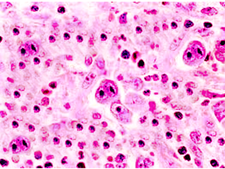
Hodgkin lymphoma characteristically presents with Hodgkin and Reed-Sternberg cells. When the cells are mononucleated, they are called Hodgkin cells; when multinucleated, they are called Reed-Sternberg cells.[1] The classic Reed-Sternberg cell is a large cell that can be >50 µm in diameter.[2] This cell is binucleated with prominent eosinophilic nuclei surrounded by abundant cytoplasm.[3]
Reed-Sternberg cells were first illustrated incorrectly by Greenfield in a paper published in 1878. Then, in 1898, Carl Sternberg published a German paper that included illustrations of the cells along with a description of their pathology. However, Sternberg believed that Hodgkin's disease was a form of tuberculosis. In 1902, Dorothy Reed described Reed-Sternberg cells in her well-known paper, "On the Pathological Changes in Hodgkin Disease, with Especial Reference to its Relation to Tuberculosis," which included a clear description of the cells and illustrations she made herself. Reed emphasized that Hodgkin's disease was unrelated to tuberculosis, and she used animal inoculation to prove her point. She also showed no immunological response to tuberculin in Hodgkin's disease.[4]
Issues of Concern
Register For Free And Read The Full Article
Search engine and full access to all medical articles
10 free questions in your specialty
Free CME/CE Activities
Free daily question in your email
Save favorite articles to your dashboard
Emails offering discounts
Learn more about a Subscription to StatPearls Point-of-Care
Issues of Concern
Many questions surround the origin of Reed-Sternberg cells. Studying these cells, as they make up 1% of the tumor tissue, is particularly difficult. Reed-Sternberg cells also depend heavily on their cellular microenvironment.[5] These cells derive from cell lineages related to macrophages, reticulum cells, and granulocytes. Experts also hypothesize that these cells result from fusions between lymphocytes, reticulum cells, and lymphocytes or cells that have had viral infections. However, the majority of the evidence supports the theory that the Reed-Sternberg cell originates from a B lymphocyte. Reed-Sternberg cells express CD15 and CD30.[6]
Causes
The presumption is that Reed-Sternberg cells derive from pre-apoptotic germinal center B-cells. How the Reed-Sternberg cells escape apoptosis is an interesting topic. First, Reed-Sternberg cells show the activity of the NF-kappa B transcription factor, and when inhibition of the factor occurs, an induction of cell death results. Other cell lines show inactivating mutations in the inhibitor of NF-kappa B, I-kappa B-alpha, or c-Rel amplifications.
Studies have also suggested that the CD30 receptor may trigger NF-kappa B activity in Reed-Sternberg cells.[7] Second, the apoptosis of germinal center B-cells is under partial control of the CD95 death receptor pathway. Reed-Sternberg cell lines still express the receptor, but many cell lines are resistant to apoptosis due to CD95 cross-linking. Expression of cFLIPL, which is an inhibitor of CD95 signaling, also occurs. CD95 gene mutations are also responsible for CD95 inactivity in a smaller subset. Third, the X-linked inhibitor of apoptosis (XIAP) inhibits the activation of caspase-3, which executes apoptosis. Reed-Sternberg cell lines have been shown to express XIAP.[1]
Anatomical Pathology
Reed-Sternberg cells are classically associated with Hodgkin lymphoma. However, large, atypical cells may have morphologic and immunophenotypic features resembling these cells and may pose a diagnostic challenge. Reed-Sternberg-like (RS-like) cells may form neoplastic sheets but can also present as scattered cells. Understanding strategies for differentiating Reed-Sternberg cells from RS-like cells is essential to overcoming these diagnostic challenges.
RS-like cells found in low-grade B-cell lymphomas are visible in follicular lymphoma, chronic lymphocytic leukemia/small lymphocytic lymphoma, or marginal zone lymphoma. In chronic lymphocytic leukemia/small lymphocytic lymphoma (CLL/SLL), RS-like cells are present amongst neoplastic cells. They resemble the immunophenotype of B-cells and may also show CD20 and CD30 expression while typically being negative for CD15. In rare cases, the RS-like cells show CD15 and CD30 expression, making them almost identical to those seen in classical Hodgkin disease. RS-like cells in CLL/SLL also show EBV positivity, which is under study as playing a role in the pathogenesis of Reed-Sternberg cells. In non-Hodgkin lymphomas, follicular lymphomas have stomal fibrosis prominently in retroperitoneal or perinephric locations. Follicular lymphoma contains neoplastic lymphoid infiltrated with small centrocytes with long nuclei, scattered centroblasts, and nucleoli. Classical Hodgkin lymphoma is also associated with fibrosis, but the inflammatory infiltrate comprises eosinophils, plasma cells, histiocytes, and small lymphocytes.
In follicular lymphomas, RS-like cells can present within or between neoplastic follicles. These cells also have indistinguishable immunoglobulin heavy chain gene rearrangements from neoplastic centroblasts and centrocytes, suggesting a common origin. RS-like cells in follicular lymphoma may express CD10, which can differentiate them from Reed-Sternberg cells. RS-like cells may also be counted mistakenly as centroblasts, so having adequately sized samples and using strict morphologic guidelines to arrive at an accurate diagnosis is essential.
B-cell lineage RS-like cells are seen in angioimmunoblastic T-cell lymphomas and rarely in peripheral T-cell lymphoma. RS-like cells express CD20, CD30, and, at times, CD15. The RS-like cells are also associated with EBV infection. Angioimmunoblastic T-cell lymphomas are diagnosable by increased vasculature. Peripheral T-cell lymphomas that contain RS-like cells are very difficult to differentiate from classical Hodgkin lymphoma because of the presence of mixed inflammatory infiltrate. However, assessing for expression of CD20, PAX-5, CD10, and BCL-6 is crucial. The absence of PAX-5 presents a strong argument against the diagnosis of classical Hodgkin lymphoma. Also, if a morphologically and immunophenotypically abnormal lymphocytic infiltrate is present, this is not characteristic of classical Hodgkin lymphoma.[8]
Clinical Pathology
Neoplastic Reed-Sternberg cells comprise only 1% of the tumor mass. Most of the infiltrate comprises non-neoplastic T-cells, B-cells, eosinophils, neutrophils, macrophages, and plasma cells. The inflammatory background is vital to the clinical behavior of Reed-Sternberg cells as bidirectional signaling occurs between the cells and their environment.[9]
Biochemical and Genetic Pathology
Advancements in the following 5 areas of cell biology have contributed to the understanding of Reed-Sternberg cells:
- Fixed tissue and cell line immunophenotyping have helped in the understanding of the Reed-Sternberg cell surface.
- Genotyping has given this information genetic context.
- Cell lines from Hodgkin Disease specimens have permitted the development of specific monoclonal antibodies.
- A relationship between Hodgkin and Epstein-Barr virus has suggested a relationship between these conditions.
- Cytokines produced in Hodgkin's Disease provide insight into the cells involved and the pathophysiology of the disease.[6]
Morphology
Classical diagnostic Reed-Sternberg cells are large (15 to 45 µm), have abundant slightly basophilic or amphophilic cytoplasm, and have at least 2 nuclear lobes or nuclei. Diagnostic Reed-Sternberg cells must have at least 2 nucleoli in 2 separate nuclear lobes. The nuclei are large and often rounded in contour with a prominent, often irregular nuclear membrane, pale chromatin, and usually 1 prominent eosinophilic nucleolus, with perinuclear clearing (halo), resembling a viral inclusion (see Image. Reed Sternberg Cells).[1]
Mononuclear variants of Reed-Sternberg cells are Hodgkin cells, characterized by a single round or oblong nucleus with large inclusion-like nucleoli. Some Reed-Sternberg cells may have condensed cytoplasm and pyknotic reddish nuclei. These variants are known as mummified cells. Reed-Sternberg cells surrounded by formalin retraction artifacts are termed ''lacunar cells''. The latter is characteristic of the nodular sclerosing Hodgkin lymphoma subtype.
Mechanisms
The first method used to isolate Reed-Sternberg cells was via suspension of fresh tissue samples. Reed-Sternberg cells were selected based on their size and negative expression of CD3, CD14, and CD20. This method did not yield consistent results and was improved upon by suspending cells onto glass slides and immunolabeling for CD30. A second technique for cell isolation used thick paraffin sections instead of fresh tissue. Enzymatic digestion and mechanical force were used to suspend cells. The suspended cells were labeled for CD30 and isolated using a hand pipette. Another technique involved the usage of hydraulically driven pipettes.[10]
Clinicopathologic Correlations
Epstein-Barr virus (EBV) establishes itself as a latent infection in B-cells. EBV infects naive B-cells that will then undergo germinal center reactions, so the virus ultimately resides in memory B-cells. Once the B-cells get infected, proliferation occurs. Lymphomas associated with EBV are of germinal center B-cell origin. They include Hodgkin lymphoma, and about 40% of the Reed-Sternberg cells are infected. The risk of Hodgkin lymphoma increases after infection with EBV. However, this association is not new. What is new is the restriction of increased risk of Hodgkin disease to EBV-positive patients without increased risk demonstrated in EBV-negative patients.[9]
Clinical Significance
Reed-Sternberg cells are the hallmark tumor cells of Hodgkin lymphoma. They represent <1% of the tumor tissue, while the majority of cells in the tissue include T-cells, B-cells, eosinophils, macrophages, and plasma cells.[11] Considering RS-like cells and their presence in tissue samples is crucial, as this may present a diagnostic conundrum. RS-like cells have been described in cases of follicular lymphoma and have been shown to be clonally related to the lymphoma population.[12] Immunochemical analyses have shown that a low CD15 and TARC (Thymus and Activation-Regulated Chemokine) expression on Reed-Sternberg cells is associated with treatment failure.[13] Additionally, PD-L1 expression in the tumor microenvironment (TME) is associated with complete remission.
A retrospective study compared CLL/SLL with isolated Reed-Sternberg cells to CLL with Hodgkin transformation and found that both groups were quite similar.[14] Experts felt that these entities could represent clinicopathologic points on a spectrum and a biological continuum and that Hodgkin therapy could be useful in both. The data suggested that Hodgkin's protocols might be favored over CLL regimens in regard to treatment.
The most well-known infectious disease with the presence of Reed-Sternberg cells is infectious mononucleosis due to infection with EBV. EBV-infected B-cells acquire the morphologic and immunophenotypic characteristics of Reed-Sternberg cells, although the mechanism of this process is still an enigma.[1]
Studies have suggested that the prognosis is proportional to the TME.[15] The TME consists of T- and B-lymphocytes, neutrophils, eosinophils, macrophages, plasma cells, and histiocytes. Hodgkin patients with a low T-cell presence and a high Reed-Sternberg cell portion portend an aggressive clinical course. About 10% to 20% of Hodkin's disease patients either relapse or are refractory. Would these patients then merit a more aggressive treatment?
A dysregulation, a mutation, of multiple signaling pathways, may also be responsible for the pathogenesis of Hodgkin disease, eg, JAK/STAT and the Nuclear Factor-KB (NF-KB).[16][17] Recent studies have shown that the JAK/STAT is overactivated in Hodgkin disease and acts as a conduit of signaling, augmentation, and support of the inflammatory TME.[18] Agents, eg, ruxolitinib (an inhibitor of Jak1 and Jak2), have shown some efficacy in Hodgkin cell models. The Anti-GSF receptor (CSF3R) may also be a surrogate marker for this JAK/STAT overactivity. NF-KB drives inflammatory cytokine production (eg, IL6, TNF-alpha, and GMCSF), affects cellular senescence, and provides cancer with prosurvival signals. Studies have also attempted to use Bortesonib (a protease inhibitor) to suppress NF-KB by preventing its IxB component degradation. Unfortunately, these studies show a failure of Bortesonib as a treatment for Hodgkin disease. The resultant belief is that there are different pathways also involved as well.
Media
(Click Image to Enlarge)

Reed Sternberg Cells. Diagnostic Reed-Sternberg cells must have at least 2 nucleoli in 2 separate nuclear lobes. The nuclei are large and often rounded in contour with a prominent, often irregular nuclear membrane, pale chromatin, and usually 1 prominent eosinophilic nucleolus, with perinuclear clearing (halo), resembling a viral inclusion.
Contributed by S Bhimji, MD
References
Küppers R, Hansmann ML. The Hodgkin and Reed/Sternberg cell. The international journal of biochemistry & cell biology. 2005 Mar:37(3):511-7 [PubMed PMID: 15618006]
Chan WC. The Reed-Sternberg cell in classical Hodgkin's disease. Hematological oncology. 2001 Mar:19(1):1-17 [PubMed PMID: 11276042]
Quintanilla-Martinez L, Fend F, Moguel LR, Spilove L, Beaty MW, Kingma DW, Raffeld M, Jaffe ES. Peripheral T-cell lymphoma with Reed-Sternberg-like cells of B-cell phenotype and genotype associated with Epstein-Barr virus infection. The American journal of surgical pathology. 1999 Oct:23(10):1233-40 [PubMed PMID: 10524524]
Level 3 (low-level) evidenceDawson PJ. Whatever happened to Dorothy Reed? Annals of diagnostic pathology. 2003 Jun:7(3):195-203 [PubMed PMID: 12808573]
Jaffe ES. The elusive Reed-Sternberg cell. The New England journal of medicine. 1989 Feb 23:320(8):529-31 [PubMed PMID: 2536895]
Haluska FG, Brufsky AM, Canellos GP. The cellular biology of the Reed-Sternberg cell. Blood. 1994 Aug 15:84(4):1005-19 [PubMed PMID: 8049419]
Steidl C. Exposing Hodgkin-Reed-Sternberg cells. Blood. 2017 Jan 5:129(1):6-7. doi: 10.1182/blood-2016-11-746701. Epub [PubMed PMID: 28057670]
Gomez-Gelvez JC, Smith LB. Reed-Sternberg-Like Cells in Non-Hodgkin Lymphomas. Archives of pathology & laboratory medicine. 2015 Oct:139(10):1205-10. doi: 10.5858/arpa.2015-0197-RAI. Epub [PubMed PMID: 26414463]
Roullet MR, Bagg A. Recent insights into the biology of Hodgkin lymphoma: unraveling the mysteries of the Reed-Sternberg cell. Expert review of molecular diagnostics. 2007 Nov:7(6):805-20 [PubMed PMID: 18020910]
Stein HS, Hummel M. Hodgkin's disease: biology and origin of Hodgkin and Reed-Sternberg cells. Cancer treatment reviews. 1999 Jun:25(3):161-8 [PubMed PMID: 10425258]
Lee IS, Kim SH, Song HG, Park SH. The molecular basis for the generation of Hodgkin and Reed-Sternberg cells in Hodgkin's lymphoma. International journal of hematology. 2003 May:77(4):330-5 [PubMed PMID: 12774919]
Ocampo Gonzalez FA, Ahmed A. Reed-Sternberg-like cells in a case of follicular lymphoma. Blood. 2022 Jul 21:140(3):290. doi: 10.1182/blood.2022016637. Epub [PubMed PMID: 35862091]
Level 3 (low-level) evidenceZijtregtop EAM, Tromp I, Dandis R, Zwaan CM, Lam KH, Meyer-Wentrup FAG, Beishuizen A. The Prognostic Value of Eight Immunohistochemical Markers Expressed in the Tumor Microenvironment and on Hodgkin Reed-Sternberg Cells in Pediatric Patients With Classical Hodgkin Lymphoma. Pathology oncology research : POR. 2022:28():1610482. doi: 10.3389/pore.2022.1610482. Epub 2022 Aug 11 [PubMed PMID: 36032657]
King RL, Gupta A, Kurtin PJ, Ding W, Call TG, Rabe KG, Kenderian SS, Leis JF, Wang Y, Schwager SM, Slager SL, Kay NE, Koehler A, Ansell SM, Inwards DJ, Habermann TM, Shi M, Hanson CA, Howard MT, Parikh SA. Chronic lymphocytic leukemia (CLL) with Reed-Sternberg-like cells vs Classic Hodgkin lymphoma transformation of CLL: does this distinction matter? Blood cancer journal. 2022 Jan 28:12(1):18. doi: 10.1038/s41408-022-00616-6. Epub 2022 Jan 28 [PubMed PMID: 35091549]
Santisteban-Espejo A, Bernal-Florindo I, Perez-Requena J, Atienza-Cuevas L, Maira-Gonzalez N, Garcia-Rojo M. Whole-slide image analysis identifies a high content of Hodgkin Reed-Sternberg cells and a low content of T lymphocytes in tumor microenvironment as predictors of adverse outcome in patients with classic Hodgkin lymphoma treated with ABVD. Frontiers in oncology. 2022:12():1000762. doi: 10.3389/fonc.2022.1000762. Epub 2022 Oct 20 [PubMed PMID: 36338756]
Küppers R, Engert A, Hansmann ML. Hodgkin lymphoma. The Journal of clinical investigation. 2012 Oct:122(10):3439-47. doi: 10.1172/JCI61245. Epub 2012 Oct 1 [PubMed PMID: 23023715]
Gopas J, Stern E, Zurgil U, Ozer J, Ben-Ari A, Shubinsky G, Braiman A, Sinay R, Ezratty J, Dronov V, Balachandran S, Benharroch D, Livneh E. Reed-Sternberg cells in Hodgkin's lymphoma present features of cellular senescence. Cell death & disease. 2016 Nov 10:7(11):e2457. doi: 10.1038/cddis.2016.185. Epub 2016 Nov 10 [PubMed PMID: 27831553]
Fernández S, Solórzano JL, Díaz E, Menéndez V, Maestre L, Palacios S, López M, Colmenero A, Estévez M, Montalbán C, Martínez Á, Roncador G, García JF. JAK/STAT blockade reverses the malignant phenotype of Hodgkin and Reed-Sternberg cells. Blood advances. 2023 Aug 8:7(15):4135-4147. doi: 10.1182/bloodadvances.2021006336. Epub [PubMed PMID: 36459489]
Level 3 (low-level) evidence