 Anatomy, Shoulder and Upper Limb, Hand Opponens Pollicis Muscle
Introduction
Motor skills of the hand result from many structures working in sync to produce certain motions for daily living. The hands are just a small part of the human body but contribute significantly to the functionality. Even though the hands are only a small part of a human, they are held together by the largest number of bones. Hand movements are influenced by hand and forearm muscles. Opposition is a small hand movement consisting of the thumb touching the tips of the other digits.
Opposition is defined as positioning something near or close to each other. In medicine, the opposition of the hand is a motion created by several muscles, bones, and nerves working in sync. The muscles that create this primary movement are the opponens muscles. There are two opponens muscles; one muscle appears in the thenar eminence and the other in the hypothenar eminence. The action of the thumb touching the tip of the fifth digit takes place from the contraction of both opponens muscles, while the action required for the thumb to touch the second, third, and fourth digits gets produced from several muscles in the hand and forearm. The lumbrical muscles, flexor digitorum superficialis muscle, and the flexor digitorum profundus muscle contract in the second, third, and fourth digits during the opposition of the hand.
Structure and Function
Register For Free And Read The Full Article
Search engine and full access to all medical articles
10 free questions in your specialty
Free CME/CE Activities
Free daily question in your email
Save favorite articles to your dashboard
Emails offering discounts
Learn more about a Subscription to StatPearls Point-of-Care
Structure and Function
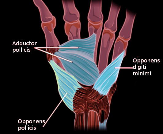
The opponens pollicis muscle is in the thenar eminence along with three other muscles that make up the thenar eminence. The opponens pollicis muscle is a muscle originates from the tubercle of the trapezium bone and the transverse carpal ligament (flexor retinaculum). The muscle then inserts onto the entire length of the first metacarpal bone. The function of the opponens pollicis muscle is to draw the thumb across the hand and touch the tips of the other digits. When the opponens pollicis muscle contracts it pulls the thumb medially towards the other digits. The medially drawn thumb is made up of flexion, internal rotation, and palmar abduction of the thumb. This combination equates to the opposition of the thumb.[1]
The movement produced from the opposition of the thumb and the other muscles allows the hand to cup objects. The pincher grip is also made possible by the opponens pollicis muscle contracting and touching the tips of the other digits.[2]
Embryology
In embryology, axial skeletal bones derive from sclerotomes and muscles are derived from myotomes.[3] The elongation of limbs occurs by endochondral ossification. This process develops the frame for the limbs by creating a cartilage model before ossifying into the bone. First, chondrocytes produce the cartilage model. Then the osteoclast and osteoblast will produce and remodel the bone. The osteoblast will lay down osteoid, and the osteoclast will break down the osteoid to shape the bone. The lengthening process for limbs and appendages are under the influence of fibroblast growth factor (FGF). As the limbs and appendages grow in length, FGF also influences the growth of the blood supply for the limbs and appendages.
The muscles in the body derive from mesenchymal tissue and neural crest cells. The mesenchymal tissue will differentiate into mesoderm. The mesoderm will further mature into myoblast. The myoblast is the immature cell of the muscle. Once the myoblast cluster and mature, it will become muscle tissue.[4]
Blood Supply and Lymphatics
Since the thenar eminence is on the radial side of the palm, it received its blood supply mainly from the superficial palmar arch. The superficial palmar arch derives from the radial artery. This arch will go onto and perfuse the opponens pollicis muscle and the other muscle in the thenar compartment. The deep palmar arch formed from the ulnar artery also provides collateral blood flow to the thenar muscles.[5]
The lymphatic drainage of the hand drains into the cubital lymph nodes and then to the axillary lymph nodes. The axillary lymph nodes will drain into the right lymphatic duct on the right upper limb. The thoracic duct will be the proximal drainage site for the left upper limb.
Nerves
The opponens pollicis muscle is innervated by spinal levels C8 and T1. These spinal levels make up the recurrent branch of the median nerve.[6] The recurrent branch of the median nerve also innervates one other muscle that is part of the thenar eminence. The other muscle innervated by the recurrent branch of the median nerve is the flexor pollicis brevis muscle.
Muscles
The thenar eminence of the hand is made up of four muscles: the opponens pollicis muscle, the adductor pollicis muscle, the abductor pollicis brevis muscle, and the flexor pollicis brevis muscle. These muscles make up what is considered the "ball" of the palm.[7][8][9]
Since the opposition of the thumb moves the first digit across the palm to touch the tips of the other digits. Other muscles perform the movement of the digits toward the thumb. The medial side of the hand has the hypothenar eminence. Within the hypothenar eminence, there is a muscle the pulls the fifth digit (little finger or pinky) across the palm towards the thumb. This muscle is called the opponens digiti minimi muscle. The opponens digiti minimi muscle's action is the opposite motion when compared to the opponens pollicis muscle, but allows the thumb to touch the tip of the fifth digit. The lumbrical muscles, flexor digitorum superficialis muscle, and the flexor digitorum profundus muscle all produce the motion that allows the thumb to touch the tip of the other fingers.
Physiologic Variants
The nerve innervation of the opponens pollicis muscle may come from the ulnar nerve in some cases. The ulnar nerve is made up of spinal roots C8 and T1. The attachment and insertion of the opponens pollicis muscle may vary from person to person, but the action of opposition is constant.[10][11]
Surgical Considerations
The blood supply and nerve innervation of the opponens pollicis muscle and the thenar eminence is essential in hand surgery. The knowledge of the blood supply and innervation allows for proper precautions to avoid damage to these structures.[12] The anatomical location of the structures in the hand is important to know during the surgical reconstruction of the hand.
The compression of the median nerve may cause atrophy of the thenar muscles and pain in the first three digits. The median nerve usually gets compressed under the flexor retinaculum for several reasons; this leads to the condition called carpal tunnel syndrome. The last resort in the treatment of carpal tunnel syndrome is surgery.
Clinical Significance
The examination of the nerve innervation of the hand is possible with the movements of the thumb. If the examiner instructs the patient to move the thumb and touch the tips of the other digits, this can determine if there an intact median nerve or a damaged median nerve. While thumb adduction (moving the thumb closer to the hand) can evaluate the functionality of the ulnar nerve. Finally, the radial nerve can undergo assessment by asking the patient to perform thumb extension.
Other Issues
The blood supply and nerve innervation to the opponens pollicis muscle originates from the recurrent branch of the median nerve. Any damage to either the nerve innervation or blood supply proximally can result in damage to the opponens pollicis muscle. Any compression to the subclavian artery or the brachial plexus can present in the hand's presentation at rest and defected hand movements. If there is damage to the median nerve, it will present as atrophy of the thenar muscles/eminence at baseline.[13][14]
Damage to the median nerve proximally will present as the "Pope's blessing." When the patient is asked to make a fist, the fourth and fifth digits will flex while the first, second, and third digits remain extended. If the damage to the median nerve is distal, it will present as the "median claw." This presentation will present when instructing the patient to extend the fingers. The fourth and fifth digits will extend while the thumb will stay in a resting position with the second and third digits flexed at the metacarpophalangeal joints.
Media
References
Adams JE, O'Brien V, Magnusson E, Rosenstein B, Nuckley DJ. Radiographic Analysis of Simulated First Dorsal Interosseous and Opponens Pollicis Loading Upon Thumb CMC Joint Subluxation: A Cadaver Study. Hand (New York, N.Y.). 2018 Jan:13(1):40-44. doi: 10.1177/1558944717691132. Epub 2017 Feb 16 [PubMed PMID: 28719976]
McDannald DW, Mansour M, Rydalch G, Bolton DAE. Motor affordance for grasping a safety handle. Neuroscience letters. 2018 Sep 14:683():131-137. doi: 10.1016/j.neulet.2018.05.040. Epub 2018 May 29 [PubMed PMID: 29857040]
Level 3 (low-level) evidenceSenthinathan B, Sousa C, Tannahill D, Keynes R. The generation of vertebral segmental patterning in the chick embryo. Journal of anatomy. 2012 Jun:220(6):591-602. doi: 10.1111/j.1469-7580.2012.01497.x. Epub 2012 Mar 28 [PubMed PMID: 22458512]
Level 3 (low-level) evidenceSagarin KA, Redgrave AC, Mosimann C, Burke AC, Devoto SH. Anterior trunk muscle shows mix of axial and appendicular developmental patterns. Developmental dynamics : an official publication of the American Association of Anatomists. 2019 Oct:248(10):961-968. doi: 10.1002/dvdy.95. Epub 2019 Aug 16 [PubMed PMID: 31386244]
Bigler MR, Buffle E, Siontis GCM, Stoller M, Grossenbacher R, Tschannen C, Seiler C. Invasive Assessment of the Human Arterial Palmar Arch and Forearm Collateral Function During Transradial Access. Circulation. Cardiovascular interventions. 2019 Jul:12(7):e007744. doi: 10.1161/CIRCINTERVENTIONS.118.007744. Epub 2019 Jul 5 [PubMed PMID: 31272228]
Katz R, Mazzocchio R, Pénicaud A, Rossi A. Distribution of recurrent inhibition in the human upper limb. Acta physiologica Scandinavica. 1993 Oct:149(2):183-98 [PubMed PMID: 8266808]
Gupta S, Michelsen-Jost H. Anatomy and function of the thenar muscles. Hand clinics. 2012 Feb:28(1):1-7. doi: 10.1016/j.hcl.2011.09.006. Epub [PubMed PMID: 22117918]
Homma T, Sakai T. [Anatomy of intrinsic hand muscles]. Kaibogaku zasshi. Journal of anatomy. 1994 Apr:69(2):123-42 [PubMed PMID: 8023676]
Homma T, Sakai T. Thenar and hypothenar muscles and their innervation by the ulnar and median nerves in the human hand. Acta anatomica. 1992:145(1):44-9 [PubMed PMID: 1414212]
Bertelli JA, Soldado F, Rodrígues-Baeza A, Ghizoni MF. Transferring the Motor Branch of the Opponens Pollicis to the Terminal Division of the Deep Branch of the Ulnar Nerve for Pinch Reconstruction. The Journal of hand surgery. 2019 Jan:44(1):9-17. doi: 10.1016/j.jhsa.2018.09.010. Epub 2018 Oct 23 [PubMed PMID: 30366737]
Dyro FM. Ulnar innervation of opponens pollicis. Electromyography and clinical neurophysiology. 1983 May-Jun:23(4):257-60 [PubMed PMID: 6872925]
Level 3 (low-level) evidenceNeth MR. Acute hand pain resulting in spontaneous thenar compartment syndrome. The American journal of emergency medicine. 2019 Mar:37(3):561.e3-561.e4. doi: 10.1016/j.ajem.2018.11.035. Epub 2018 Nov 24 [PubMed PMID: 30527915]
Jupiter JB, Nunez FA Jr, Nunez F Sr, Fernandez DL, Shin AY. Current Perspectives on Complex Wrist Fracture-Dislocations. Instructional course lectures. 2018 Feb 15:67():155-174 [PubMed PMID: 31411409]
Level 3 (low-level) evidenceLin CP, Chen IJ, Chang KV, Wu WT, Özçakar L. Utility of Ultrasound Elastography in Evaluation of Carpal Tunnel Syndrome: A Systematic Review and Meta-analysis. Ultrasound in medicine & biology. 2019 Nov:45(11):2855-2865. doi: 10.1016/j.ultrasmedbio.2019.07.409. Epub 2019 Aug 9 [PubMed PMID: 31402226]
Level 1 (high-level) evidence