 Subacute Cutaneous Lupus Erythematosus
Introduction
Lupus erythematosus is an inflammatory connective tissue disorder characterized by pathogenic autoantibody production and immune complex formation and deposition resulting from a loss of immune tolerance. Dermatological manifestations of lupus constitute one of the diagnostic criteria for systemic lupus erythematosus (SLE). Cutaneous manifestations in lupus cover a broad spectrum and may or may not occur in the context of SLE.[1]
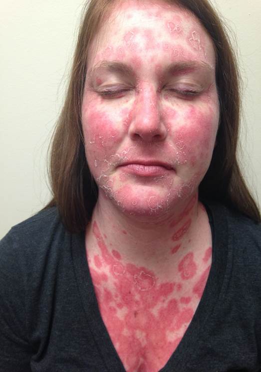
The lesions may be described as lupus erythematosus-specific and lupus erythematosus-nonspecific.[2] Lupus erythematosus-specific manifestations include various subtypes of cutaneous lupus erythematosus (CLE) and are subdivided into 3 different categories described by Gillian et al. These categories are acute, subacute, and chronic CLE.[3] This activity provides updated information on subacute CLE (SCLE), which typically presents as a symmetric, nonscarring, photosensitive erythematous rash over sun-exposed areas such as the face, neck, arms, upper back, and shoulders (see Image. Subacute Cutaneous Lupus Erythematosus Face and Neck Lesions).
Etiology
Register For Free And Read The Full Article
Search engine and full access to all medical articles
10 free questions in your specialty
Free CME/CE Activities
Free daily question in your email
Save favorite articles to your dashboard
Emails offering discounts
Learn more about a Subscription to StatPearls Point-of-Care
Etiology
The etiology of SLE and CLE is not well-defined. The classical precipitating factor is sunlight exposure in a patient with an abnormal milieu of genetic predisposition and immune dysregulation. Drug-induced SCLE has been reported as well. Commonly used drugs that have been associated with SCLE are angiotensin-converting enzyme inhibitors, anticonvulsants, β-blockers, and immune modulators, including biologics, chemotherapeutic agents, and immune checkpoint inhibitors.[4][5][6] SCLE has been reported to develop in association with certain malignancies.
Epidemiology
SCLE primarily occurs in young to middle-aged female individuals, with female cohorts 3 to 4 times more likely to develop the lesions than male individuals. The condition usually presents in the 3rd or 4th decade of life. The mean age of onset of SCLE described by Bizar et al ranged from 50 to 52 years. SCLE is highly photosensitive, with 48% to 90% of patients meeting the American College of Rheumatology's definition of increased photosensitivity. The 2 morphologic variants of SCLE are annular and papulosquamous. Drug-induced forms may be seen in either sex and at older ages of onset.
Pathophysiology
The pathogenesis is multifactorial. SCLE is thought to develop due to genetics, environmental triggers, or immunologic factors. Recent data suggests some potential candidate genes involved in the evolution of SCLE, including HLA1, B8, DR3, HLA1, B8, DR3, DQ2, DRw52, DR3, and C4 null ancestral haplotype.[7][8] Moreover, deficiencies of the C2 and C4 complement components have been associated with SCLE.[9] Environmental factors, such as UV light and certain drugs, contribute to the development of SCLE. Studies have shown that both innate and cell-mediated immunity, along with dysfunction of the T helper (Th) cells, Th1, Th2, and Th17, and abnormal expression of various cytokines and adhesion molecules, are involved in the development of SCLE.[10][11]
Anti-Ro/SS-A antibodies and apoptotic keratinocytes, including UVB-irradiated keratinocytes, have been implicated in SCLE pathogenesis, as studies have shown deposition of autoantibodies, immunoglobulins, and complement at the dermoepidermal junction. Antibody-dependent cell-mediated cytotoxicity, CD8+ cytotoxicity, and complement-mediated cytolysis are all involved in the destruction of keratinocytes.[12][13][14]
Histopathology
Histologically, lupus erythematosus-specific cutaneous findings share many features, but SCLE has some distinct attributes. The most characteristic findings are moderate hyperkeratosis with focal disorientation, interface dermatitis, and periappendageal mononuclear cell infiltrates confined to the superficial dermis. Basement membrane thickening is usually not seen in SCLE, although dermal edema may be noted. The inflammatory infiltrates of interface dermatitis consist mainly of activated T cells and macrophages. Immunofluorescence studies show the deposition of immunoglobulin G (IgG) or complement components in a granular pattern at the dermoepidermal junction in about 60% of patients with SCLE. Anti-SS-A/Ro and anti-SS-B/La autoantibodies have been strongly associated with SCLE.[15]
History and Physical
SCLE lesions typically appear in a characteristic distribution, presenting as either papulosquamous or annular lesions with central clearing. These 2 forms can coexist, and the lesions generally heal without causing atrophy or scarring. Patients may have hypopigmentation or telangiectasia. However, the skin returns to normal in most. Individuals with SCLE frequently have a mild illness with musculoskeletal complaints and serologic abnormalities.[16] SCLE has a predilection for sun-exposed areas like the neck, shoulders, chest, and extensor surfaces of the arms, but it usually spares the face.
Evaluation
SCLE, along with other forms of CLE, is mainly a clinical diagnosis. Confirmatory histopathologic examination with skin biopsy is indicated when the diagnosis is uncertain. Laboratory studies like anti-SSA/Ro and anti-SSB/La are warranted as well. Direct immunofluorescence may be used to help support the diagnosis if histopathology is inconclusive. Immunofluorescence shows a granular pattern of deposition of immunoglobulin at the dermopidermal junction.
Treatment / Management
Management of SCLE includes lifestyle modification and pharmacological therapy. Important preventive measures include physical protection, such as wearing broad-brimmed hats and sun-protective clothing, as well as properly applying sunscreen. Educating patients about their disease, avoidance of potential triggers like excessive sun exposure, and strict sunscreen adherence with chemical or physical blocking agents is essential. A broad-spectrum sunscreen with a sun protection factor of at least 50 should be applied in sufficient amounts about 20 to 30 minutes before expected exposure.[17]
Tobacco is phototoxic and enhances the responsiveness of toll-like receptor 9, leading to the increased production of type 1 interferons in plasmacytoid dendritic cells and the upregulation of the expression of metalloproteinases 1 through 8.[18] Hence, smoking cessation education is very important.
For the pharmacological approach, topical corticosteroids, as well as the topical calcineurin inhibitors tacrolimus 0.1% and pimecrolimus 0.3%, are usually the first-line agents. Intralesional corticosteroids may be used in localized areas. Systemic therapy may be considered when a patient with SCLE does not respond appropriately to topical modalities or the cutaneous disease is widespread.
All patients should be started on antimalarials, particularly hydroxychloroquine, chloroquine, or quinacrine, due to their photoprotective and anti-inflammatory properties. Dosage recommendations are as follows:
- Hydroxychloroquine 6.0 to 6.5 mg/kg of ideal body weight
- Chloroquine 3.5 to 4 mg/kg of ideal body weight
- Quinacrine 100 mg/day
Immunosuppressive agents may be considered in patients unresponsive to the standard initial treatment. Immunosuppressants that have been used include methotrexate (MTX), dapsone, mycophenolate (MMF), azathioprine, and thalidomide. MTX was introduced in 1965 and is considered a 2nd line of therapy. Studies have shown that the use of MTX leads to a significant reduction in autoantibodies in patients with lupus compared to the control group.[19] The typical dosage ranges from 7.5 mg to 25 mg, administered once weekly via oral, intravenous, or subcutaneous routes.(A1)
MMF is used in lesions refractory to antimalarials and other immunomodulatory agents. MMF acts on both T and B cells by inducing T cell apoptosis and preventing B cells from producing antibodies. The dosing for MMF ranges from 1.0 to 3.0 g/day, with renal dosage adjustments if required. Thalidomide is administered at a dose of 400 mg daily, while lenalidomide is given at 5 to 10 mg/day. These agents inhibit tumor necrosis factor-α synthesis.
Other treatment options include systemic steroids, belimumab, dapsone, and anifrolumab. Belimumab is an IgG1 monoclonal antibody that targets the protein B lymphocyte stimulator and has shown efficacy in treating SCLE. This agent is particularly effective for managing cutaneous symptoms when used in conjunction with standard therapies.[20] Dapsone is an antibiotic that blocks the myeloperoxidase enzyme and has anti-inflammatory and immunomodulatory effects. Dapsone is started at 50 mg daily, with a maximum dosage of 200 mg daily. Anifrolumab, a human monoclonal antibody against type I interferon receptor subunit 1, is now used in the treatment of cutaneous and systemic lupus.[21][22][23](B2)
The use of intravenous immunoglobulin and rituximab has also been documented in case reports. Rituximab is a chimeric anti-CD20 monoclonal antibody. This agent induces B-cell lysis through antibody-dependent cellular toxicity. Rituximab is the first choice for severe autoimmune diseases resistant to conventional treatment, including severe subacute lupus cases.[24] Intravenous immunoglobulin has also shown some promising results in refractory cases of SCLE.[25](B3)
Differential Diagnosis
The differential diagnosis of SCLE includes the following:
- Psoriasis
- Tinea corporis
- Nummular eczema
- Dermatomyositis
- Pityriasis rubra pilaris
- Sarcoidosis
- Cutaneous T-cell lymphoma
- Drug eruptions [26]
Careful clinical investigation and diagnostic test selection can differentiate SCLE from other conditions, guiding management appropriately.
Prognosis
Since SCLE is a photosensitive rash, around 50% of patients with SCLE meet the criteria for classification as SLE. However, systemic disease is usually mild, with arthralgia, myalgia, oral ulcers, positivity for antinuclear antibody and anti-dsDNA, and low complement levels being the most common findings.[27][28] Severe systemic disease is uncommon. Central nervous system involvement, vasculitis, and nephritis are seen in around 10% of patients.[29] Renal disease has been associated with papulosquamous variants of SCLE.[30]
Complications
Patients may develop vitamin D deficiency due to sun protection. SCLE rarely may involve large body surface areas, causing excessive discomfort and impairing a patient's quality of life. Systemic manifestations of SCLE may lead to vital organ involvement and result in complications.
Consultations
Consultations with dermatology and rheumatology are recommended. Other specialties may also need to get involved, such as nephrology, pulmonary, and neurology, depending on what other organs are affected and the severity of tissue damage and symptoms.
Deterrence and Patient Education
Patients need to be counseled and educated about sun protection, smoking cessation, and vitamin D replacement. Affected individuals should also be advised to avoid any drugs that can lead to SCLE exacerbation or worsening of lesions.
Pearls and Other Issues
The diagnosis of SCLE involves recognizing characteristic skin lesions and confirming the presence of the relevant autoantibodies. Treatment typically includes topical corticosteroids, immunomodulators, and strict sun protection measures. Smoking cessation and correction of vitamin D deficiency are other aspects of management. Regular follow-up is essential to monitor disease progression and adjust treatment as needed.
Enhancing Healthcare Team Outcomes
Early recognition of SLE and SCLE can lead to prompt initiation of treatment to avoid complications. Communication between primary care providers and specialists is key to limiting morbidity in patients with diseases like SCLE or SLE.
Primary care providers can help reinforce lifestyle changes, including sun protection and smoking cessation, which can improve long-term outcomes. Dermatology nurses assist with patient education and arrange for follow-up appointments and laboratory tests. Pharmacists review prescriptions, check for drug interactions, and inform patients about the importance of compliance and potential side effects. These professionals may also consult with the clinician regarding optimal agent selection and dosing. Both nurses and pharmacists need to alert the clinician if they encounter any issues of concern. These examples of interprofessional coordination can help drive better patient outcomes for SLE.
Media
(Click Image to Enlarge)
References
Parodi A, Caproni M, Cardinali C, Bernacchi E, Fuligni A, De Panfilis G, Zane C, Papini M, Veller FC, Vaccaro M, Fabbri P. Clinical, histological and immunopathological features of 58 patients with subacute cutaneous lupus erythematosus. A review by the Italian group of immunodermatology. Dermatology (Basel, Switzerland). 2000:200(1):6-10 [PubMed PMID: 10681606]
Level 2 (mid-level) evidenceBiazar C, Sigges J, Patsinakidis N, Ruland V, Amler S, Bonsmann G, Kuhn A, EUSCLE co-authors. Cutaneous lupus erythematosus: first multicenter database analysis of 1002 patients from the European Society of Cutaneous Lupus Erythematosus (EUSCLE). Autoimmunity reviews. 2013 Jan:12(3):444-54. doi: 10.1016/j.autrev.2012.08.019. Epub 2012 Sep 18 [PubMed PMID: 23000206]
Level 2 (mid-level) evidenceMillard TP, Kondeatis E, Cox A, Wilson AG, Grabczynska SA, Carey BS, Lewis CM, Khamashta MA, Duff GW, Hughes GR, Hawk JL, Vaughan RW, McGregor JM. A candidate gene analysis of three related photosensitivity disorders: cutaneous lupus erythematosus, polymorphic light eruption and actinic prurigo. The British journal of dermatology. 2001 Aug:145(2):229-36 [PubMed PMID: 11531784]
Level 2 (mid-level) evidenceFigueredo Zamora E, Callen JP, Schadt CR. Drug-induced subacute cutaneous lupus erythematosus associated with abatacept. Lupus. 2021 Apr:30(4):661-663. doi: 10.1177/0961203320981146. Epub 2020 Dec 22 [PubMed PMID: 33349110]
Khorasanchi A, Korman AM, Manne A, Meara A. Immune checkpoint inhibitor-induced subacute cutaneous lupus erythematosus: a case report and review of the literature. Frontiers in medicine. 2024:11():1334718. doi: 10.3389/fmed.2024.1334718. Epub 2024 Feb 1 [PubMed PMID: 38362536]
Level 3 (low-level) evidenceCollada Sánchez VL, Álvarez Criado J, Martinez Martin V, Zamora Auñon P, Espinosa Arranz E, Mayor Ibarguren A, Nacher Jimenez I, Herrero Ambrosio A. Subacute cutaneous lupus erythematosus following ribociclib therapy for metastatic breast cancer. Journal of oncology pharmacy practice : official publication of the International Society of Oncology Pharmacy Practitioners. 2023 Oct:29(7):1793-1796. doi: 10.1177/10781552231185855. Epub 2023 Jul 16 [PubMed PMID: 37455486]
Sontheimer RD, Stastny P, Gilliam JN. Human histocompatibility antigen associations in subacute cutaneous lupus erythematosus. The Journal of clinical investigation. 1981 Jan:67(1):312-6 [PubMed PMID: 7451656]
Watson RM, Talwar P, Alexander E, Bias WB, Provost TT. Subacute cutaneous lupus erythematosus-immunogenetic associations. Journal of autoimmunity. 1991 Feb:4(1):73-85 [PubMed PMID: 2031665]
Callen JP, Hodge SJ, Kulick KB, Stelzer G, Buchino JJ. Subacute cutaneous lupus erythematosus in multiple members of a family with C2 deficiency. Archives of dermatology. 1987 Jan:123(1):66-70 [PubMed PMID: 3467658]
Level 3 (low-level) evidenceBennion SD, Norris DA. Ultraviolet light modulation of autoantigens, epidermal cytokines and adhesion molecules as contributing factors of the pathogenesis of cutaneous LE. Lupus. 1997:6(2):181-92 [PubMed PMID: 9061667]
Robinson ES, Werth VP. The role of cytokines in the pathogenesis of cutaneous lupus erythematosus. Cytokine. 2015 Jun:73(2):326-34. doi: 10.1016/j.cyto.2015.01.031. Epub 2015 Mar 9 [PubMed PMID: 25767072]
Norris DA, Lee LA. Pathogenesis of cutaneous lupus erythematosus. Clinics in dermatology. 1985 Jul-Sep:3(3):20-35 [PubMed PMID: 2463862]
Norris DA, Lee LA. Antibody-dependent cellular cytotoxicity and skin disease. The Journal of investigative dermatology. 1985 Jul:85(1 Suppl):165s-175s [PubMed PMID: 3874244]
Level 3 (low-level) evidenceFurukawa F, Kashihara-Sawami M, Lyons MB, Norris DA. Binding of antibodies to the extractable nuclear antigens SS-A/Ro and SS-B/La is induced on the surface of human keratinocytes by ultraviolet light (UVL): implications for the pathogenesis of photosensitive cutaneous lupus. The Journal of investigative dermatology. 1990 Jan:94(1):77-85 [PubMed PMID: 2132545]
Lee LA, Roberts CM, Frank MB, McCubbin VR, Reichlin M. The autoantibody response to Ro/SSA in cutaneous lupus erythematosus. Archives of dermatology. 1994 Oct:130(10):1262-8 [PubMed PMID: 7944507]
Bangert JL, Freeman RG, Sontheimer RD, Gilliam JN. Subacute cutaneous lupus erythematosus and discoid lupus erythematosus. Comparative histopathologic findings. Archives of dermatology. 1984 Mar:120(3):332-7 [PubMed PMID: 6703733]
Level 2 (mid-level) evidenceNutan F, Ortega-Loayza AG. Cutaneous Lupus: A Brief Review of Old and New Medical Therapeutic Options. The journal of investigative dermatology. Symposium proceedings. 2017 Oct:18(2):S64-S68. doi: 10.1016/j.jisp.2017.02.001. Epub [PubMed PMID: 28941497]
Ortiz A, Grando SA. Smoking and the skin. International journal of dermatology. 2012 Mar:51(3):250-62. doi: 10.1111/j.1365-4632.2011.05205.x. Epub [PubMed PMID: 22348557]
Miyawaki S, Nishiyama S, Aita T, Yoshinaga Y. The effect of methotrexate on improving serological abnormalities of patients with systemic lupus erythematosus. Modern rheumatology. 2013 Jul:23(4):659-66. doi: 10.1007/s10165-012-0707-9. Epub 2012 Jul 19 [PubMed PMID: 23011357]
Level 1 (high-level) evidenceManzi S, Sánchez-Guerrero J, Merrill JT, Furie R, Gladman D, Navarra SV, Ginzler EM, D'Cruz DP, Doria A, Cooper S, Zhong ZJ, Hough D, Freimuth W, Petri MA, BLISS-52 and BLISS-76 Study Groups. Effects of belimumab, a B lymphocyte stimulator-specific inhibitor, on disease activity across multiple organ domains in patients with systemic lupus erythematosus: combined results from two phase III trials. Annals of the rheumatic diseases. 2012 Nov:71(11):1833-8. doi: 10.1136/annrheumdis-2011-200831. Epub 2012 May 1 [PubMed PMID: 22550315]
Morand EF, Furie R, Tanaka Y, Bruce IN, Askanase AD, Richez C, Bae SC, Brohawn PZ, Pineda L, Berglind A, Tummala R, TULIP-2 Trial Investigators. Trial of Anifrolumab in Active Systemic Lupus Erythematosus. The New England journal of medicine. 2020 Jan 16:382(3):211-221. doi: 10.1056/NEJMoa1912196. Epub 2019 Dec 18 [PubMed PMID: 31851795]
Flouda S, Emmanouilidou E, Karamanakos A, Koumaki D, Katsifis-Nezis D, Repa A, Bertsias G, Boumpas D, Fanouriakis A. Anifrolumab for systemic lupus erythematosus with multi-refractory skin disease: A case series of 18 patients. Lupus. 2024 Oct:33(11):1248-1253. doi: 10.1177/09612033241273023. Epub 2024 Aug 4 [PubMed PMID: 39098049]
Level 2 (mid-level) evidenceJafari AJ, McGee C, Klimas N, Hebert AA. Monoclonal Antibodies for the Management of Cutaneous Lupus Erythematosus: An Update on the Current Treatment Landscape. Clinical and experimental dermatology. 2024 Sep 7:():. pii: llae374. doi: 10.1093/ced/llae374. Epub 2024 Sep 7 [PubMed PMID: 39243383]
Kieu V, O'Brien T, Yap LM, Baker C, Foley P, Mason G, Prince HM, McCormack C. Refractory subacute cutaneous lupus erythematosus successfully treated with rituximab. The Australasian journal of dermatology. 2009 Aug:50(3):202-6. doi: 10.1111/j.1440-0960.2009.00539.x. Epub [PubMed PMID: 19659984]
Level 3 (low-level) evidenceGoodfield M, Davison K, Bowden K. Intravenous immunoglobulin (IVIg) for therapy-resistant cutaneous lupus erythematosus (LE). The Journal of dermatological treatment. 2004 Jan:15(1):46-50 [PubMed PMID: 14754650]
Ziemer M, Milkova L, Kunz M. Lupus erythematosus. Part II: clinical picture, diagnosis and treatment. Journal der Deutschen Dermatologischen Gesellschaft = Journal of the German Society of Dermatology : JDDG. 2014 Apr:12(4):285-301; quiz 302. doi: 10.1111/ddg.12254. Epub 2014 Jan 15 [PubMed PMID: 24423191]
Cohen MR, Crosby D. Systemic disease in subacute cutaneous lupus erythematosus: a controlled comparison with systemic lupus erythematosus. The Journal of rheumatology. 1994 Sep:21(9):1665-9 [PubMed PMID: 7799346]
Level 2 (mid-level) evidenceTiao J, Feng R, Carr K, Okawa J, Werth VP. Using the American College of Rheumatology (ACR) and Systemic Lupus International Collaborating Clinics (SLICC) criteria to determine the diagnosis of systemic lupus erythematosus (SLE) in patients with subacute cutaneous lupus erythematosus (SCLE). Journal of the American Academy of Dermatology. 2016 May:74(5):862-9. doi: 10.1016/j.jaad.2015.12.029. Epub 2016 Feb 18 [PubMed PMID: 26897388]
Sontheimer RD. Subacute cutaneous lupus erythematosus. Clinics in dermatology. 1985 Jul-Sep:3(3):58-68 [PubMed PMID: 3880024]
Callen JP, Kulick KB, Stelzer G, Fowler JF. Subacute cutaneous lupus erythematosus. Clinical, serologic, and immunogenetic studies of forty-nine patients seen in a nonreferral setting. Journal of the American Academy of Dermatology. 1986 Dec:15(6):1227-37 [PubMed PMID: 3543071]
