Introduction
Laugier-Hunziker syndrome, also known as Laugier-Gerbig-Hunziker syndrome, Laugier-Hunziker-Baran syndrome, or idiopathic lenticular mucocutaneous pigmentation, is a hereditary pigmentary disorder characterized by a unique expression of pigmentation over the mucosal, nail, and acral sites.[1][2] The condition is known to be benign; nevertheless, a few associations with esophageal melanocytosis, actinic lichen planus, hypocellular bone marrow, and thrombocytopenia have been reported.[3] Due to its close resemblance to more serious conditions such as Addison's disease, Peutz-Jeghers syndrome, Cronkhite-Canada syndrome, and lentiginosis profuse, this is usually classified as an exclusion diagnosis.
Etiology
Register For Free And Read The Full Article
 Search engine and full access to all medical articles
- 10 free questions in your specialty
- Free CME/CE Activities
- Free daily question in your email
- Save favorite articles to your dashboard
- Emails offering discounts
Learn more about a Subscription to StatPearls Point-of-Care
Etiology
The most plausible mechanism for this syndrome is the presence of altered melanocytes in the epidermis.[4] The description is that of L-3,4 dihydroxyphenylalanine reactive melanocytes seen as large dendritic melanocytes. These cells are then capable of increasing melanogenesis.
Epidemiology
Laugier-Hunziker syndrome has more frequently been reported in the Asian population and displays a higher incidence in the Chinese population.[5][6] Cases have also been reported in European regions such as France and Italy. Based on gender predilection, a significant female preponderance has been described. Familial cases usually follow autosomal dominant as well as recessive traits, while sporadic cases are not uncommon.[6][7]
Histopathology
Histopathology of lesions shows increased pigmentation in the basal layer with a few dermal melanophages. Electron microscopy reveals multiple mature melanosomes within keratinocytes and melanophages.[4]
History and Physical
There have been juvenile and adult cases that carry specific disease presentation features but show increased severity in adult form. Juvenile cases have been reported between the ages of 10 and 22 years, while most adult cases are seen between 43 and 55 years of age. The unique features are better described according to the sites affected. Mucosal involvement is characterized by well-defined light brown to brown-black macules, usually 0.1 to 0.5 cm in size, over the oral and genital mucosa. Oral lesions are present over labial mucosa, buccal mucosa, hard palate, tongue, and posterior pharyngeal mucosa while genital lesions are seen on the glans and shaft of the penis in males and vulva in females. A few cases of isolated tongue pigmentation have been reported. Acral or cutaneous involvement manifests as sharply marginated light brown to black lenticular macules and patches approximately 0.5 to 1.5 cm in size, specifically seen over distal two-thirds of all digits of the upper limb and bilateral plantar surfaces (see Image. Laugier-Hunziker Syndrome). Besides this, the lesions extend dorsally by involving the medial and lateral borders of digits.[2][8]
Nail involvement is seen in two-thirds of cases and can be divided into 4 types based on the extent of pigmentation:
- Single 1 to 2 mm longitudinal streaks
- Double 2 to 3 mm longitudinal streaks on the lateral parts
- Homogenous pigmentation involving radial or ulnar half
- Complete pigmentation
However, 1 or all types may be seen in the same patient. One striking feature of nail involvement is nail fold pigmentation, termed pseudo-Hutchinson’s sign.[9] Rare reports of varied pigmentation include isolated tongue pigmentation, conjunctival pigmentation, neck and trunk pigmentation, and diffuse pigmentation, while the common finding of oral and acral involvements is more commonly encountered.[10][9][3][11][12]
Evaluation
Owing to the insidious, although asymptomatic nature of Laugier-Hunziker syndrome, delayed attendance years after onset is usually seen. A thorough history and clinical examination for signs of fatigue, weight loss, gastrointestinal involvement, and drug intake are necessary.
Testing to be performed to exclude other conditions include:
- Corticotrophin/adrenocorticotrophic hormone
- Serum cortisol
- Electrolytes
- Liver function tests
- Endoscopy
- Colonoscopy
- Ultrasound
- Thyroid function test
- HIV testing
- Radiographic barium studies[5]
Recent reports of associated malignancies have suggested cancer screening, particularly in adult cases.[13]
Treatment / Management
The goal of therapy is purely cosmetic in cases of Laugier-Hunziker syndrome. Treatment options include cryotherapy, Q-switched Nd:YAG laser, Q-switched alexandrite laser, erbium:YAG laser, CO2 laser, and diode laser.[14][15][16](A1)
Differential Diagnosis
The differential diagnosis and their differentiating features include:
- Addison’s disease: Described as primary or secondary results from inadequate levels of adrenocorticotrophic hormone. Primary Addison disease results in hyperpigmentation, which is described as more generalized, with a predilection for sun-exposed areas and recent scars. A few reports of coexistent vitiligo have been described. Cutaneous lesions seem to precede systemic features of fatigue, lethargy, myalgia, nausea, personality changes, and hypotension.
- Peutz-Jeghers syndrome: An autosomal dominant condition characterized by intestinal polyposis and increased susceptibility to malignancies. Mucosal pigmentation differs from Laugier-Hunziker syndrome by crossing the vermilion border. Nail pigmentation is not seen in Peutz-Jeghers syndrome.
- McCune-Albright syndrome: Manifests with café-au-lait macules and not lentiginous lesions, as seen in Laugier-Hunziker syndrome. Other features are polyostotic fibrous dysplasia and precocious puberty.
- Cronkhite-Canada syndrome: This sporadic disorder manifests with gastrointestinal polyposis, anosmia, and dysgeusia. Hyperpigmentation is described with more proximal involvement (arms, legs) than in Laugier-Hunziker syndrome.
- Lentiginosis profusa and Leopard syndrome: Autosomal dominant syndrome characterized by multiple lentigines, hypertelorism, deafness, and cardiac conduction defects.
- Carney syndrome: An autosomal dominant syndrome comprising of lentiginous pigmentation, endocrinopathy, and malignancies.
- Bandler syndrome: Hyperpigmentation resembles Laugier-Hunziker syndrome; systemic involvement is seen as intestinal vascular malformations.
- Acquired immunodeficiency syndrome (AIDS): A diffuse hyperpigmentation may develop in advanced cases.
Other disorders include lichen planus, Smoker’s melanosis, Benign racial pigmentation, melanonychia striata, post-inflammatory hyperpigmentation, Nutritional deficiency (vitamin B12 and folate), and heavy metal poisoning (lead, arsenic, mercury, gold, bismuth, and silver).[17][18][19] Generalized hypermelanosis can be seen with minocycline, phenothiazine, antimalarials, zidovudine, amiodarone, oral contraceptives, clofazimine, and chemotherapeutic agents. Disorders that display the pseudo-Hutchinson sign include Peutz-Jeghers syndrome, subungual hematoma, Bowen disease, and AIDS, while the true Hutchinson sign is specific to melanoma.
Prognosis
The pigmentary lesions of Laugier-Hunziker syndrome usually respond poorly to therapy, and recurrence is high. Of particular note is sun avoidance following successful therapy, which has demonstrated lower recurrence rates.[14]
Consultations
Reports of esophageal melanocytosis, actinic lichen planus, hypocellular bone marrow, and thrombocytopenia have been reported; however, these disorders are more likely coincident findings unrelated to Laugier-Hunziker syndrome.[3] A recent report of pancreatic malignancy warrants evaluation in suspected cases.[13]
Pearls and Other Issues
Key facts to keep in mind about Laugier-Hunziker syndrome include:
- Laugier-Hunziker syndrome is a benign pigmentary condition that can be familial or sporadic.
- The absence of systemic features has usually led it to be named Laugier-Hunziker pigmentation.
- Classically presents as lenticular macules involving oral mucosa and palmoplantar skin.
- Nail pigmentation with the pseudo-Hutchinson sign is a common finding.
- Lesions are usually resistant to treatment and display high rates of recurrence.
Enhancing Healthcare Team Outcomes
Laugier-Hunziker syndrome is a rare disease, and diagnosis may be challenging. A coordinated team approach between primary care providers and dermatologists is necessary to provide the best care for patients with this condition.
Media
(Click Image to Enlarge)
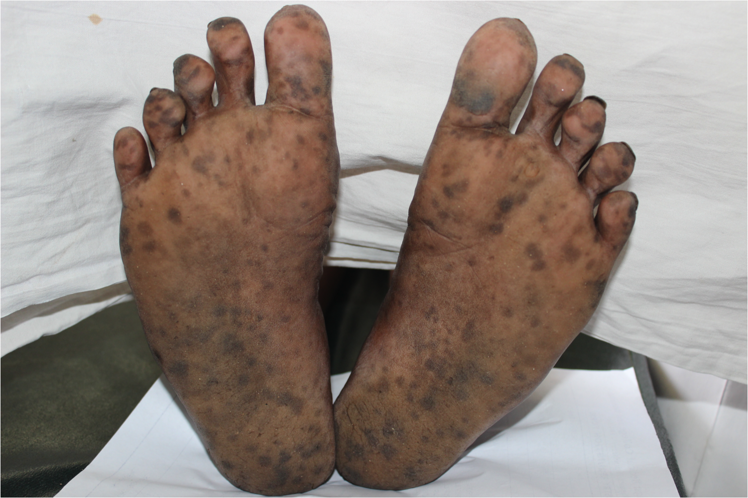
Laugier-Hunziker Syndrome. Multiple well-defined lentiginous macules over plantar aspect of bilateral feet.
Contributed by S Aboobacker, MD
References
Wei Z, Li GY, Ruan HH, Zhang L, Wang WM, Wang X. Laugier-Hunziker syndrome: A case report. Journal of stomatology, oral and maxillofacial surgery. 2018 Apr:119(2):158-160. doi: 10.1016/j.jormas.2017.12.003. Epub 2017 Dec 12 [PubMed PMID: 29246753]
Level 3 (low-level) evidenceDupré A, Viraben R. Laugier's disease. Dermatologica. 1990:181(3):183-6 [PubMed PMID: 2269375]
Montebugnoli L, Grelli I, Cervellati F, Misciali C, Raone B. Laugier-hunziker syndrome: an uncommon cause of oral pigmentation and a review of the literature. International journal of dentistry. 2010:2010():525404. doi: 10.1155/2010/525404. Epub 2010 Jul 7 [PubMed PMID: 20671949]
Level 3 (low-level) evidenceMoore RT,Chae KA,Rhodes AR, Laugier and Hunziker pigmentation: a lentiginous proliferation of melanocytes. Journal of the American Academy of Dermatology. 2004 May [PubMed PMID: 15097932]
Level 3 (low-level) evidenceNayak RS, Kotrashetti VS, Hosmani JV. Laugier-Hunziker syndrome. Journal of oral and maxillofacial pathology : JOMFP. 2012 May:16(2):245-50. doi: 10.4103/0973-029X.99079. Epub [PubMed PMID: 22923898]
Sachdeva S, Sachdeva S, Kapoor P. Laugier-hunziker syndrome: a rare cause of oral and acral pigmentation. Journal of cutaneous and aesthetic surgery. 2011 Jan:4(1):58-60. doi: 10.4103/0974-2077.79199. Epub [PubMed PMID: 21572687]
Level 3 (low-level) evidenceMakhoul EN, Ayoub NM, Helou JF, Abadjian GA. Familial Laugier-Hunziker syndrome. Journal of the American Academy of Dermatology. 2003 Aug:49(2 Suppl Case Reports):S143-5 [PubMed PMID: 12894104]
Level 3 (low-level) evidenceDuan N,Zhang YH,Wang WM,Wang X, Mystery behind labial and oral melanotic macules: Clinical, dermoscopic and pathological aspects of Laugier-Hunziker syndrome. World journal of clinical cases. 2018 Sep 26 [PubMed PMID: 30283795]
Level 3 (low-level) evidenceLampe AK, Hampton PJ, Woodford-Richens K, Tomlinson I, Lawrence CM, Douglas FS. Laugier-Hunziker syndrome: an important differential diagnosis for Peutz-Jeghers syndrome. Journal of medical genetics. 2003 Jun:40(6):e77 [PubMed PMID: 12807976]
Level 3 (low-level) evidenceWang WM, Wang X, Duan N, Jiang HL, Huang XF. Laugier-Hunziker syndrome: a report of three cases and literature review. International journal of oral science. 2012 Dec:4(4):226-30. doi: 10.1038/ijos.2012.60. Epub 2012 Nov 23 [PubMed PMID: 23174847]
Level 3 (low-level) evidenceAsati DP, Tiwari S. Laugier-Hunziker syndrome. Indian journal of dermatology, venereology and leprology. 2011 Jul-Aug:77(4):536. doi: 10.4103/0378-6323.82422. Epub [PubMed PMID: 21727718]
Level 3 (low-level) evidenceJabbari A,Gonzalez ME,Franks AG Jr,Sanchez M, Laugier Hunziker syndrome. Dermatology online journal. 2010 Nov 15 [PubMed PMID: 21163174]
Level 3 (low-level) evidenceWondratsch H, Feldmann R, Steiner A, Breier F. Laugier-hunziker syndrome in a patient with pancreatic cancer. Case reports in dermatology. 2012 May:4(2):174-6. doi: 10.1159/000342070. Epub 2012 Aug 16 [PubMed PMID: 22949900]
Level 3 (low-level) evidenceErgun S, Saruhanoğlu A, Migliari DA, Maden I, Tanyeri H. Refractory Pigmentation Associated with Laugier-Hunziker Syndrome following Er:YAG Laser Treatment. Case reports in dentistry. 2013:2013():561040. doi: 10.1155/2013/561040. Epub 2013 Dec 3 [PubMed PMID: 24367727]
Level 3 (low-level) evidenceAbduljabbar T, Vohra F, Akram Z, Ghani SMA, Al-Hamoudi N, Javed F. Efficacy of surgical laser therapy in the management of oral pigmented lesions: A systematic review. Journal of photochemistry and photobiology. B, Biology. 2017 Aug:173():353-359. doi: 10.1016/j.jphotobiol.2017.06.016. Epub 2017 Jun 15 [PubMed PMID: 28641206]
Level 1 (high-level) evidencePereira PM,Rodrigues CA,Lima LL,Reyes SA,Mariano AV, Do you know this syndrome? Anais brasileiros de dermatologia. 2010 Sep-Oct [PubMed PMID: 21152810]
Level 3 (low-level) evidenceMiličević T, Žaja I, Tešanović D, Radman M. Laugier-Hunziker syndrome in endocrine clinical practice. Endocrinology, diabetes & metabolism case reports. 2018:2018():. pii: EDM180025. doi: 10.1530/EDM-18-0025. Epub 2018 Jul 26 [PubMed PMID: 30087778]
Niiyama S, Katsuoka K. Laugier-Hunziker syndrome. European journal of dermatology : EJD. 2013 Apr 1:23(2):284-5. doi: 10.1684/ejd.2013.2000. Epub [PubMed PMID: 23567753]
Level 3 (low-level) evidenceBarman PD, Das A, Mondal AK, Kumar P. Laugier-Hunziker Syndrome Revisited. Indian journal of dermatology. 2016 May-Jun:61(3):338-9. doi: 10.4103/0019-5154.182429. Epub [PubMed PMID: 27293265]