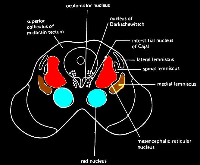
Introduction
The interstitial nucleus of Cajal is a prominent group of cells within the medial longitudinal fasciculus of the brainstem that is responsible for maintaining oculomotor control, head posture, and vertical eye movement. The interstitial nucleus of Cajal is essential in integrating velocity signals from the gaze control system into position signals when maintaining eye and head posture.
Structure and Function
Register For Free And Read The Full Article
Search engine and full access to all medical articles
10 free questions in your specialty
Free CME/CE Activities
Free daily question in your email
Save favorite articles to your dashboard
Emails offering discounts
Learn more about a Subscription to StatPearls Point-of-Care
Structure and Function
The interstitial nucleus of Cajal is lateral to the Edinger-Westphal nucleus and ventrolateral to the periaqueductal gray in the rostral part of the midbrain.[1] It is the most prominent cell group within the medial longitudinal fasciculus.[2] It contains small and medium-sized cells with large neurons that project to the vestibular nuclei, including the medial and descending vestibular nuclei, the superior vestibular nucleus, and the lateral vestibular nucleus. These cells belong to at least two different classes: large pyramidal or multipolar neurons and small or medium pyramidal, fusiform, and round cells.[2] Previous studies have demonstrated that stimulating the interstitial nucleus of Cajal can influence a large portion of the vestibular nuclei neurons that are related to horizontal and vertical semicircular canals.[2][3] Bilateral lesions close to the interstitial nucleus of Cajal have also been shown to result in an inability to maintain eccentric vertical eye position after saccades, phase advance, and a smaller response of the vestibulo-ocular reflex.[4] Lesions within the interstitial nucleus of Cajal have been shown to not allow monkeys and cats from maintaining eccentric gaze and impede their vertical vestibulo-ocular responses.[4] Since the interstitial nucleus of Cajal has many neurons that are related to vertical eye movement during saccades, the vestibulo-ocular response, smooth pursuit, and optokinetic eye movements, the interstitial nucleus of Cajal is essential for vertical eye movement.[3] However, the interstitial nucleus of Cajal has also been shown not to be able to generate eye position signals alone but has to collaborate with other regions during vertical eye movement.[5] The interstitial nucleus of Cajal also incorporates velocity commands from the gaze control system into position signals for eye and head posture. The right interstitial nucleus of Cajal encodes clockwise-up and clockwise-down eye and head movements while the left interstitial nucleus of Cajal encodes counterclockwise-up and counterclockwise-down movements. Neurons of the interstitial nucleus of Cajal also usually encode the vertical position of the eyes.[2]
Embryology
Previous studies have demonstrated that in the growth of the central nervous system in vertebrates, axons from the interstitial nucleus of Cajal along with the mesencephalic trigeminal nucleus contribute to the formation of the trajectories of the medial longitudinal fasciculus and the lateral longitudinal fasciculus.[6] Neurons from the interstitial nucleus of Cajal arise from a region near the border between the diencephalon and the mesencephalon.[7] They initially move ventrally toward the midline and turn caudally to generate the medial longitudinal fasciculus that goes toward the spinal cord.[8]
Blood Supply and Lymphatics
The medial longitudinal fasciculus and the interstitial nucleus of Cajal both receive blood supply from the basilar artery. The basilar artery supplies the brainstem and cerebellum and provides circulation to the posterior part of the brain via the posterior cerebral arteries.[9]
Nerves
The medial longitudinal fasciculus, the tract which ascends to the interstitial nucleus of Cajal, is closely associated with the oculomotor nerve, trochlear nerve, and abducens nerve. These nerves form the extraocular muscle system because they help move the eyes in parallel to have normal conjugate gaze. For the horizontal portion of the oculomotor system, rightward movements get generated by stimulating nuclei of the interstitial nucleus of Cajal to the right of the midline and leftward movements are generated by stimulating nuclei of the interstitial nucleus of Cajal to the left of the midline.[10] For the vertical component of the oculomotor system, nuclei of the interstitial nucleus of Cajal on the right of the midline encode clockwise-up and clockwise-down movements while nuclei of the interstitial nucleus of Cajal on the left encode counterclockwise-up and counterclockwise-down eye movements.[10]
Several projection systems originate from the interstitial nucleus of Cajal. The first projection system goes past the posterior commissure that later transmits many terminal fields in the interstitial nucleus of Cajal on the opposite side of the body, the oculomotor nucleus, and the trochlear nucleus.[10] The second system relays terminal fields within pontine and medullary nuclei on the same side of the body and the ventral segment of the cervical spinal nerves. Finally, the third system sends terminal fields within the mesencephalic and diencephalic structures on the same side of the body. Maintaining the structure of the first projection system is essential for integrating position signals normally from velocity signals in the vertical axis.[10] This is because lesions after the posterior commissure can cause the eyes to lose their ability to maintain eccentric eye movements in the vertical axis and decrease their vertical vestibulo-ocular response.
Surgical Considerations
Previous studies suggested stimulation of the interstitial nucleus of Cajal and its cerebellar inputs may function as a future strategy to treat cervical dystonia (also known as spasmodic torticollis).[11] Cervical dystonia is a rare disorder in which one starts to constrict their neck muscles without control, causing the head to lean forward or backward without restraint. For instance, Hassler and Hess used monopolar electrical stimulation of the interstitial nucleus of Cajal to elicit head movements in humans.[12] Hassler later targeted the efferent portion of the interstitial nucleus of Cajal to treat cervical dystonia and the prestitial nucleus for retrocollis.[13] Even though they abandoned these procedures because the results were unpredictable, they provide an avenue for clinicians to develop more refined techniques that might lead to more effective surgical treatments for cervical dystonia.
Clinical Significance
As stated previously, the interstitial nucleus of Cajal is essential in the vertical eye movement during saccades, the vestibulo-ocular reflex, smooth pursuit, and optokinetic eye movements.[3] The interstitial nucleus of Cajal is essential in maintaining a steady vertical eccentric gaze. Lesions in this structure can result in nystagmus in upward or downward gaze. Inactivation of the interstitial nucleus of Cajal on the right side has also been shown to result in drifting of the head in a counterclockwise direction while inactivation of the interstitial nucleus of Cajal on the left side has been shown to result in drifting of the head in a clockwise direction.[10] The interstitial nucleus of Cajal also incorporates velocity commands from the gaze control system into position signals for eye and head posture. The right interstitial nucleus of Cajal encodes clockwise-up and clockwise-down eye and head movements while the left interstitial nucleus of Cajal encodes counterclockwise-up and counterclockwise-down movements.[10] Any lesion of the interstitial nucleus of Cajal can cause abnormal turning and twisting of the head. The interstitial nucleus of Cajal also incorporates velocity commands from the gaze control system into position signals for eye and head posture. Moreover, neurons of the interstitial nucleus of Cajal usually encode the vertical position of the eyes. Stimulating the interstitial nucleus of Cajal and its cerebellar inputs may also lead to more refined techniques that could help treat cervical dystonia in the future.[11]
Media
References
Fukushima K. The interstitial nucleus of Cajal and its role in the control of movements of head and eyes. Progress in neurobiology. 1987:29(2):107-92 [PubMed PMID: 3108957]
Level 3 (low-level) evidenceDalezios Y, Scudder CA, Highstein SM, Moschovakis AK. Anatomy and physiology of the primate interstitial nucleus of Cajal. II. Discharge pattern of single efferent fibers. Journal of neurophysiology. 1998 Dec:80(6):3100-11 [PubMed PMID: 9862908]
Level 3 (low-level) evidenceFukushima K, Takahashi K, Fukushima J, Ohno M, Kimura T, Kato M. Effects of lesion of the interstitial nucleus of Cajal on vestibular nuclear neurons activated by vertical vestibular stimulation. Experimental brain research. 1986:64(3):496-504 [PubMed PMID: 3803487]
Level 3 (low-level) evidenceFukushima K. The interstitial nucleus of Cajal in the midbrain reticular formation and vertical eye movement. Neuroscience research. 1991 Apr:10(3):159-87 [PubMed PMID: 1650435]
Level 3 (low-level) evidenceKokkoroyannis T, Scudder CA, Balaban CD, Highstein SM, Moschovakis AK. Anatomy and physiology of the primate interstitial nucleus of Cajal I. efferent projections. Journal of neurophysiology. 1996 Feb:75(2):725-39 [PubMed PMID: 8714648]
Level 3 (low-level) evidenceMolle KD, Chédotal A, Rao Y, Lumsden A, Wizenmann A. Local inhibition guides the trajectory of early longitudinal tracts in the developing chick brain. Mechanisms of development. 2004 Feb:121(2):143-56 [PubMed PMID: 15037316]
Level 3 (low-level) evidenceKlier EM, Wang H, Constantin AG, Crawford JD. Midbrain control of three-dimensional head orientation. Science (New York, N.Y.). 2002 Feb 15:295(5558):1314-6 [PubMed PMID: 11847347]
Level 3 (low-level) evidenceLakke EA. The projections to the spinal cord of the rat during development: a timetable of descent. Advances in anatomy, embryology, and cell biology. 1997:135():I-XIV, 1-143 [PubMed PMID: 9257458]
Level 3 (low-level) evidenceAdigun OO, Reddy V, Sevensma KE. Anatomy, Head and Neck: Basilar Artery. StatPearls. 2023 Jan:(): [PubMed PMID: 29083786]
Klier EM, Wang H, Crawford JD. Interstitial nucleus of cajal encodes three-dimensional head orientations in Fick-like coordinates. Journal of neurophysiology. 2007 Jan:97(1):604-17 [PubMed PMID: 17079347]
Level 3 (low-level) evidenceShaikh AG, Zee DS, Crawford JD, Jinnah HA. Cervical dystonia: a neural integrator disorder. Brain : a journal of neurology. 2016 Oct:139(Pt 10):2590-2599 [PubMed PMID: 27324878]
HASSLER R, HESS WR. [Experimental and anatomical findings in rotatory movements and their nervous apparatus]. Archiv fur Psychiatrie und Nervenkrankheiten, vereinigt mit Zeitschrift fur die gesamte Neurologie und Psychiatrie. 1954:192(5):488-526 [PubMed PMID: 13229347]
Hassler R, Dieckmann G. Stereotactic treatment of different kinds of spasmodic torticollis. Confinia neurologica. 1970:32(2):135-43 [PubMed PMID: 4926178]
