Introduction
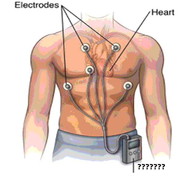
A Holter monitor is an ambulatory electrocardiographic system discovered by Dr Norman J Holter and his team in 1957 (see Image. Holter Monitor). Little did they know that their device would change how we track cardiac rhythms outside the hospital.
Historically electrocardiography began in 1893 with the work of Einthovens string galvanometer.[1] The Holter monitor is a device that works on Galvanometer's principle to record electrocardiographic signals from an individual going about his daily activities, like continuous ambulatory electrocardiography. From 1961, various other modalities and advanced gadgets have been manufactured for this purpose.[2] Since then, it has formed the backbone of rhythm detection and analysis in Cardiac Electrophysiology.
Indications
Register For Free And Read The Full Article
Search engine and full access to all medical articles
10 free questions in your specialty
Free CME/CE Activities
Free daily question in your email
Save favorite articles to your dashboard
Emails offering discounts
Learn more about a Subscription to StatPearls Point-of-Care
Indications
No relevant recommendations are available in the literature to distinguish patients who may benefit from the application of ambulatory electrocardiogram monitoring. The present proposals are as follows:
- To establish the link between palpitations and abnormal heart rhythms
- To diagnose the cause of syncope or near syncope
- To evaluate transient episodes of cardiac arrhythmias or myocardial ischemia
- Patients with neurologic events when transient atrial fibrillation or flutter is suspected
- To monitor the efficacy and safety of pharmacological or nonpharmacological therapies
- To detect proarrhythmic responses to antiarrhythmic therapy in patients at high-risk
- To analyze the function of pacemakers or other implantable devices
- To evaluate prognosis
- To stratify the risk of sudden cardiac death [3]
The selection between the common 2 to 3-lead or the 12-lead Holter ECG monitoring depends mostly on the desired goal. 2 to 3 leads are sufficient to monitor heart rate and rhythm. In contrast, if the purpose is to establish the origin of premature beats/ dysrhythmias or tachycardia, then a 12-lead Holter electrocardiography is preferred.
The selection of which monitor to use depends on the frequency of symptoms. When symptoms are continuous, a routine 12-lead electrocardiogram is sufficient to make a diagnosis. Usually, Cardiologists use the Holter monitor for intermittent symptoms. If symptoms rarely occur, devices for longer duration, like an implantable loop recorder (ILR) or event monitor, can be used.[4]
A 12-lead Holter monitor is very accurate and can instantly diagnose supraventricular tachycardia (SVT), ventricular tachycardia (VT), atrial flutter, atrial fibrillation, monomorphic or Polymorphic VTs, long QT syndrome, supraventricular premature complexes, ventricular premature complexes, dominant atrioventricular accessory pathways, atrioventricular block, right and left bundle branch block, and left anterior and posterior fascicular block.[5] Current European guidelines endorse the use of an intracardiac monitor for patients diagnosed with a cryptogenic stroke.
Contraindications
The Holter monitor is contraindicated when:
- It delays urgent treatment, hospitalization, or a procedure. For example, it should not be part of the initial investigation for angina, where a stress test would be more appropriate.
- The patient has syncope and high-risk factors, at which time inpatient management is mandatory.
- The patient has symptoms such as syncope, near-syncope, episodic dizziness, or palpitation in which other clear causes have been identified by history, physical examination, or laboratory tests.
- The ACC/AHA guidelines discouraged using ambulatory ECG for either arrhythmia detection or analysis of heart rhythm variability for risk assessment in patients without arrhythmia symptoms, even if they had cardiovascular conditions such as left ventricular hypertrophy or valvular heart disease.
- A patient who refuses to undergo further therapy once arrhythmia is established
- Routine screening of asymptomatic patients.
Equipment
The initial Holter monitor was about the size of a briefcase and comprised an amplifier, tape recorder, electrodes attached to the individual's chest, playback unit, and analyzing unit. It consisted of 10 electrocardiogram leads, including 6 standard precordial and 4 torso electrodes. The function of the torso leads is to prevent signal disturbances. However, torso leads can show variations. To overcome these variations, investigators proposed mathematical algorithms.[6] While initially consisting of 12 leads, recent models have comprised as little as 2 to 3 leads.[7]
After continuous improvement and progress, the Holter is now the size of a small cell phone and gives 2 types of primary data to analyze. One is the QRS complex, and the other is the R-R interval. It continuously records until detached from the patient or runs out of power, although it is usually used for 24 to 48 hours. The power supply lasts 80-100 hours with a tape recording capacity of 10 hours.
Personnel
The physician explains the procedure and should educate the patient about how to record the event if it happens. A physician should inform patients regarding serious events like chest pain, shortness of breath, or lightheadedness. The device should be interrogated within 24 hours of the event to prevent further events and to take necessary actions if needed.
Preparation
Unlike invasive procedures, no preparation is needed, but the patient should be advised to keep the monitor away from other electrical devices while wearing the device. The physician should recommend not putting lotion or moisturizer on the chest as it affects the attachment of leads.[8]
Technique or Treatment
Holter monitoring continuously records an Electrocardiogram (EKG) tracing on 3 channels for 12 to 48 hours. The patient activates a button to correlate the timing of symptoms with the EKG. Cardiac technicians and skilled physicians examine the entire recorded EKG for abnormalities.[9]
In children, Holter monitor use is limited because of wires and cables. In 2011, the Federal Drug Administration approved a wireless, water-resistant device that allows up to 14 days of continuous single-channel rhythm monitoring. The patients can trigger symptoms by pressing a button to start the recording and correlate symptoms with concurrent rhythm. A study that compared a new wireless device with a standard Holter monitor found that the new wireless adhesive monitoring patch diagnosed more arrhythmic events than the Holter monitor.[10][11]
Complications
The device stays in a pocket outside of the body close to the patient's chest, usually in the form of a neck sling or the patient's vest pocket. So theoretically, there aren't any complications, but the surface electrodes can irritate the underlying skin and, if left in place, cause skin ulceration. However, this rarely happens as the device technicians remove them when they remove the machine from the patient.
Clinical Significance
Utilization of the Holter monitor has significantly increased, especially in the detection of occult atrial fibrillation as a cause of a cryptogenic stroke. Anticoagulation is always superior to antiplatelet in the secondary prevention of stroke due to atrial fibrillation. So, the diagnosis of occult atrial fibrillation to initiate anticoagulation with the help of the Holter monitor can prevent recurrent strokes.[12] Patients with LV systolic dysfunction and presumed arrhythmic events, as well as those with symptoms supposed to be due to transient second or third-degree heart blocks, also form a significant number of people who undergo the test.
Enhancing Healthcare Team Outcomes
Holter monitors are excellent resources to look for and diagnose cardiac arrhythmias in the appropriate patient populace. Clinicians and physician assistants should be aware of the possibility of arrhythmias as they usually present with non-specific symptoms. Hence, a high index of suspicion and prompt referral/ recommendation is essential in this regard.
With the advent of highly effective pharmacotherapy and interventional treatment of cardiovascular ailments, the pool of survivors and life expectancy is ever-growing. With this, the potential substrate for arrhythmias is also increasing daily, so Holter monitors can provide more information when appropriately utilized.
Media
(Click Image to Enlarge)
References
HOLTER NJ, GENERELLI JA. Remote recording of physiological data by radio. Rocky Mountain medical journal. 1949 Sep:46(9):747-51 [PubMed PMID: 18137532]
Pevnick JM, Birkeland K, Zimmer R, Elad Y, Kedan I. Wearable technology for cardiology: An update and framework for the future. Trends in cardiovascular medicine. 2018 Feb:28(2):144-150. doi: 10.1016/j.tcm.2017.08.003. Epub 2017 Aug 9 [PubMed PMID: 28818431]
Giada F, Bartoletti A. Value of Ambulatory Electrocardiographic Monitoring in Syncope. Cardiology clinics. 2015 Aug:33(3):361-6. doi: 10.1016/j.ccl.2015.04.004. Epub [PubMed PMID: 26115822]
Diemberger I, Gardini B, Martignani C, Ziacchi M, Corzani A, Biffi M, Boriani G. Holter ECG for pacemaker/defibrillator carriers: what is its role in the era of remote monitoring? Heart (British Cardiac Society). 2015 Aug:101(16):1272-8. doi: 10.1136/heartjnl-2015-307614. Epub 2015 May 22 [PubMed PMID: 26001846]
Wang R, Blackburn G, Desai M, Phelan D, Gillinov L, Houghtaling P, Gillinov M. Accuracy of Wrist-Worn Heart Rate Monitors. JAMA cardiology. 2017 Jan 1:2(1):104-106. doi: 10.1001/jamacardio.2016.3340. Epub [PubMed PMID: 27732703]
Jakicic JM, Davis KK, Rogers RJ, King WC, Marcus MD, Helsel D, Rickman AD, Wahed AS, Belle SH. Effect of Wearable Technology Combined With a Lifestyle Intervention on Long-term Weight Loss: The IDEA Randomized Clinical Trial. JAMA. 2016 Sep 20:316(11):1161-1171. doi: 10.1001/jama.2016.12858. Epub [PubMed PMID: 27654602]
Level 1 (high-level) evidenceMâsse LC, Fuemmeler BF, Anderson CB, Matthews CE, Trost SG, Catellier DJ, Treuth M. Accelerometer data reduction: a comparison of four reduction algorithms on select outcome variables. Medicine and science in sports and exercise. 2005 Nov:37(11 Suppl):S544-54 [PubMed PMID: 16294117]
Prabhu S, Taylor AJ, Costello BT, Kaye DM, McLellan AJA, Voskoboinik A, Sugumar H, Lockwood SM, Stokes MB, Pathik B, Nalliah CJ, Wong GR, Azzopardi SM, Gutman SJ, Lee G, Layland J, Mariani JA, Ling LH, Kalman JM, Kistler PM. Catheter Ablation Versus Medical Rate Control in Atrial Fibrillation and Systolic Dysfunction: The CAMERA-MRI Study. Journal of the American College of Cardiology. 2017 Oct 17:70(16):1949-1961. doi: 10.1016/j.jacc.2017.08.041. Epub 2017 Aug 27 [PubMed PMID: 28855115]
Chai PR. Wearable Devices and Biosensing: Future Frontiers. Journal of medical toxicology : official journal of the American College of Medical Toxicology. 2016 Dec:12(4):332-334 [PubMed PMID: 27352082]
George L, Gargiulo GD, Lehmann T, Hamilton TJ. Concept Design for a 1-Lead Wearable/Implantable ECG Front-End: Power Management. Sensors (Basel, Switzerland). 2015 Nov 19:15(11):29297-315. doi: 10.3390/s151129297. Epub 2015 Nov 19 [PubMed PMID: 26610497]
Lobodzinski SS. ECG patch monitors for assessment of cardiac rhythm abnormalities. Progress in cardiovascular diseases. 2013 Sep-Oct:56(2):224-9. doi: 10.1016/j.pcad.2013.08.006. Epub [PubMed PMID: 24215754]
Albers GW, Bernstein RA, Brachmann J, Camm J, Easton JD, Fromm P, Goto S, Granger CB, Hohnloser SH, Hylek E, Jaffer AK, Krieger DW, Passman R, Pines JM, Reed SD, Rothwell PM, Kowey PR. Heart Rhythm Monitoring Strategies for Cryptogenic Stroke: 2015 Diagnostics and Monitoring Stroke Focus Group Report. Journal of the American Heart Association. 2016 Mar 15:5(3):e002944. doi: 10.1161/JAHA.115.002944. Epub 2016 Mar 15 [PubMed PMID: 27068633]
