Introduction
Progressive collapsing foot deformity, previously known as adult-acquired flatfoot or posterior tibial tendon dysfunction, is a complex pathology defined by the collapse of the medial longitudinal arch of the foot with continued progressive deformity of the foot and ankle (see Image. Progressive Collapsing Foot Deformity).[1] Progressive collapsing foot deformity is a debilitating condition that affects up to 5 million people in the United States.[2]
The anatomy of the foot and ankle are complex, with multiple structures involved in the stability and function needed to walk and bear weight. The posterior tibial tendon is a structure principally engaged in the development of progressive collapsing foot deformity. In addition to plantar flexion, the posterior tibial tendon is the primary inverter of the foot. This tendon inserts principally on the navicular tuberosity but has lesser insertions on other tarsal and metatarsal structures.[3] The spring and deltoid ligaments are crucial to the stability of the foot and ankle. The spring ligament, the most frequently involved in progressive collapsing foot deformity, supports the ankle by connections from the sustentaculum tali of the calcaneus to the navicular. The spring ligament supports the head of the talus.[4] The deltoid ligament is usually affected later in the progression of the adult-acquired flatfoot. The superficial deltoid ligament has a wide insertion on the navicular to the posterior tibiotalar capsule. This ligament is the primary support against tibiotalar valgus angulation. The deep deltoid ligament prevents axial rotation of the talus, where the ligament inserts from the origin on the intercollicular groove and posterior colliculus. The deltoid ligament is critical in supporting the articulating surfaces of the ankle and the spring ligament.[5]
Acquired flatfoot grading utilizes the Johnson and Strom classification system, which has classification grades of I to III.[6][7] Myerson added a fourth grade in 1997. The classification system aids clinicians in identifying the severity and can also guide treatment plans. Characteristically, stage I disease presents with posterior tibial tendon tenosynovitis with no arch collapse. Patients with stage II progressive collapsing foot deformity will have foot collapse and will be unable to perform a single-leg heel rise. This stage is further subcategorized into stages IIa and IIb. Stage IIa is foot collapse with valgus deformity of the hindfoot but no midfoot abduction, while in stage IIb, midfoot abduction is present. Patients with stage III progressive collapsing foot deformity will have fixed deformity with hindfoot valgus and forefoot abduction. Patients with stage IV deformity will have ankle valgus secondary to deltoid ligament attenuation.
The classification systems enable clinicians to identify the severity of the condition and can also guide treatment plans. However, over the past few years, a better understanding of the biomechanics of the medial longitudinal arch and the progression of flatfoot led to a new classification system. The newly accepted terminology of progressive collapsing foot deformity acknowledges the involvement of soft tissue structures and joint alignment of the midfoot, rearfoot, and ankle.[7]
Etiology
Register For Free And Read The Full Article
 Search engine and full access to all medical articles
- 10 free questions in your specialty
- Free CME/CE Activities
- Free daily question in your email
- Save favorite articles to your dashboard
- Emails offering discounts
Learn more about a Subscription to StatPearls Point-of-Care
Etiology
Acquired flatfoot was historically attributed to posterior tibial tendon deficiency. However, now the understanding is that the ligamentous structures of the ankle play a role in foot deformity development.[1][7] The dysfunction of the posterior tibial tendon is a multifactorial process. In many patients, preexisting flatfoot is common, and many patients are also obese. Another factor is the relative hypovascularity of the tendon during an abrupt turn posterior to the medial malleolus; this predisposes the tendon to rupture due to insufficient repair.[8] Episodes of previous trauma, corticosteroid injections, arthritis, neuromuscular conditions, and diabetes all predispose to the development of progressive collapsing foot deformity.[9]
Epidemiology
Acquired flatfoot is a common orthopedic condition, although the paucity of literature on the epidemiology regarding progressive collapsing foot deformity is apparent. 5 million people in the United States are affected by the condition. The estimated prevalence in the United Kingdom is over 3% in women over 40.[2][10] Posterior tibial tendon issues are prevalent in older populations, with 10% of patients affected. Ikpeze et al postulated that the geriatric population might be predisposed to more severe progressive collapsing foot deformity due to the degeneration of muscle mass and bone structure.[11] Patients with chronic vascular diseases are at increased risk; this includes patients with diabetes or hypertension.[9]
Pathophysiology
The posterior tibial tendon is critical in maintaining the appropriate gait and function of the foot. Contraction of the tibialis posterior causes the inversion of the foot and locking of the transverse tarsal joints, granting stability for push-off. Deficiency of the posterior tibial tendon leads to an unstable transverse tarsal joint and forefoot abduction, allowing unopposed action of the peroneus brevis. The ensuing abduction of the forefoot and transverse talar joints also displace the force of the calcaneal tendon laterally, accentuating the valgus defect of the foot.[12] The continued strain on the static stabilizing ligaments eventually leads to their attenuation. Most commonly, the spring ligament is affected, and failure leads to medial and plantar subluxation of the talar head relative to the navicular. Failure of the deltoid ligament leads to a valgus position of the talus within the ankle mortise.[1][3][13][9] These events may occur stepwise, corresponding to the Johnson and Strom classification system. However, some patients develop a valgus talar tilt without a fixed flatfoot deformity.[5] In addition, metabolic and genetic conditions can predispose the disorganization of collagen fibers and contribute to polymorphism of collagen types III, V, and matrix metalloproteinases 1, 8, and 13. The pathology contributes to tendon thickening and impaired healing.[14][15]
History and Physical
Evaluating the patient with suspected progressive collapsing foot deformity during weight-bearing is essential. When the patient is standing, excessive forefoot abduction can be noted by the “too many toes” sign. This test is positive if the clinician can see more than the fifth and part of the fourth toe. Inspection and palpation over the posterior tibial tendon at the area of the medial malleolus may demonstrate swelling or pain. Pain may be elicited on the foot's lateral aspect if ligamentous structures are impinged.[16]
Single and double heel rise tests may be performed to assess the strength of the posterior tibial tendon. Failure to invert the foot on heel rise or perform heel rise indicates posterior tibial tendon dysfunction. Ankle range of motion requires assessment. Efforts should be concerted to correct valgus deformity in a range of motion exercise to evaluate for fixed deformity. A fixed deformity of the subtalar joint or fixed abduction of the forefoot has implications in treatment.[3]
Evaluation
The gold standard for evaluating progressive collapsing foot deformity is weight-bearing radiographs. However, weight-bearing computed tomography is a reliable imaging tool.[17] Radiographs are necessary in the anteroposterior, lateral, and hindfoot views. These views will assess the degree of arch collapse, particularly by measuring the lateral first tarsometatarsal angle and forefoot abduction at the talonavicular joint. Talar head exposure can also be measured in the setting of stage IV disease by the lateral talonavicular angle (see Image. Radiograph of Progressive Collapsing Foot Deformity). Lateral views of the foot can also display naviculocuneiform and first tarsometatarsal collapse. Arthritis should be carefully assessed on all radiographic films as this may have implications for treatment options.[16]
Magnetic resonance imaging (MRI) is typically unnecessary to evaluate progressive collapsing foot deformity; however, imaging may benefit some patients with ligamentous involvement to alter surgical treatment planning. This is particularly relevant for patients with medial peritalar instability, who have improved functional outcomes with deltoid-spring ligament reconstruction.[18] In 2020, MRI was voted for removal in the new classification system.[19] Several studies indicate that ultrasound may be useful in assessing the posterior tibial tendon compared to the more time-consuming and costly MRI. Results from an ultrasonographic evaluation of the posterior tibial tendon were equivalent to MRI in 87% to 94% of patients.[20][21]
Treatment / Management
Treating progressive collapsing foot deformity is complex. Multiple treatment options are available, and the treatment of foot deformity has been the focus of most recent research. However, first-line therapy for the condition remains non-invasive. Treatment with orthotic devices, such as low-articulating ankle-foot orthosis, cast-boot walkers, and other ankle-foot orthoses, in conjunction with nonsteroidal anti-inflammatory drugs (NSAIDs) and physical therapy, have a resolution of symptoms in 87% of patients, according to one study. Other studies have demonstrated results with success rates of 67% to 90% using conservative measures.[16]
Surgical treatment is indicated in patients who have attempted conservative therapy and are not satisfied with their results. Surgical treatment depends on the stage of the disease, as well as other factors, including medical comorbidities, functional status, and use of tobacco. Surgical management of stage I disease is uncommon, but if required, patients should undergo posterior tibial tendon tenosynovectomy, debridement, or flexor digitorum longus tendon autograft. Repair of the posterior tibial tendon may cause complications with long-term failure; therefore, surgical treatment should be carefully considered before any intervention.[13]
Stage II disease treatment has succeeded by medializing calcaneal osteotomy and a flexor digitorum longus transfer. Results from one study showed 87% patient satisfaction with this treatment after a mean follow-up of 15 years.[22] Stage III disease is more challenging to treat due to the fixed nature of the defect. Arthrodesis is the standard of care, with common double and triple arthrodesis. Double arthrodesis entails a fusion of the subtalar and talonavicular joints. Triple arthrodesis involves the calcaneocuboid joint and the 2 joints mentioned above. Double arthrodesis has the advantage of reduced arthritic complications and reduced joint stiffness associated with calcaneocuboid fusion. Unfortunately, joint fusion has an inherent loss of mobility, and patients may struggle on uneven ground. Risk of nonunion, deltoid insufficiency, and ankle valgus is also present.
Stage IV disease management depends on whether rigid flatfoot has developed. Some patients progress to stage IV disease without rigid disease due to the failure of the deltoid ligament. In this setting, patients may have treatment with a deltoid ligament repair. Patients with rigid stage IV disease will require an ankle fusion, which is associated with significant morbidity. Ankle replacement is an alternative to arthrodesis.[3][23](B2)
A recent meta-analysis of surgical treatments for progressive collapsing foot deformity was conducted to study therapy efficacy further. This study analyzed different radiographic angles to measure the effectiveness of each procedure, including medial calcaneal osteotomies, lateral column lengthening, and double and triple arthrodesis. The results suggested that all treatment types result in significant improvements for the patient.[24](A1)
The orthopedic consensus agreed on 3 statements concerning the operative treatment of progressive collapsing foot deformity: mobility and range of motion should always be prioritized, especially in younger patients. Talonavicular joint fusion should be considered only in arthritic joints, important sagittal plane sagging, severe deformity, inadequate correction of talonavicular abduction, and patients with higher body mass index will generally do worse with reconstructive osteotomies when compared to arthrodesis.[25](B3)
Differential Diagnosis
While the diagnosis of progressive collapsing foot deformity is relatively straightforward, the treating clinician needs to rule out several diagnoses when acquired flatfoot is suspected. These include adult flexible flatfoot, tarsal coalition, Charcot foot, neuromuscular flatfoot, and arthritic, post-traumatic, and iatrogenic deformity. A careful history and physical can rule out most of these etiologies.[26]
Staging
Johnson and Strom Classification (1989) Modified by Myerson (1997):
Stage 1: Normal radiographs, able to perform single-heel raise and mild tenosynovitis
Stage 2A: Arch collapse on a radiograph, unable to perform single-heel raise, and a flexible flatfoot deformity
Stage 2B: Arch collapse and talonavicular exposure (over 40%) on a radiograph, unable to perform single heel raise, flexible flatfoot deformity, and characteristic forefoot abduction or “too many toes” sign
Stage 3: Subtalar arthritis on a radiograph, unable to perform single heel raise, flatfoot deformity with rigid forefoot abduction, and hindfoot valgus
Stage 4: Valgus deformity of talus in the ankle mortise visualized on an anterior-posterior radiograph of the ankle-talar tilt due to deltoid ligament compromise, subtalar arthritis on radiographs, unable to perform single heel raise, flatfoot deformity with rigid forefoot abduction, and hindfoot valgus [19]
Orthopedic Consensus Group (2020 Classifications)
Stage 1 is flexible, and stage 2 is rigid. Both stages 1 and 2 can be subdivided into classes A to E listed below:
Class A: Hindfoot valgus deformity
- Hindfoot valgus alignment
- Increased hindfoot moment arm, hindfoot alignment angle, foot and ankle offset
Class B: Midfoot/forefoot abduction deformity
- Decreased talar head coverage
- Increased talonavicular coverage angle
- Presence of sinus tarsi impingement
Class C: Forefoot varus deformity/medial column instability
- Increased talus-first metatarsal angle
- Plantar gapping first tarsometatarsal joint or naviculocuneiform joints
- Clinical forefoot varus
Class D: Peritalar subluxation/dislocation
- Significant subtalar joint subluxation/subfibular impingement
Class E: Ankle instability
- Valgus tilting of the ankle joint
Prognosis
The prognosis for most patients with progressive collapsing foot deformity is positive. Most patients do not require surgery. Approximately 10% of patients who require surgery can expect favorable outcomes.[16] The higher-grade diseases require more extensive surgery, with stage IV progressive collapsing foot deformity requiring ankle fusion or total ankle arthroplasty.[3]
Complications
If left untreated, progressive collapsing foot deformity can progress to more severe stages that require more invasive treatments. Early diagnosis is critical and can even help avoid surgery in some patients. As the disease progresses into the late stages, the need for the patient to get an ankle fusion increases. With an ankle fusion, the patient essentially has an immobile ankle joint, and many recreational activities are affected.
Consultations
The following consults are recommended for progressive collapsing foot deformity:
- Orthopedic surgeon
- Podiatric surgeon or podiatrist
- Physical therapist
- Orthotist
Deterrence and Patient Education
Progressive collapsing foot deformity appears to be associated with obesity, diabetes, hypertension, and other diseases that are treatable with a healthy lifestyle and weight reduction. Patients may benefit from the treatment of their medical comorbidities. Additionally, patients may prevent further disease progression if an appropriate orthotic device is prescribed. Most patients who use an orthotic device with physical therapy or medications will not need surgical intervention.[16]
Enhancing Healthcare Team Outcomes
Progressive collapsing foot deformity is a common disease that, when recognized early, is highly treatable with excellent outcomes. All healthcare team members can play a role in the identification, treatment, and follow-up. This team may include family clinicians, specialty-trained nurses, orthopedic and foot and ankle surgeons, physical therapists, and radiologists. As with all orthopedic surgeries, a physical therapist must work closely with surgeons to ensure adequate recovery and slow progression to full activity.
Media
(Click Image to Enlarge)
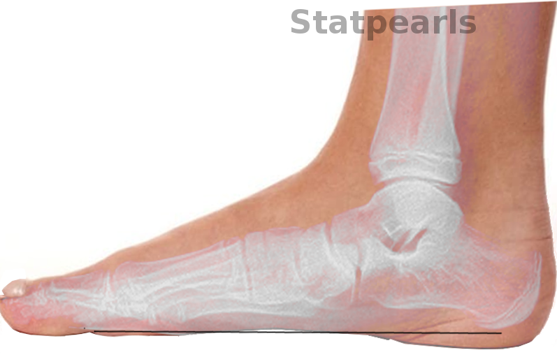
Progressive Collapsing Foot Deformity. A complex pathology defined by the collapse of the medial longitudinal arch of the foot with continued progressive deformity of the foot and ankle.
Contributed by S Bhimji, MD
(Click Image to Enlarge)

Radiograph of Progressive Collapsing Foot Deformity (PCFD). The radiograph demonstrates PCFD with increased talar head exposure and forefoot abduction.
Contributed by MA Dreyer, DPM, FACFAS
References
Deland JT. Adult-acquired flatfoot deformity. The Journal of the American Academy of Orthopaedic Surgeons. 2008 Jul:16(7):399-406 [PubMed PMID: 18611997]
Hadfield MH, Snyder JW, Liacouras PC, Owen JR, Wayne JS, Adelaar RS. Effects of medializing calcaneal osteotomy on Achilles tendon lengthening and plantar foot pressures. Foot & ankle international. 2003 Jul:24(7):523-9 [PubMed PMID: 12921356]
Abousayed MM, Alley MC, Shakked R, Rosenbaum AJ. Adult-Acquired Flatfoot Deformity: Etiology, Diagnosis, and Management. JBJS reviews. 2017 Aug:5(8):e7. doi: 10.2106/JBJS.RVW.16.00116. Epub [PubMed PMID: 28806265]
Bastias GF, Dalmau-Pastor M, Astudillo C, Pellegrini MJ. Spring Ligament Instability. Foot and ankle clinics. 2018 Dec:23(4):659-678. doi: 10.1016/j.fcl.2018.07.012. Epub 2018 Sep 22 [PubMed PMID: 30414659]
Smith JT, Bluman EM. Update on stage IV acquired adult flatfoot disorder: when the deltoid ligament becomes dysfunctional. Foot and ankle clinics. 2012 Jun:17(2):351-60. doi: 10.1016/j.fcl.2012.03.011. Epub 2012 Apr 10 [PubMed PMID: 22541531]
Johnson KA, Strom DE. Tibialis posterior tendon dysfunction. Clinical orthopaedics and related research. 1989 Feb:(239):196-206 [PubMed PMID: 2912622]
Abousayed MM, Tartaglione JP, Rosenbaum AJ, Dipreta JA. Classifications in Brief: Johnson and Strom Classification of Adult-acquired Flatfoot Deformity. Clinical orthopaedics and related research. 2016 Feb:474(2):588-93 [PubMed PMID: 26472584]
Petersen W, Hohmann G, Stein V, Tillmann B. The blood supply of the posterior tibial tendon. The Journal of bone and joint surgery. British volume. 2002 Jan:84(1):141-4 [PubMed PMID: 11837820]
Holmes GB Jr, Mann RA. Possible epidemiological factors associated with rupture of the posterior tibial tendon. Foot & ankle. 1992 Feb:13(2):70-9 [PubMed PMID: 1349292]
Level 2 (mid-level) evidenceKohls-Gatzoulis J, Woods B, Angel JC, Singh D. The prevalence of symptomatic posterior tibialis tendon dysfunction in women over the age of 40 in England. Foot and ankle surgery : official journal of the European Society of Foot and Ankle Surgeons. 2009:15(2):75-81. doi: 10.1016/j.fas.2008.08.003. Epub 2008 Oct 1 [PubMed PMID: 19410173]
Ikpeze TC, Brodell JD Jr, Chen RE, Oh I. Evaluation and Treatment of Posterior Tibialis Tendon Insufficiency in the Elderly Patients. Geriatric orthopaedic surgery & rehabilitation. 2019:10():2151459318821461. doi: 10.1177/2151459318821461. Epub 2019 Jan 24 [PubMed PMID: 30719400]
Brodsky JW. Preliminary gait analysis results after posterior tibial tendon reconstruction: a prospective study. Foot & ankle international. 2004 Feb:25(2):96-100 [PubMed PMID: 14992709]
Smyth NA, Aiyer AA, Kaplan JR, Carmody CA, Kadakia AR. Adult-acquired flatfoot deformity. European journal of orthopaedic surgery & traumatology : orthopedie traumatologie. 2017 May:27(4):433-439. doi: 10.1007/s00590-017-1945-5. Epub 2017 Mar 21 [PubMed PMID: 28324203]
Diniz-Fernandes T, Godoy-Santos AL, Santos MC, Pontin P, Pereira CAA, Jardim YJ, Velosa APP, Maffulli N, Teodoro WR, Capelozzi VL. Matrix metalloproteinase-1 (MMP-1) and (MMP-8) gene polymorphisms promote increase and remodeling of the collagen III and V in posterior tibial tendinopathy. Histology and histopathology. 2018 Sep:33(9):929-936. doi: 10.14670/HH-11-982. Epub 2018 Mar 13 [PubMed PMID: 29532899]
de Araujo Munhoz FB, Baroneza JE, Godoy-Santos A, Fernandes TD, Branco FP, Alle LF, de Souza RL, Dos Santos MC. Posterior tibial tendinopathy associated with matrix metalloproteinase 13 promoter genotype and haplotype. The journal of gene medicine. 2016 Nov:18(11-12):325-330. doi: 10.1002/jgm.2934. Epub [PubMed PMID: 27886420]
Vulcano E, Deland JT, Ellis SJ. Approach and treatment of the adult acquired flatfoot deformity. Current reviews in musculoskeletal medicine. 2013 Dec:6(4):294-303. doi: 10.1007/s12178-013-9173-z. Epub [PubMed PMID: 23765382]
Barbachan Mansur NS, Lalevée M, Lee HY, Ehret A, Fayed A, Mann TS, de Carvalho KAM, de Cesar Netto C. Influence of Weightbearing Computed Tomography in the Progressive Collapsing Foot Deformity Classification System. Foot & ankle international. 2023 Feb:44(2):125-129. doi: 10.1177/10711007221141898. Epub 2023 Jan 13 [PubMed PMID: 36639923]
Brodell JD Jr, MacDonald A, Perkins JA, Deland JT, Oh I. Deltoid-Spring Ligament Reconstruction in Adult Acquired Flatfoot Deformity With Medial Peritalar Instability. Foot & ankle international. 2019 Jul:40(7):753-761. doi: 10.1177/1071100719839176. Epub 2019 Mar 22 [PubMed PMID: 30902021]
Myerson MS, Thordarson DB, Johnson JE, Hintermann B, Sangeorzan BJ, Deland JT, Schon LC, Ellis SJ, de Cesar Netto C. Classification and Nomenclature: Progressive Collapsing Foot Deformity. Foot & ankle international. 2020 Oct:41(10):1271-1276. doi: 10.1177/1071100720950722. Epub 2020 Aug 28 [PubMed PMID: 32856474]
Arnoldner MA, Gruber M, Syré S, Kristen KH, Trnka HJ, Kainberger F, Bodner G. Imaging of posterior tibial tendon dysfunction--Comparison of high-resolution ultrasound and 3T MRI. European journal of radiology. 2015 Sep:84(9):1777-81. doi: 10.1016/j.ejrad.2015.05.021. Epub 2015 May 21 [PubMed PMID: 26037267]
Harish S, Kumbhare D, O'Neill J, Popowich T. Comparison of sonography and magnetic resonance imaging for spring ligament abnormalities: preliminary study. Journal of ultrasound in medicine : official journal of the American Institute of Ultrasound in Medicine. 2008 Aug:27(8):1145-52 [PubMed PMID: 18645072]
Level 3 (low-level) evidenceChadwick C, Whitehouse SL, Saxby TS. Long-term follow-up of flexor digitorum longus transfer and calcaneal osteotomy for stage II posterior tibial tendon dysfunction. The bone & joint journal. 2015 Mar:97-B(3):346-52. doi: 10.1302/0301-620X.97B3.34386. Epub [PubMed PMID: 25737518]
Ketz J, Myerson M, Sanders R. The salvage of complex hindfoot problems with use of a custom talar total ankle prosthesis. The Journal of bone and joint surgery. American volume. 2012 Jul 3:94(13):1194-200. doi: 10.2106/JBJS.K.00421. Epub [PubMed PMID: 22760387]
Level 2 (mid-level) evidenceTao X, Chen W, Tang K. Surgical procedures for treatment of adult acquired flatfoot deformity: a network meta-analysis. Journal of orthopaedic surgery and research. 2019 Feb 21:14(1):62. doi: 10.1186/s13018-019-1094-0. Epub 2019 Feb 21 [PubMed PMID: 30791933]
Level 1 (high-level) evidenceSangeorzan BJ, Hintermann B, de Cesar Netto C, Day J, Deland JT, Ellis SJ, Johnson JE, Myerson MS, Schon LC, Thordarson DB. Progressive Collapsing Foot Deformity: Consensus on Goals for Operative Correction. Foot & ankle international. 2020 Oct:41(10):1299-1302. doi: 10.1177/1071100720950759. Epub 2020 Aug 27 [PubMed PMID: 32851848]
Level 3 (low-level) evidenceLee MS, Vanore JV, Thomas JL, Catanzariti AR, Kogler G, Kravitz SR, Miller SJ, Gassen SC, Clinical Practice Guideline Adult Flatfoot Panel. Diagnosis and treatment of adult flatfoot. The Journal of foot and ankle surgery : official publication of the American College of Foot and Ankle Surgeons. 2005 Mar-Apr:44(2):78-113 [PubMed PMID: 15768358]
Level 1 (high-level) evidence