Introduction
Surgical airway techniques have been described for thousands of years, evolving significantly over time. Hieroglyphics indicate that ancient Egyptian surgeons may have practiced some form of this intervention. In 100 BC, Asclepiades of Bithynia completed the first documented elective surgical airway, though the term “tracheotomy” was not introduced until 1649 by Thomas Fienus.[1]
Despite its 5,000-year history, the surgical airway remained an informal practice until the 20th century. In 1909, Dr. Chevalier Jackson, a laryngologist at Jefferson Medical School in Philadelphia, described a procedure he termed “high tracheostomy.”[2] The method bore similarities to cricothyroidotomy and was used for patients with inflammatory airway conditions such as diphtheria.[3][2] After reviewing nearly 200 cases of tracheal stenosis, Dr. Jackson ultimately discouraged the use of his technique, leading to its decline in practice.[4]
In the 1970s, cricothyroidotomy returned to mainstream practice when Brantigan and Grow published a series involving 655 patients undergoing elective cricothyroidotomy. The review demonstrated a low complication rate, with only 0.01% of patients developing subglottic stenosis during prolonged mechanical ventilation.[5] Emergency cricothyroidotomy currently remains the preferred surgical rescue technique for adolescents and adults.
Over the last 100 years, various methods have been developed to establish airway control through the cricothyroid membrane (CTM). Three primary approaches are presently in use.
Jet ventilation involves the percutaneous insertion of a small-caliber cannula, such as an intravenous angiocatheter, through the CTM. High-pressure oxygen is then insufflated into the trachea. However, because this technique relies on an unobstructed upper airway for passive expiration, it does not prevent hypercapnia and is unsuitable for prolonged ventilation.
The Seldinger technique utilizes commercially available kits containing large-caliber cannulas, typically at least 4 mm in internal diameter, which are inserted percutaneously over a guidewire. These devices allow for low-pressure ventilation and are available from various manufacturers.
The open surgical approach, specifically the rapid "scalpel-finger-bougie" technique, is the preferred method in emergency medicine. This technique requires minimal equipment and is readily available in the emergency department.[6][7] The procedure involves making an incision through the CTM with a scalpel, inserting a finger into the trachea as a placeholder, and advancing a bougie to guide the placement of a cannula.
The incidence of surgical airway placement in prehospital and emergency department settings has declined over time. Recent data estimate cricothyroidotomy rates in prehospital care between 0.06% and 0.72%, while rates in emergency departments range from 0.14% to 1.4%.
Anatomy and Physiology
Register For Free And Read The Full Article
Search engine and full access to all medical articles
10 free questions in your specialty
Free CME/CE Activities
Free daily question in your email
Save favorite articles to your dashboard
Emails offering discounts
Learn more about a Subscription to StatPearls Point-of-Care
Anatomy and Physiology
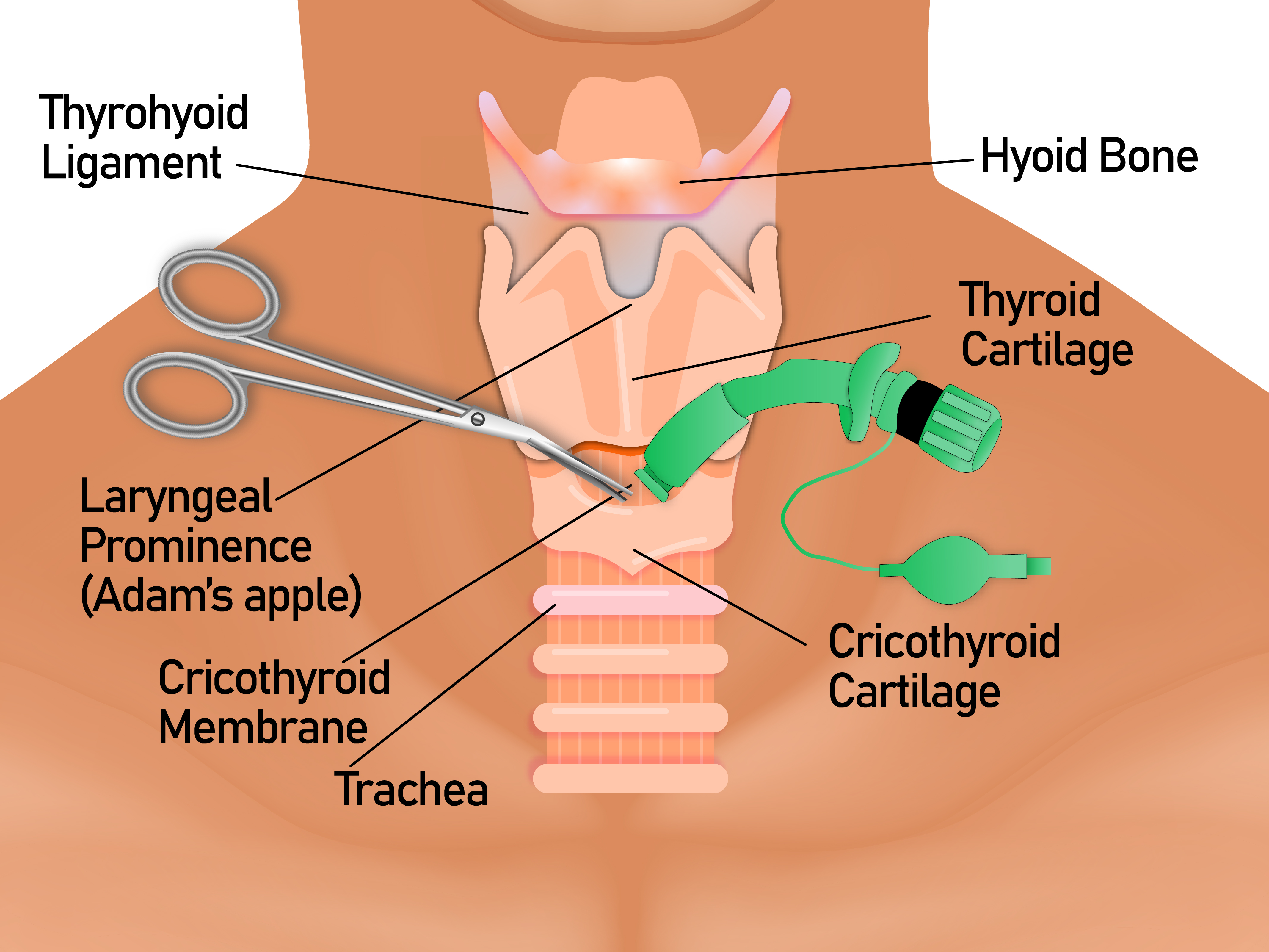
Cricothyroidotomy involves inserting a tube through an incision in the CTM. This membrane is bordered superiorly by the thyroid cartilage, inferiorly by the cricoid cartilage, and laterally by the cricothyroideus muscles. Located approximately 2 cm below the laryngeal prominence, the CTM can be identified by palpating the depression just superior to the cricoid cartilage (see Image. Anatomy and Airway Framework of the Neck).
Several critical vascular structures are in proximity to the CTM. The internal jugular vein and common carotid artery course laterally to the trachea, cricoid cartilage, and CTM. Additionally, the cricothyroid arteries and veins, branches of the superior thyroid artery and vein, overlay and supply blood to the CTM.
Indications
Emergency cricothyroidotomy is the final step in the airway management algorithm. When endotracheal (ET) intubation fails, and a cannot-intubate, cannot-oxygenate (CICO) scenario arises, the immediate establishment of a surgical airway is critical. Delayed recognition and intervention in a CICO situation can quickly lead to hypoxemia and death.[8]
Emergency cricothyroidotomy is indicated in any CICO scenario.[9] This situation may occur in the following cases:
- Neck, oral, or maxillofacial trauma (reported in 28% to 95% of patients who had a cricothyroidotomy)
- Cervical spine trauma
- Profuse oral hemorrhage
- Copious emesis
- Orofacial anatomic abnormalities
- Oropharyngeal swelling
These conditions obstruct mouth opening or prevent visualization of the vocal cords through standard airway management techniques like direct laryngoscopy.
Contraindications
In a CICO situation, emergent cricothyroidotomy has no absolute contraindications. However, relative contraindications include a history of tracheal surgery, laryngeal fracture, laryngotracheal disruption, and age below 5 to 12 years. Patients with acute laryngeal disease face a higher risk of subglottic stenosis, making elective cricothyroidotomy relatively contraindicated.[10]
The use of cricothyroidotomy in CICO crises in younger children is further limited due to the funnel-shaped airway and theoretical risk of subglottic stenosis. Airway rescue options in this group include rigid bronchoscopy and front-of-neck access via surgical or needle cricothyroidotomy and surgical tracheotomy. Guidelines favor surgical tracheotomy for children aged 1 to 8 years when an otolaryngologist is available. However, most CICO emergencies occur in infants younger than 1, where no consensus exists on the optimal front-of-neck access approach. Transtracheal jet ventilation and extracorporeal membrane oxygenation (ECMO) may be considered in select cases.[11][12]
Equipment
Equipment and materials considered essential for performing a cricothyroidotomy using the scalpel-finger-tube technique include the following (see Image. Cricothyroidotomy, Equipment):
- Yankauer suction
- Scalpel (preferably #20 blade)
- Gum elastic bougie
- Cuffed 6.0 to 7.0 tracheostomy tube
- 10-cc syringe
- Securement device
- Ventilator and tubing
- Antiseptic solution (eg, chlorhexidine orpovidone-iodine)
- Sterile gloves
- Local anesthetic (eg, lidocaine with epinephrine)
Personnel
Members of the interprofessional team whose presence is necessary for performing cricothyroidotomy include the physician, nurse, and respiratory therapist. Effective collaboration facilitates rapid execution, minimizes complications, and optimizes patient outcomes.
Preparation
When a difficult airway is anticipated and circumstances allow, obtaining informed consent for surgical airway placement is ideal. However, the urgency of an emergent cricothyroidotomy often precludes a detailed discussion of the risks, benefits, and potential complications. In a true CICO situation, securing the airway should not be delayed to obtain consent.
To enhance institutional protocols and training, cognitive aids such as standardized checklists can improve performance in high-risk procedures like cricothyroidotomy. In prehospital and emergency settings, structured airway management checklists enhance safety, increase first-attempt success rates, and reduce delays in recognizing a CICO scenario. Integrating hard stops and prompts for surgical cricothyroidotomy into existing difficult airway protocols and simulation-based training ensures provider readiness and reinforces best practices, ultimately improving patient outcomes.
Technique or Treatment
Identification of the Cricothyroid Membrane
Early identification of the CTM is essential when managing a difficult airway that may require cricothyroidotomy. The ability to accurately palpate the CTM depends on patient sex, positioning, and body habitus, with misidentification being more common in female patients.[13] Anatomical landmarks may be challenging to locate in patients with obesity, neck trauma, or a history of radiation or surgery to the neck. Extending the neck can aid in identifying these structures.
The Difficult Airway Society (DAS) recommends a 3-step method called the “laryngeal handshake” for identifying the CTM. However, the modified upward laryngeal handshake technique has demonstrated greater accuracy than other palpation methods. This approach involves stepwise palpation from the sternal notch to the CTM. Using the nondominant hand, the provider places fingers on one side of the trachea and the thumb on the other, advancing superiorly until the cricoid cartilage and cricothyroid junction are reached. The middle finger then rests on the cricoid cartilage while the index finger palpates the CTM.[14]
The DAS recommends ultrasound guidance in identifying the trachea and CTM during preoperative evaluations if palpation is insufficient. Airway ultrasound offers advantages over traditional surface-based airway assessments, as it helps predict difficult airways, confirm tracheal intubation, and evaluate vocal cord function. However, the role of this modality in improving emergency cricothyroidotomy success rates remains uncertain.[15]
Still, airway ultrasound is portable and allows for immediate assessment at the bedside, making it potentially useful for identifying the CTM during emergency cricothyroidotomy. Ultrasound may be helpful for patients with severe obesity and anatomical abnormalities of the neck. In these individuals, anterior neck creases do not reliably indicate the CTM's location and should not be used as primary landmarks. The DAS recommends considering this imaging tool during emergencies, but it should not delay airway access.[16][17]
Cricothyroidotomy Techniques
Among the various techniques for emergent surgical airway placement, the scalpel-finger-bougie technique, also known as the 3-step method, is preferred. This straightforward approach and resemblance to other core emergency and critical care procedures enhance provider confidence and success in CICO scenarios.[18][19][20] The scalpel-finger-bougie technique consists of the following steps:
- The CTM is identified using the index finger, while the larynx is stabilized between the thumb and middle finger of the nondominant hand.
- A 4-cm vertical incision is made through the skin overlying the CTM.
- The subcutaneous tissue is bluntly dissected until the CTM becomes identifiable. The procedure should not be discontinued if bleeding occurs.
- The CTM is punctured with a scalpel, and a horizontal incision is made.
- A finger is inserted through the incision.
- A gum elastic bougie is slid through the incision and guided inferiorly into the trachea. Advancing the bougie at an excessively shallow angle should be avoided to prevent the creation of a false tract.
- A 6.0 cuffed ET tube is passed over the bougie until the balloon is no longer visible and the cuff is inflated.
- Placement is confirmed using a bag-valve mask (BVM) and end-tidal capnography.
- The ET tube is secured in place.[21]
The standard open technique differs from the scalpel-finger-bougie approach, as it relies on mechanical dilation rather than a bougie for tube guidance. This method includes the following steps:
- The larynx is immobilized, and the CTM is identified by palpation with the index finger of the nondominant hand.
- A vertical incision, approximately 3 to 5 cm in length, is made in the midline of the neck through the skin overlying the CTM while the larynx is stabilized.
- After the vertical skin incision is created, the CTM is palpated, and a horizontal incision is made through the membrane. The scalpel is directed caudally to avoid the vocal cords, and care is taken to prevent injury to the posterior tracheal wall.
- The tip of the index finger is kept in the incision through the CTM while a tracheal hook is inserted under the thyroid cartilage. Upward traction is applied to the thyroid cartilage.
- A trousseau dilator is inserted to extend the horizontal incision vertically.
- A tracheostomy tube is inserted through the trousseau dilator and advanced caudally into the trachea. Advancement at an excessively shallow angle is avoided to prevent the creation of a false tract.
- The trousseau dilator and tracheal hook are removed.
- The obturator of the tracheostomy tube is afterward removed.
- The inner cannula of the tracheostomy tube is inserted.
- The balloon is inflated.
- The tube is secured in place.[22]
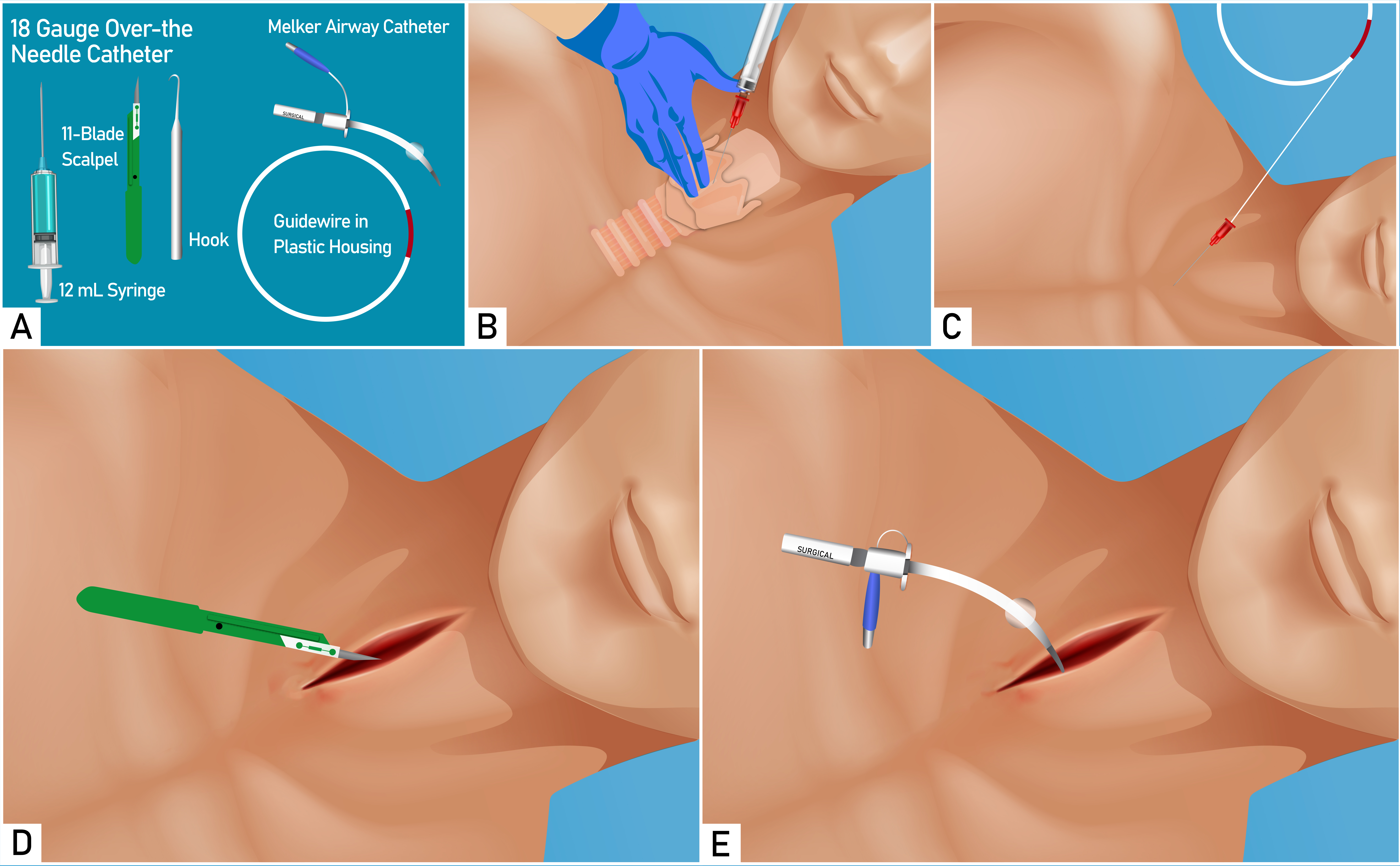
Needle cricothyroidotomy differs from the previously described methods as it uses a percutaneous approach that relies on a hollow needle or catheter rather than a scalpel and open dissection. The steps include the following:
- The larynx is stabilized, and tension is created over the CTM by using the thumb and middle finger of the nondominant hand to stretch the skin in a vertical direction. The CTM is palpated with the index finger.
- A 3- to 10-mL syringe filled with 50% saline is attached to an angiocatheter. With the dominant hand, the skin is punctured at the inferior margin of the CTM, directing the needle caudally at a 30°-45° angle. Continuous negative pressure is applied to the syringe while advancing the needle. The presence of air bubbles within the syringe confirms tracheal placement.
- The catheter is advanced over the needle until the hub abuts the skin, and the needle is removed.
- The saline syringe is reattached to the catheter, and intratracheal placement is confirmed by aspirating air.
- The catheter is connected to high-pressure tubing or a bag-valve mask with 100% oxygen. Ventilation is provided at a rate of 10 to 12 breaths per minute, using an inspiratory-to-expiratory ratio of approximately 1:4.[23]
- The catheter is held in place at all times, as sutures should not be relied upon for secure positioning.
Ultrasound guidance can aid in identifying the CTM and facilitating needle cricothyroidotomy, particularly in patients with anatomic abnormalities of the neck. A study comparing static and real-time ultrasound techniques found that real-time ultrasound improved accuracy but had a success rate of only 60%, suggesting limitations in emergency settings. However, anesthesiologists achieved a higher success rate (78%), indicating potential clinical feasibility for trained providers. While ultrasound enhances CTM identification and needle placement, further studies are needed to determine its role in optimizing overall cricothyroidotomy outcomes.[24]
Monitoring
After proper placement is confirmed, ventilation and oxygenation should be continuously assessed using capnography and pulse oximetry.[25] Standard monitoring should also include noninvasive blood pressure and heart rate assessment. Electrocardiography should be considered when available.[26]
Complications
Early Complications
Early complications of cricothyroidotomy include bleeding, endobronchial intubation, laceration or fracture of local structures, posterior tracheal injury, unintentional tracheostomy, false tract formation, and hypoxia.[27][28] Bleeding is the most commonly reported complication, though some hemorrhage is expected during CTM incision and dissection due to the presence of several overlying or adjacent blood vessels. Significant hemorrhage occurs in up to 50% of cases and, in rare instances, may lead to asphyxiation or aspiration. Bleeding in the cricothyroid area can also reduce visibility during airway management.
If severe bleeding occurs, direct pressure or packing at the site can help control the sources. In such cases, the provider may need to rely on palpation and tactile feedback rather than direct visualization. For example, detecting the "clicking" sensation of a bougie or ET tube as it passes over the tracheal rings can help confirm correct placement.
The American Heart Association recommends hemostatic dressings as an adjunct to direct manual pressure for managing life-threatening bleeding, as these materials promote faster clotting and reduce blood loss more effectively than pressure alone. First aid providers can use these dressings to enhance bleeding control, though current evidence does not support one specific type over another.[29] Hemostatic dressings have been used to control traumatic junctional zone injuries where tourniquets are inappropriate, such as those in the neck, and direct pressure may be insufficient.[30][31]
Right mainstem bronchus intubation can occur when the tube is too long or advanced too far. This complication arises in up to 46% of cricothyrodotomies and is more frequent with ET than tracheostomy tubes due to their greater length. To prevent misplacement, the ET tube should be advanced only until the balloon crosses the CTM incision, reducing the likelihood of endobronchial intubation.
Laceration of local structures, including the cricoid and thyroid cartilages and tracheal rings, may result from misplaced incisions. This risk may be minimized by accurately identifying the CTM through palpation or ultrasound before making an incision. If misplacement occurs, the incision may be extended superiorly or inferiorly, depending on its location, to expose the CTM properly.[32] Cricoid and thyroid cartilage fractures may arise if an oversized tube is forced through the CTM. If placing an oversized tube is not possible, a bougie should be inserted through the tract into the airway, followed by railroading a smaller cannula over the bougie. (Source: Mazza et al, 2021)
Posterior tracheal perforation or injury can occur during needle cricothyroidotomy if the needle is advanced too far dorsally after penetrating the CTM. Posterior tracheal injury may lead to more severe complications, including esophageal damage and mediastinal bleeding, infection, or free air collection, all of which may significantly impact patient outcomes. Using a shorter needle, which limits excessive insertion depth, along with precise technique and careful control of the needle, can help minimize this risk.[33] Additionally, uniquely shaped incision tools that incorporate depth guards, such as the Cric-Guide, may help ensure proper insertion depth and reduce the likelihood of posterior tracheal damage. (Source: Vanner et al, 2023)
Unintentional tracheostomy occurs when the incision is made too low or in an incorrect location, causing tracheal damage. Careful identification of the CTM and precise technique are essential to avoid this complication. If significant tracheal damage occurs or if a stable airway cannot be maintained due to incorrect placement, conversion to a formal tracheostomy may be necessary.[34]
A false tract forms when an instrument deviates from the tracheal lumen, often due to poor visualization, incorrect technique, or difficult anatomy. False tracts may cause failed ventilation, subcutaneous emphysema, hemorrhage, infection, and esophageal perforation. This complication may be prevented by ensuring proper identification of the CTM before incision and using a midline, caudally directed approach when inserting a scalpel or needle. In a scalpel-first approach, the incision site should be maintained using a tracheal hook, scalpel handle, pinky finger, or bougie after the horizontal stab incision. In a needle-first technique, a shallow caudal angle should be avoided. If a false tract is suspected, withdrawing the instrument and reassessing the airway landmarks before reattempting the procedure can help ensure correct placement.
Hypoxia during cricothyroidotomy can occur due to delays in establishing a surgical airway, improper technique, or complications like tube obstruction or malposition. Rapid execution, preoxygenation, and proper technique are crucial to minimize the risk of hypoxia. If hypoxia develops, immediate actions like confirming or adjusting tube placement, suctioning obstructions, and adjusting ventilation parameters are necessary to restore oxygenation.
Infection following cricothyroidotomy may present either early or late. Antibiotic coverage should be tailored to the clinical presentation, taking into account the depth and timing of the infection, as well as potential antibiotic resistance.[35]
Late Complications
Late complications of cricothyroidotomy may include scarring, fistula formation, subglottic stenosis, dysphagia, and voice changes. Subglottic stenosis, fistulas, and scarring often necessitate surgical treatment. Dysphagia and voice changes may be managed conservatively through therapy if mild, but severe cases may require surgical intervention.[36][37]
In the context of a CICO scenario, an emergent cricothyroidotomy offers life-saving benefits that far outweigh the potential risks. Reported complication rates vary, ranging from 0% to 54%, influenced by clinical factors, the level of provider expertise, and the procedure’s setting.
Clinical Significance
Cricothyroidotomy is the final step in the difficult airway algorithm when encountering a CICO scenario. Readiness and ability to perform a cricothyroidotomy at any time are critical, given the time-sensitive and life-saving nature of this procedure. While potential complications exist, the benefits of performing a cricothyroidotomy in a life-threatening situation far outweigh these risks. With careful technique and the use of appropriate tools, many of these complications can be effectively prevented or managed, ensuring the best possible outcome.
Enhancing Healthcare Team Outcomes
Successful management of difficult airways requires close collaboration among interprofessional team members, including physicians, prehospital providers, advanced practice providers, nurses, and respiratory therapists. Regular review of advanced airway techniques, including surgical airway procedures, is essential for interprofessional contributors. Team confidence and competence may be enhanced through practice with simulator models (Source: Hauglum et al, 2024).
The physician overseeing airway management should clearly verbalize each step of the airway plan, particularly when a cricothyroidotomy or surgical airway is necessary. This communication ensures the team is prepared with the appropriate equipment and addresses potential cognitive barriers to performing the procedure. Premarking anatomical landmarks on the neck may also prove beneficial. Open communication among all team members is crucial as the patient’s condition evolves. Effective interprofessional coordination is key to achieving positive patient outcomes.
Media
(Click Image to Enlarge)

Anatomy and Airway Framework of the Neck. This image illustrates the anatomy and the framework of the airway in the neck region. The cricothyroid membrane is located between the thyroid cartilage superiorly and the cricoid cartilage inferiorly. The cricothyroid membrane must be identified by palpation of the surrounding cartilaginous structures.
Contributed by A Tariq, MD
(Click Image to Enlarge)
References
Patel SA, Meyer TK. Surgical airway. International journal of critical illness and injury science. 2014 Jan:4(1):71-6. doi: 10.4103/2229-5151.128016. Epub [PubMed PMID: 24741501]
Brantigan CO, Grow JB Sr. Cricothyroidotomy: elective use in respiratory problems requiring tracheotomy. The Journal of thoracic and cardiovascular surgery. 1976 Jan:71(1):72-81 [PubMed PMID: 1249960]
Aminov RI. A brief history of the antibiotic era: lessons learned and challenges for the future. Frontiers in microbiology. 2010:1():134. doi: 10.3389/fmicb.2010.00134. Epub 2010 Dec 8 [PubMed PMID: 21687759]
Cole RR, Aguilar EA 3rd. Cricothyroidotomy versus tracheotomy: an otolaryngologist's perspective. The Laryngoscope. 1988 Feb:98(2):131-5 [PubMed PMID: 3276995]
Level 3 (low-level) evidenceBrantigan CO, Grow JB Sr. Subglottic stenosis after cricothyroidotomy. Surgery. 1982 Feb:91(2):217-21 [PubMed PMID: 7058500]
Level 3 (low-level) evidencePaix BR, Griggs WM. Emergency surgical cricothyroidotomy: 24 successful cases leading to a simple 'scalpel-finger-tube' method. Emergency medicine Australasia : EMA. 2012 Feb:24(1):23-30. doi: 10.1111/j.1742-6723.2011.01510.x. Epub 2011 Dec 7 [PubMed PMID: 22313556]
Level 3 (low-level) evidenceLacy AJ, Kim MJ, Li JL, Croft A, Kane EE, Wagner JC, Walker PW, Brent CM, Brywczynski JJ, Mathews AC, Long B, Koyfman A, Svancarek B. Prehospital Cricothyrotomy: A Narrative Review of Technical, Educational, and Operational Considerations for Procedure Optimization. The Journal of emergency medicine. 2024 Sep 3:():. pii: S0736-4679(24)00284-1. doi: 10.1016/j.jemermed.2024.08.018. Epub 2024 Sep 3 [PubMed PMID: 39915151]
Level 3 (low-level) evidenceHamaekers AE, Henderson JJ. Equipment and strategies for emergency tracheal access in the adult patient. Anaesthesia. 2011 Dec:66 Suppl 2():65-80. doi: 10.1111/j.1365-2044.2011.06936.x. Epub [PubMed PMID: 22074081]
Schroeder AA. Cricothyroidotomy: when, why, and why not? American journal of otolaryngology. 2000 May-Jun:21(3):195-201 [PubMed PMID: 10834555]
Level 3 (low-level) evidenceKress TD, Balasubramaniam S. Cricothyroidotomy. Annals of emergency medicine. 1982 Apr:11(4):197-201 [PubMed PMID: 7073035]
Scrase I, Woollard M. Needle vs surgical cricothyroidotomy: a short cut to effective ventilation. Anaesthesia. 2006 Oct:61(10):962-74 [PubMed PMID: 16978312]
Level 3 (low-level) evidenceBerger-Estilita J, Wenzel V, Luedi MM, Riva T. A Primer for Pediatric Emergency Front-of-the-Neck Access. A&A practice. 2021 Apr 6:15(4):e01444. doi: 10.1213/XAA.0000000000001444. Epub 2021 Apr 6 [PubMed PMID: 33821828]
Campbell M, Shanahan H, Ash S, Royds J, Husarova V, McCaul C. The accuracy of locating the cricothyroid membrane by palpation - an intergender study. BMC anesthesiology. 2014:14():108. doi: 10.1186/1471-2253-14-108. Epub 2014 Nov 22 [PubMed PMID: 25844061]
Chang JE, Kim H, Won D, Lee JM, Kim TK, Min SW, Hwang JY. Comparison of the Conventional Downward and Modified Upward Laryngeal Handshake Techniques to Identify the Cricothyroid Membrane: A Randomized, Comparative Study. Anesthesia and analgesia. 2021 Nov 1:133(5):1288-1295. doi: 10.1213/ANE.0000000000005744. Epub [PubMed PMID: 34517392]
Level 1 (high-level) evidenceNakazawa H, Uzawa K, Tokumine J, Lefor AK, Motoyasu A, Yorozu T. Airway ultrasound for patients anticipated to have a difficult airway: Perspective for personalized medicine. World journal of clinical cases. 2023 Mar 26:11(9):1951-1962. doi: 10.12998/wjcc.v11.i9.1951. Epub [PubMed PMID: 36998948]
Level 3 (low-level) evidenceFrerk C, Mitchell VS, McNarry AF, Mendonca C, Bhagrath R, Patel A, O'Sullivan EP, Woodall NM, Ahmad I, Difficult Airway Society intubation guidelines working group. Difficult Airway Society 2015 guidelines for management of unanticipated difficult intubation in adults. British journal of anaesthesia. 2015 Dec:115(6):827-48. doi: 10.1093/bja/aev371. Epub 2015 Nov 10 [PubMed PMID: 26556848]
Higgs A, McGrath BA, Goddard C, Rangasami J, Suntharalingam G, Gale R, Cook TM, Difficult Airway Society, Intensive Care Society, Faculty of Intensive Care Medicine, Royal College of Anaesthetists. Guidelines for the management of tracheal intubation in critically ill adults. British journal of anaesthesia. 2018 Feb:120(2):323-352. doi: 10.1016/j.bja.2017.10.021. Epub 2017 Nov 26 [PubMed PMID: 29406182]
George N, Consunji G, Storkersen J, Dong F, Archambeau B, Vara R, Serrano J, Hajjafar R, Tran L, Neeki MM. Comparison of emergency airway management techniques in the performance of emergent Cricothyrotomy. International journal of emergency medicine. 2022 May 30:15(1):24. doi: 10.1186/s12245-022-00427-3. Epub 2022 May 30 [PubMed PMID: 35637444]
Quick JA, MacIntyre AD, Barnes SL. Emergent surgical airway: comparison of the three-step method and conventional cricothyroidotomy utilizing high-fidelity simulation. The Journal of emergency medicine. 2014 Feb:46(2):304-7. doi: 10.1016/j.jemermed.2013.08.065. Epub 2013 Nov 1 [PubMed PMID: 24188608]
Bye R, St Clair T, Delorenzo A, Bowles KA, Smith K. Needle Cricothyroidotomy by Intensive Care Paramedics. Prehospital and disaster medicine. 2022 Oct:37(5):625-629. doi: 10.1017/S1049023X22001157. Epub 2022 Aug 12 [PubMed PMID: 35959773]
Braude D, Webb H, Stafford J, Stulce P, Montanez L, Kennedy G, Grimsley D. The bougie-aided cricothyrotomy. Air medical journal. 2009 Jul-Aug:28(4):191-4. doi: 10.1016/j.amj.2009.02.001. Epub [PubMed PMID: 19573767]
Level 3 (low-level) evidenceBramwell KJ, Davis DP, Cardall TV, Yoshida E, Vilke GM, Rosen P. Use of the Trousseau dilator in cricothyrotomy. The Journal of emergency medicine. 1999 May-Jun:17(3):433-6 [PubMed PMID: 10338233]
Level 1 (high-level) evidencePatel RG. Percutaneous transtracheal jet ventilation: a safe, quick, and temporary way to provide oxygenation and ventilation when conventional methods are unsuccessful. Chest. 1999 Dec:116(6):1689-94 [PubMed PMID: 10593796]
Watanabe H, Nakazawa H, Tokumine J, Yorozu T. Real-time vs. static ultrasound-guided needle cricothyroidotomy: a randomized crossover simulation trial. Scientific reports. 2025 Mar 8:15(1):8112. doi: 10.1038/s41598-025-92684-4. Epub 2025 Mar 8 [PubMed PMID: 40057614]
Level 1 (high-level) evidenceAvva U, Lata JM, Hendrix JM, Kiel J. Airway Management. StatPearls. 2025 Jan:(): [PubMed PMID: 29262130]
Andresen ÅEL, Kramer-Johansen J, Kristiansen T. Emergency cricothyroidotomy in difficult airway simulation - a national observational study of Air Ambulance crew performance. BMC emergency medicine. 2022 Apr 9:22(1):64. doi: 10.1186/s12873-022-00624-6. Epub 2022 Apr 9 [PubMed PMID: 35397493]
Level 2 (mid-level) evidenceBair AE, Panacek EA, Wisner DH, Bales R, Sakles JC. Cricothyrotomy: a 5-year experience at one institution. The Journal of emergency medicine. 2003 Feb:24(2):151-6 [PubMed PMID: 12609644]
Level 2 (mid-level) evidenceErlandson MJ, Clinton JE, Ruiz E, Cohen J. Cricothyrotomy in the emergency department revisited. The Journal of emergency medicine. 1989 Mar-Apr:7(2):115-8 [PubMed PMID: 2661666]
Level 2 (mid-level) evidencePellegrino JL, Charlton NP, Carlson JN, Flores GE, Goolsby CA, Hoover AV, Kule A, Magid DJ, Orkin AM, Singletary EM, Slater TM, Swain JM. 2020 American Heart Association and American Red Cross Focused Update for First Aid. Circulation. 2020 Oct 27:142(17):e287-e303. doi: 10.1161/CIR.0000000000000900. Epub 2020 Oct 21 [PubMed PMID: 33084370]
Simpson C, Tucker H, Hudson A. Pre-hospital management of penetrating neck injuries: a scoping review of current evidence and guidance. Scandinavian journal of trauma, resuscitation and emergency medicine. 2021 Sep 16:29(1):137. doi: 10.1186/s13049-021-00949-4. Epub 2021 Sep 16 [PubMed PMID: 34530879]
Level 2 (mid-level) evidenceShina A, Lipsky AM, Nadler R, Levi M, Benov A, Ran Y, Yitzhak A, Glassberg E. Prehospital use of hemostatic dressings by the Israel Defense Forces Medical Corps: A case series of 122 patients. The journal of trauma and acute care surgery. 2015 Oct:79(4 Suppl 2):S204-9. doi: 10.1097/TA.0000000000000720. Epub [PubMed PMID: 26406432]
Level 2 (mid-level) evidenceHessert MJ, Bennett BL. Optimizing emergent surgical cricothyrotomy for use in austere environments. Wilderness & environmental medicine. 2013 Mar:24(1):53-66. doi: 10.1016/j.wem.2012.07.003. Epub 2012 Oct 10 [PubMed PMID: 23062323]
Katayama A, Watanabe K, Tokumine J, Lefor AK, Nakazawa H, Jimbo I, Yorozu T. Cricothyroidotomy needle length is associated with posterior tracheal wall injury: A randomized crossover simulation study (CONSORT). Medicine. 2020 Feb:99(9):e19331. doi: 10.1097/MD.0000000000019331. Epub [PubMed PMID: 32118765]
Level 1 (high-level) evidenceRaimonde AJ,Westhoven N,Winters R, Tracheostomy 2020 Jan; [PubMed PMID: 32644550]
Zabaglo M, Leslie SW, Sharman T. Postoperative Wound Infections. StatPearls. 2025 Jan:(): [PubMed PMID: 32809368]
Spaite DW, Joseph M. Prehospital cricothyrotomy: an investigation of indications, technique, complications, and patient outcome. Annals of emergency medicine. 1990 Mar:19(3):279-85 [PubMed PMID: 2310067]
Level 2 (mid-level) evidenceRugnath N, Rexrode LE, Kurnutala LN. Unanticipated Difficult Airway During Elective Surgery: A Case Report and Review of Literature. Cureus. 2022 Dec:14(12):e32996. doi: 10.7759/cureus.32996. Epub 2022 Dec 27 [PubMed PMID: 36712753]
Level 3 (low-level) evidence