 Cerebral Cavernous Malformations
Introduction
Cerebral cavernous malformations (CCMs) are abnormally large collections of "low flow" vascular channels without brain parenchyma intervening between the sinusoidal vessels.[1][2] McCormick (1966) recognized CCMs as one of the four classes of cerebral vascular malformations which include arteriovenous malformations (AVM), developmental venous anomalies (DVA), and capillary telangiectasia. Clinically, CCMs are highly variable in both symptomatic presentation and natural history. Adding to the confusion, CCM has assumed a variety of names in the medical literature including cavernomas, cavernous angiomas, and cavernous hemangiomas, though CCM is the preferred nomenclature.[2] CCMs range in size from punctate to several centimeters in diameter and may occur anywhere in the central nervous system with up to 20% located in the brainstem.[3]
CCM may be diagnosed in both young children and adults and may develop de novo or even regress spontaneously during a patient’s lifetime. A thorough understanding of the natural history of this entity is of paramount importance to avoid unnecessary and potentially morbid interventions. Given the heterogeneity of this condition, the ontogenesis, diagnosis, management strategies for CCMs are subjects of ongoing debate among neuroscientists and treatment paradigms continue to evolve.
Etiology
Register For Free And Read The Full Article
Search engine and full access to all medical articles
10 free questions in your specialty
Free CME/CE Activities
Free daily question in your email
Save favorite articles to your dashboard
Emails offering discounts
Learn more about a Subscription to StatPearls Point-of-Care
Etiology
Experts do not fully understand the pathogenesis of CCMs, but the genetic underpinnings have been clarified in recent years. The majority of CCMs are sporadic, but up to 20% follow a familial, autosomal dominant inheritance pattern characterized by the presence of multiple CCMs in a single patient.[4][5] This has led to the identification of three homologically distinct genes responsible for CCM development: CCM1, CCM2, and CCM3.[6] Mutations in any one of these genes can result in multifocal CCM and all three show relatively high genetic penetrance. Many authors have proposed a "two-hit" hypothesis of familial CCM wherein epigenetic or environmental exposure (the second hit) results in CCM gene loss-of-function and may account for the proclivity of these lesions to accumulate over time and with exposure to radiation.[7] Studies of sporadic CCM support a common pathway involving de novo mutations of CCM genes.[6]
CCM protein products interact with each other and other cellular machinery responsible for a range of functions including cell-cell communication and angiogenesis. The most critical dysfunction found in CCM mutants is endothelial junction permeability, an effect mediated by Notch1 and Rho kinase activity.[8] This correlates with the characteristic histopathological appearance of CCM which lacks mature vessel wall architecture and mature blood-brain-barrier.[9] CCMs are distinguished from other cerebral vascular malformations by the absence of direct arteriovenous communication and lack of intervening brain parenchyma.
Epidemiology
CCMs are the second most common incidental vascular finding – after aneurysms – on brain magnetic resonance imaging (MRI), with a prevalence of 1 in 625 neurologically asymptomatic people.[10][11][12][13] Clinical presentation is bimodal with a significant number of cases detected in both adolescents and middle-aged adults. There is no discernible sex difference in prevalence; however, there is conflicting research as to whether prognosis is different among men and women.[14] Familial CCM is notably prevalent among persons of northern Mexican ancestry, an effect that has been traced to a common founder mutation.[1] The incidence of incidentally detected CCM has increased substantially due to the widespread use of magnetic resonance imaging (MRI).[15] The majority (approximately 75%) of CCMs are found in the supratentorial compartment in predictable proportion to the volume of neural tissue present.[16]
Pathophysiology
The propensity for intra-lesional and extra-lesional hemorrhage is the chief mechanism underlying the clinical manifestations of CCM. Sluggish blood flow through dysplastic channels results in recurrent thrombosis, calcification, and deposition of hemosiderin along the margins of the lesion. Hemorrhage into adjacent brain parenchyma can produce focal neurologic deficits (FND), seizure, or a headache prompting the patient to present for evaluation. Clinical and lifestyle risk factors for a first symptomatic episode of CCM hemorrhage are unknown, but risk factors for re-hemorrhage are well-studied.[2] The pathogenesis of CCM-related epilepsy has been attributed to peri-lesional reactive gliosis due to clinically silent micro-hemorrhages which alters conduction adjacent white matter pathways. The observation that seizure-free outcomes are improved when the entire lesion, including the surrounding hemosiderin rim, is resected, supports this.[17]
Histopathology
Histopathologically, CCMs are well-circumscribed, multilobate vascular lesions consisting of sinusoidal channels lined by a single layer of epithelium, devoid of smooth muscle and lacking intervening brain parenchyma. On gross inspection, cavernous malformations appear "mulberry-like."[16]
History and Physical
While the clinical presentation of symptomatic cavernous malformations varies by location, the most common clinical manifestations are seizures (50%), intracranial hemorrhage (25%), and focal neurological deficits (FND) without radiographic evidence of recent hemorrhage (25%).[2][18] Supratentorial lesions most commonly present with seizures whereas FND or ataxia is the most common presentation in patients with infratentorial lesions.[19] Up to 20% to 50% of patients are asymptomatic and diagnosed incidentally on brain MRI.[2][11][15] When CCM is diagnosed, clinicians should perform a thorough history for evidence of prior symptomatic hemorrhage and a comprehensive neurologic exam to assess for the deficit which may otherwise have been unrecognized. Headaches are common in patients with CCM and determining a causal relationship may be difficult. Similarly, CCM-related epilepsy (CRE) can present a diagnostic challenge as seizure focus may be challenging to localize. The criteria for CRE have been defined by expert consensus and can be broadly categorized as "definite," "probable," or "unrelated to CCM" based proximity of localized seizure focus to the CCM.[20] Given the high prevalence of familial CCM, the Angioma Alliance advocates obtaining a detailed, 3 generation family history when MRI diagnoses new CCM. When multiple CCM are present, or family history is positive, genetic screening for CCM1, CCM2, and CCM3 should be considered. Given the autosomal dominant nature of CCM inheritance, appropriate counseling regarding familial risk is warranted, and risk-benefit discussion regarding testing asymptomatic relatives should be offered.[2]
Evaluation
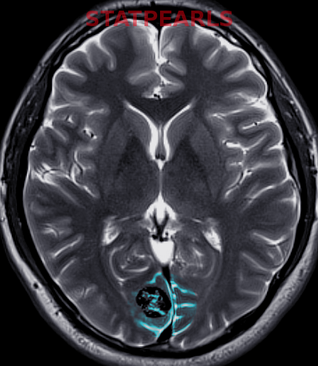
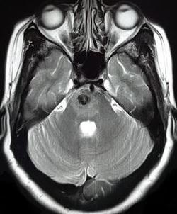
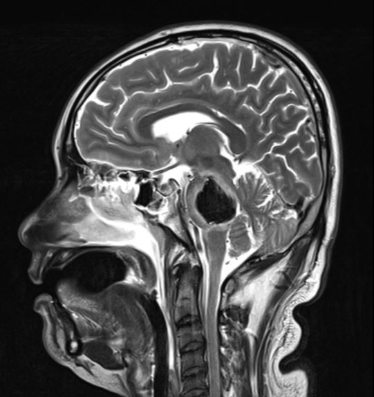
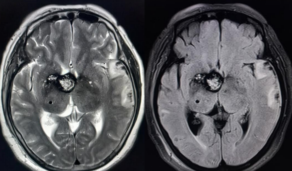
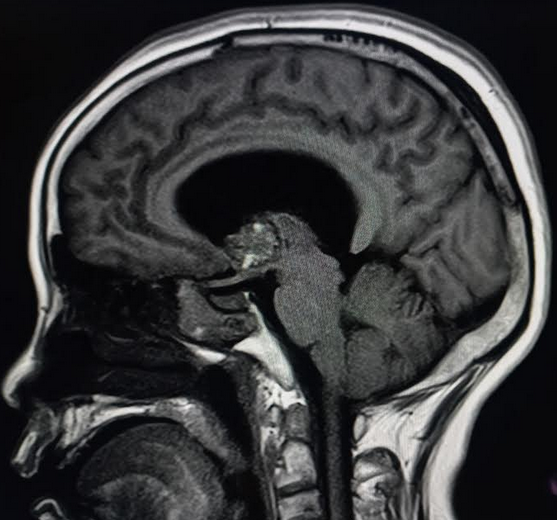
The American College of Radiology (ACR) Appropriateness Criteria provide expert consensus recommendations for acute neurologic symptoms including a headache, FND, altered consciousness. [21] When parenchymal hemorrhage is diagnosed, follow-up imaging with contrast-enhanced MRI is indicated to assess for an underlying vascular lesion. Whether symptomatic or incidentally detected, the majority of CCMs are diagnosed by MRI which has been shown to have nearly 100% sensitivity.[5][15][1][22] MRI is particularly valuable in identifying multiple lesions in the case of familial CCM.[1] T2 weighted MRI typically demonstrates a characteristic mixed signal "popcorn" core with a hypointense rim.[16]
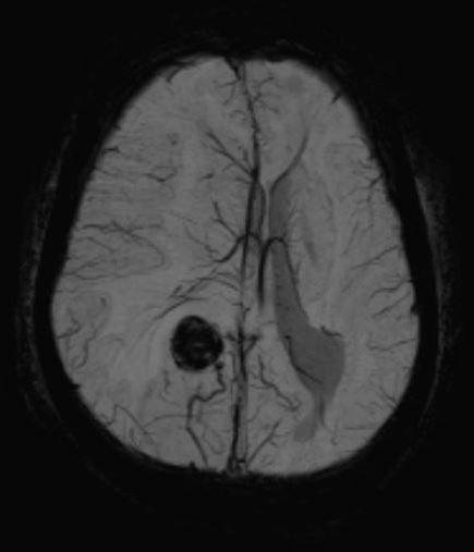
The hemosiderin rim lining the margins of CCMs generates a profound signal void on gradient-echo MRI due to the ferromagnetic dephasing of proton spins. The effect is a striking signal void or "blooming" artifact on gradient echo or susceptibility-weighted sequences (SWI) which is easily detected but can overestimate the actual size of the lesion. MRI has, therefore, become the gold standard tool for the diagnosis and staging of CCM. Guidelines for imaging follow-up of known CCM are not well-established, but it is generally recommended that new symptoms warrant repeat imaging to assess for acute or subacute hemorrhage.[2][5]
CCMs are characteristically angiographically occult lesions due to the slow transit of blood via the dysplastic channels save for the frequently associated developmental venous anomalies (DVA). CT angiography and digital subtraction angiography are therefore of limited utility in the workup of CCM however they may show indirect evidence of CCM by highlighting adjacent DVAs which typically enhance and opacify briskly on angiogram.[23]
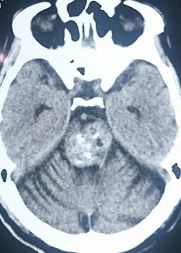
CCM may present on non-contrast head CT (NCCT) as amorphous calcifications, but further imaging with MRI is warranted to confirm the diagnosis unless contraindicated. The principal role of NCCT is the identification of hemorrhage in symptomatic patients. The Angioma Alliance has defined standard definitions of CCM-related hemorrhage and recommended reporting criteria to improve diagnostic consistency and accuracy among neuroimagers and clinicians.[5] These guidelines define CCM hemorrhage by the temporal concordance of neurologic symptoms and quantitative imaging biomarkers seeking to avoid misattribution of symptoms to clinically silent CCM.
Advanced MR imaging is playing an increasingly important role in the management of surgically complex CCM. Diffusion tensor imaging (DTI) has been used to identify critical white matter tracts in preoperative planning for brainstem CCM.[24] Functional MRI techniques such as blood oxygen level-dependent (BOLD) task-activation mapping of language function are highly accurate and non-invasive tools that have proven useful in the preoperative workup of CCM.[25] Emerging work using high field strength SWI may provide detailed information on the angioarchitecture of CCM, potentially identifying at-risk lesions.[26]
Treatment / Management
Location is the most important factor that determines the natural history of CCM.[16] Because the clinical course of CCM is highly variable and location-dependent, management and therapeutic decision-making require multidisciplinary discussion and a thorough evaluation of the patient risk tolerance profile. When feasible, surgical resection is the preferred treatment option for symptomatic CCM. In select cases, targeted radiotherapy is used to treat lesions that are surgically inaccessible. There is no role for endovascular therapy in CCM.
Surgical excision is the only definitive treatment for CCM but the decision to operate remains challenging as postoperative morbidity may approach or exceed the complications of the untreated disease.[27] Conservative management and observation are therefore favored for all patients with solitary CCM who are asymptomatic.[2] For supratentorial lesions in non-eloquent regions, surgical excision can be curative with high success rates and relatively low complications.[28] Surgery should, therefore, be considered in this patient population for patients who present with symptomatic hemorrhage. In cases of medically refractory epilepsy, early surgery is favored for amenable lesions, especially if there is high confidence that a solitary CCM is an epileptogenic source.[2][17][20](A1)
CCMs located in the deep gray nuclei and brainstem pose a much greater challenge. Studies using diffusion tensor imaging and diffusion tensor tractography have shown that up to 82% of patients with brainstem lesions have involvement of corticospinal tract and other major fiber tracts[29] highlighting the extreme difficulty neurosurgeons face with patient and approach selection. While good outcomes may be achieved in surgically resected brainstem lesions at high-volume, specialized centers, complication rates are high, and new postoperative neurologic deficits are expected (53% of cases).[30] Long-term outcomes are better for lesions with a pial presentation which may facilitate resection and minimal collateral injury.[31] Aggressive intervention in the brainstem is therefore reserved only for patients who have suffered a single disabling bleed, or with long life expectancy which may pose a higher cumulative risk for future hemorrhage.[2][30][31] With the utilization of image guidance, appropriate patient and approach selection, and detailed knowledge of the intrinsic brainstem anatomy, these lesions can be safely resected with good outcomes.[32](A1)
The technical goal of surgery should be, at a minimum, complete lesionectomy.[33] Seizure outcomes seem to be better if the surrounding hemosiderin-stained gliotic brain is also resected.[17] With selective adjuncts such as frameless stereotactic guidance, electrocorticography, and intraoperative MRI, microsurgical resection of CCMs has become much safer and more complete. If the lesion is not adjacent to eloquent areas, resecting the hemosiderin-laden gliotic 'pseudocapsule' surrounding the lesion should be strongly considered esp. if treatment indication is intractable seizures. CCM associated developmental venous anomalies (DVA) should be spared during microsurgical resection as they typically drain the surrounding normal brain. Each case demands a tailored resection technique. Large lesions can be tackled by piecemeal excision while smaller ones may be amenable for en-bloc resection.[16](B2)
Stereotactic radiosurgery (SRS) has long served as an alternative to surgery in symptomatic patients with anatomically foreboding lesions or unfavorable risk profile. SRS is highly accurate and allows targeted delivery of high-dose radiation (typically 11 to 15 Gy) with sparing of adjacent, healthy brain parenchyma. The mechanism of therapeutic SRS is uncertain; lesion size may decrease, remain stable, or even increase and there is no reliable imaging biomarker for successful CCM obliteration as with metastasis and high-flow vascular lesions.[34] Several series on SRS for CMs suggest that the greatest risk reduction in hemorrhage usually happens after a 2-year latency period. Hasegawa et al. showed that for patients with high-risk, symptomatic, diencephalic or brainstem CCM, radiotherapy reduced re-hemorrhage rates from 33% to 12.3% in the 2-year post-treatment period with a further reduction in annualized hemorrhage rate to less than 1% after 2 years.[35] A recent meta-analysis found a modest reduction in hemorrhage rates with a substantial incidence of radiation-related complications (11%) including new focal neurologic deficits, hydrocephalus, and painful paresthesia.[34] SRS is also strongly linked to the development of de novo CCM, although such cases rarely become symptomatic.[36] Because radiation-induced damage in the brainstem can be devastating, SRS is not recommended as a treatment option for brainstem CMs.[32](A1)
CCM is a surgical disease, and the role of medical management is limited but the subject of much ongoing laboratory research. Preclinical animal model studies have recently shown that both statins and targeted Rho-kinase inhibitors may reduce symptoms and lesion progression.[37] The medical complications of CCM should be managed following evidence-based guidelines. Intraparenchymal hemorrhage due to CCM should follow standard evidence-based guidelines and protocols which have been well-described by expert consensus.[38] Recent prospective data found no increase in hemorrhage or other adverse events in patients with CCM who are treated with anticoagulant or antiplatelet medications for other purposes.[39](B2)
The risks of surgical morbidity should be weighed against the natural history of the disease. While microsurgical resection is curative for intractable cases, most patients with supratentorial cavernous malformations are managed conservatively either with radiographic and clinical observation alone or in addition to anti-epileptic drugs, as the current first-line management strategy.[16] Radiosurgery is still seen with an eye of suspicion as its benefit is not conferred for at least 2 to 3 years after treatment, concurrent with the period of temporal hemorrhage clustering. Early results from the use of MRI-guided laser interstitial thermal therapy (LIIT) for cavernous malformation-related epilepsy are promising.[40] Further use and expansion of such minimally invasive therapies seem assured.[16]
Differential Diagnosis
Classic CCM rarely poses a diagnostic dilemma as the radiographic differential diagnosis for isolated T2* artifact, a non-enhancing lesion on MRI is limited. When numerous small CCM are present, as is often the case with familial CCM, the differential diagnosis is broad and includes other etiologies of diffuse cerebral microbleeds including cerebral amyloid angiopathy, chronic hypertension, and hemorrhagic or previously treated metastases, among others. Lesion calcification, which can be detected on routine non-contrast head CT, favors the diagnosis of CCM over other types of microbleed. Finally, the co-existence of a developmental venous anomaly (DVA) strongly supports the diagnosis of CCM.[23]
Staging
The most widely accepted tool for grading CCM was derived from early observations by Zabramski et al. on the MR manifestations of hemorrhage within the lesion.[41] This classification schema defines type I lesions by the presence of subacute hemorrhage (high T1 signal, days to weeks from ictus), type II lesions as "popcorn" lesions with central T2* blooming, type III as chronic hemorrhage (low T1 and T2 signal, more than 1-month-old) and lastly, type IV CCM corresponding to multiple punctate microhemorrhages visualized on gradient-echo sequences (T2*-weighted).
Prognosis
The natural history of CCM has been characterized in several large studies.[4][14][41][18] The overall annualized hemorrhage rate in untreated CCM is estimated at 2.4% with a predicted cumulative 5-year risk of hemorrhage 15.8% from the time of diagnosis.[22][13] For patients with incidentally detected CCM, the risk of hemorrhage is substantially lower, estimated to be 0.33% per year.[4] Rates of epilepsy in incidental lesions are similarly low at 1% to 2%.[20] Conversely, patients who have a documented history of CCM hemorrhage are at significantly greater risk of repeat hemorrhage (23% 5-year rate), a finding which has been replicated in multiple large case series and meta-analyses to date.[4][15][22][42] CCMs display a phenomenon termed temporal clustering wherein re-hemorrhage tends to occur, within the first 2 to 3 years after a prior hemorrhage. After this initial clustering of hemorrhage events, a relatively quiescent period where no overt hemorrhages occur may be seen.[16][43] Prior hemorrhage is a significant risk factor for future hemorrhagic events.[16] Several factors have been associated with CCM rupture including lesion location, size, multiplicity, and the presence of an associated DVA.[44][42][22] Studies have shown that supratentorial lobar CCMs have a much more benign prognosis than deep lesions in the thalamus, basal ganglia, or posterior fossa. In one study the event rate for superficial lesions was 0% per year while that for deep lesions was 10.6% per year (p = 0.0001).[16][45] Brainstem CCMs are the most dangerous and have a high relative event rate (4- to 7-times more likely to rupture than isolated supratentorial lesions).[42][30] In one meta-analysis, non-brainstem hemorrhage rates were reported to be 0.3% per year vs. 2.8% per year for brainstem lesions.[42] Also of note, the initial presentation of patients with intracranial hemorrhage (ICH) or focal neurological deficit and brainstem location was independently associated with a hemorrhage over the 5 years after the initial diagnosis.[13][16] Female gender as a risk factor for hemorrhage remains a topic of debate.[14][13] In familial CCM, more aggressive CCM behavior has been observed in CCM3 mutants in contrast with a more benign clinical course in CCM1 deletions.[46][6]
Complications
The risks and benefits of surgical or radiotherapeutic intervention must be assessed on a case-by-case basis, and the prospective risks of untreated CCM must be balanced with the anticipated morbidity of intervention. The overall mortality associated with CCM hemorrhage is low, estimated at 2.2%, but progressive neurologic deficits can accumulate and reduce a patient's quality of life.[42] In the hands of experienced surgeons with appropriate patient selection, postoperative morbidity can be quite low with one recent estimate of 1.5%.[31] Nonetheless, when feasible, conservative management may be favorable as shown in one recent prospective study in which CCM excision worsened short-term disability and increased risk of neurologic deficit or recurrent hemorrhage.[27]
Postoperative and Rehabilitation Care
Although there are no guidelines on the role of anti-epileptics following surgical resection of CCM, patients are typically maintained on anti-epileptic monotherapy following surgery.[47] Seizure-free outcomes following surgery are dependent on various factors such as pre-operative seizure frequency, the extent of CCM resection, the extent of perilesional "hemosiderin-ring" resection, and timing of surgery relative to the initial presentation.[48][49] Anti-epileptic drug withdrawal following surgery should be planned with appropriate dose tapering to reduce the risk of seizure recurrence.[50][51]
Deterrence and Patient Education
Patients with CCM are encouraged to explore the official website of the multidisciplinary Angioma Alliance (AA). The Angioma Alliance is dedicated to providing up-to-date patient resources including educational videos.
Information is provided regarding genetic testing, participation in ongoing clinical research, and tissue banking. The Angioma Alliance also provides social support via online forums and social media sites allowing patients and family members to support one another and share their experiences with CCM.
Pearls and Other Issues
RhoA/Rho kinase pathway is seen as a potential target for the pharmacotherapeutic treatment of cavernous malformations. Normally, CCM2 and CCM1 act together to suppress RhoA. CCM1 and CCM2 deficiency lead to constitutively active Rho-kinase (ROCK), which destabilizes endothelial cell junctions and vascular permeability.[52] ROCK suppressants have been experimentally shown to enable vasculogenesis in CCM1-, CCM2-, and CCM3-deficient cells.[53] Fasudil, a ROCK inhibitor has been shown to decrease the lesion burden in CCM1-deficient mice.[54]
While CCM1, CCM2 or CCM3 deficiencies have been shown to activate bone morphogenic protein (BMP) and transforming growth factor-beta (TGF-beta) causing an endothelial-to-mesenchymal transition (EndoMT), inhibiting either BMP or TGF-beta was found to decrease the lesion burden in CCM-1 deficient mice representing another avenue of research in CCM therapy.[55] Similarly, suppressing Beta-catenin was also found to reduce the number and size of cavernous malformations in a CCM3-deficient mice model.[56] These findings demonstrate the importance of a thorough understanding of the molecular biology underpinning CCM.
Enhancing Healthcare Team Outcomes
Comprehensive care for CCM requires an interprofessional effort involving neurosurgeons, epileptologists, neuroradiologists, mental health professionals, specialty certified nurses, and genetic counselors.
Given the complexity of this disease and its frequent presentation as an incidental finding, patient counseling and risk profile assessment should be performed at the time of diagnosis. Familial CCM follows an autosomal dominant inheritance pattern; thus, there is consensus agreement that detailed family history/pedigree and consideration of genetic screening for common mutations should be considered (Class I/Level C).[2] Ancillary care from social workers and mental health providers can address patient anxiety, and support groups such as the Angioma Alliance provide additional support mechanisms.
Neuroradiology reports should adhere to established reporting guidelines for CCM which clarify the clinical-radiologic correlation of symptoms and imaging findings.[5] An accurate radiographic description is critical for determining if patients are candidates for conservative or surgical treatment. Brain MRI with gradient echo and susceptibility-weighted sequences are the preferred tools for both diagnosis and follow-up imaging (Class 1/Level C).[2]
Evidence-based consensus guidelines for management have been described and are described in detail elsewhere (see references [2][20]). Conservative observation is favored for asymptomatic CCM, and curative surgical resection for CCM with recurrent hemorrhage or refractory epilepsy is supported by Class II/Level A evidence. For CCM with solitary hemorrhage, case-by-case consideration is warranted. The role of radiosurgery is the subject of ongoing debate for deep or brainstem CCM or lesions involving the eloquent cortex.
Media
References
Rigamonti D, Hadley MN, Drayer BP, Johnson PC, Hoenig-Rigamonti K, Knight JT, Spetzler RF. Cerebral cavernous malformations. Incidence and familial occurrence. The New England journal of medicine. 1988 Aug 11:319(6):343-7 [PubMed PMID: 3393196]
Akers A, Al-Shahi Salman R, A Awad I, Dahlem K, Flemming K, Hart B, Kim H, Jusue-Torres I, Kondziolka D, Lee C, Morrison L, Rigamonti D, Rebeiz T, Tournier-Lasserve E, Waggoner D, Whitehead K. Synopsis of Guidelines for the Clinical Management of Cerebral Cavernous Malformations: Consensus Recommendations Based on Systematic Literature Review by the Angioma Alliance Scientific Advisory Board Clinical Experts Panel. Neurosurgery. 2017 May 1:80(5):665-680. doi: 10.1093/neuros/nyx091. Epub [PubMed PMID: 28387823]
Level 3 (low-level) evidenceOtten P, Pizzolato GP, Rilliet B, Berney J. [131 cases of cavernous angioma (cavernomas) of the CNS, discovered by retrospective analysis of 24,535 autopsies]. Neuro-Chirurgie. 1989:35(2):82-3, 128-31 [PubMed PMID: 2674753]
Level 2 (mid-level) evidenceFlemming KD, Link MJ, Christianson TJ, Brown RD Jr. Prospective hemorrhage risk of intracerebral cavernous malformations. Neurology. 2012 Feb 28:78(9):632-6. doi: 10.1212/WNL.0b013e318248de9b. Epub 2012 Feb 1 [PubMed PMID: 22302553]
Level 2 (mid-level) evidenceAl-Shahi Salman R, Berg MJ, Morrison L, Awad IA, Angioma Alliance Scientific Advisory Board. Hemorrhage from cavernous malformations of the brain: definition and reporting standards. Angioma Alliance Scientific Advisory Board. Stroke. 2008 Dec:39(12):3222-30. doi: 10.1161/STROKEAHA.108.515544. Epub 2008 Oct 30 [PubMed PMID: 18974380]
Level 1 (high-level) evidenceLabauge P, Denier C, Bergametti F, Tournier-Lasserve E. Genetics of cavernous angiomas. The Lancet. Neurology. 2007 Mar:6(3):237-44 [PubMed PMID: 17303530]
Jain R, Robertson PL, Gandhi D, Gujar SK, Muraszko KM, Gebarski S. Radiation-induced cavernomas of the brain. AJNR. American journal of neuroradiology. 2005 May:26(5):1158-62 [PubMed PMID: 15891176]
Level 2 (mid-level) evidenceFischer A, Zalvide J, Faurobert E, Albiges-Rizo C, Tournier-Lasserve E. Cerebral cavernous malformations: from CCM genes to endothelial cell homeostasis. Trends in molecular medicine. 2013 May:19(5):302-8. doi: 10.1016/j.molmed.2013.02.004. Epub 2013 Mar 15 [PubMed PMID: 23506982]
Level 3 (low-level) evidenceZawistowski JS, Stalheim L, Uhlik MT, Abell AN, Ancrile BB, Johnson GL, Marchuk DA. CCM1 and CCM2 protein interactions in cell signaling: implications for cerebral cavernous malformations pathogenesis. Human molecular genetics. 2005 Sep 1:14(17):2521-31 [PubMed PMID: 16037064]
Level 3 (low-level) evidenceGreving JP, Wermer MJ, Brown RD Jr, Morita A, Juvela S, Yonekura M, Ishibashi T, Torner JC, Nakayama T, Rinkel GJ, Algra A. Development of the PHASES score for prediction of risk of rupture of intracranial aneurysms: a pooled analysis of six prospective cohort studies. The Lancet. Neurology. 2014 Jan:13(1):59-66. doi: 10.1016/S1474-4422(13)70263-1. Epub 2013 Nov 27 [PubMed PMID: 24290159]
Level 2 (mid-level) evidenceMorris Z, Whiteley WN, Longstreth WT Jr, Weber F, Lee YC, Tsushima Y, Alphs H, Ladd SC, Warlow C, Wardlaw JM, Al-Shahi Salman R. Incidental findings on brain magnetic resonance imaging: systematic review and meta-analysis. BMJ (Clinical research ed.). 2009 Aug 17:339():b3016. doi: 10.1136/bmj.b3016. Epub 2009 Aug 17 [PubMed PMID: 19687093]
Level 1 (high-level) evidenceVernooij MW, Ikram MA, Tanghe HL, Vincent AJ, Hofman A, Krestin GP, Niessen WJ, Breteler MM, van der Lugt A. Incidental findings on brain MRI in the general population. The New England journal of medicine. 2007 Nov 1:357(18):1821-8 [PubMed PMID: 17978290]
Horne MA, Flemming KD, Su IC, Stapf C, Jeon JP, Li D, Maxwell SS, White P, Christianson TJ, Agid R, Cho WS, Oh CW, Wu Z, Zhang JT, Kim JE, Ter Brugge K, Willinsky R, Brown RD Jr, Murray GD, Al-Shahi Salman R, Cerebral Cavernous Malformations Individual Patient Data Meta-analysis Collaborators. Clinical course of untreated cerebral cavernous malformations: a meta-analysis of individual patient data. The Lancet. Neurology. 2016 Feb:15(2):166-173. doi: 10.1016/S1474-4422(15)00303-8. Epub 2015 Dec 2 [PubMed PMID: 26654287]
Level 1 (high-level) evidenceDel Curling O Jr, Kelly DL Jr, Elster AD, Craven TE. An analysis of the natural history of cavernous angiomas. Journal of neurosurgery. 1991 Nov:75(5):702-8 [PubMed PMID: 1919691]
Level 2 (mid-level) evidenceMoore SA, Brown RD Jr, Christianson TJ, Flemming KD. Long-term natural history of incidentally discovered cavernous malformations in a single-center cohort. Journal of neurosurgery. 2014 May:120(5):1188-92. doi: 10.3171/2014.1.JNS131619. Epub 2014 Mar 14 [PubMed PMID: 24628608]
Level 2 (mid-level) evidenceEllis JA, Barrow DL. Supratentorial cavernous malformations. Handbook of clinical neurology. 2017:143():283-289. doi: 10.1016/B978-0-444-63640-9.00027-8. Epub [PubMed PMID: 28552151]
Baumann CR, Schuknecht B, Lo Russo G, Cossu M, Citterio A, Andermann F, Siegel AM. Seizure outcome after resection of cavernous malformations is better when surrounding hemosiderin-stained brain also is removed. Epilepsia. 2006 Mar:47(3):563-6 [PubMed PMID: 16529622]
Level 2 (mid-level) evidenceAl-Shahi Salman R, Hall JM, Horne MA, Moultrie F, Josephson CB, Bhattacharya JJ, Counsell CE, Murray GD, Papanastassiou V, Ritchie V, Roberts RC, Sellar RJ, Warlow CP, Scottish Audit of Intracranial Vascular Malformations (SAIVMs) collaborators. Untreated clinical course of cerebral cavernous malformations: a prospective, population-based cohort study. The Lancet. Neurology. 2012 Mar:11(3):217-24. doi: 10.1016/S1474-4422(12)70004-2. Epub 2012 Jan 31 [PubMed PMID: 22297119]
Washington CW, McCoy KE, Zipfel GJ. Update on the natural history of cavernous malformations and factors predicting aggressive clinical presentation. Neurosurgical focus. 2010 Sep:29(3):E7. doi: 10.3171/2010.5.FOCUS10149. Epub [PubMed PMID: 20809765]
Level 2 (mid-level) evidenceRosenow F, Alonso-Vanegas MA, Baumgartner C, Blümcke I, Carreño M, Gizewski ER, Hamer HM, Knake S, Kahane P, Lüders HO, Mathern GW, Menzler K, Miller J, Otsuki T, Ozkara C, Pitkänen A, Roper SN, Sakamoto AC, Sure U, Walker MC, Steinhoff BJ, Surgical Task Force, Commission on Therapeutic Strategies of the ILAE. Cavernoma-related epilepsy: review and recommendations for management--report of the Surgical Task Force of the ILAE Commission on Therapeutic Strategies. Epilepsia. 2013 Dec:54(12):2025-35. doi: 10.1111/epi.12402. Epub 2013 Oct 17 [PubMed PMID: 24134485]
Expert Panel on Neurologic Imaging:, Salmela MB, Mortazavi S, Jagadeesan BD, Broderick DF, Burns J, Deshmukh TK, Harvey HB, Hoang J, Hunt CH, Kennedy TA, Khalessi AA, Mack W, Patel ND, Perlmutter JS, Policeni B, Schroeder JW, Setzen G, Whitehead MT, Cornelius RS, Corey AS. ACR Appropriateness Criteria(®) Cerebrovascular Disease. Journal of the American College of Radiology : JACR. 2017 May:14(5S):S34-S61. doi: 10.1016/j.jacr.2017.01.051. Epub [PubMed PMID: 28473091]
Gross BA, Lin N, Du R, Day AL. The natural history of intracranial cavernous malformations. Neurosurgical focus. 2011 Jun:30(6):E24. doi: 10.3171/2011.3.FOCUS1165. Epub [PubMed PMID: 21631226]
Level 3 (low-level) evidencePetersen TA, Morrison LA, Schrader RM, Hart BL. Familial versus sporadic cavernous malformations: differences in developmental venous anomaly association and lesion phenotype. AJNR. American journal of neuroradiology. 2010 Feb:31(2):377-82. doi: 10.3174/ajnr.A1822. Epub 2009 Oct 15 [PubMed PMID: 19833796]
Level 2 (mid-level) evidenceLi D, Jiao YM, Wang L, Lin FX, Wu J, Tong XZ, Wang S, Cao Y. Surgical outcome of motor deficits and neurological status in brainstem cavernous malformations based on preoperative diffusion tensor imaging: a prospective randomized clinical trial. Journal of neurosurgery. 2019 Jan 1:130(1):286-301. doi: 10.3171/2017.8.JNS17854. Epub 2018 Mar 16 [PubMed PMID: 29547081]
Level 1 (high-level) evidencePouratian N, Bookheimer SY, Rex DE, Martin NA, Toga AW. Utility of preoperative functional magnetic resonance imaging for identifying language cortices in patients with vascular malformations. Neurosurgical focus. 2002 Oct 15:13(4):e4 [PubMed PMID: 15771403]
Dammann P, Wrede K, Zhu Y, Matsushige T, Maderwald S, Umutlu L, Quick HH, Hehr U, Rath M, Ladd ME, Felbor U, Sure U. Correlation of the venous angioarchitecture of multiple cerebral cavernous malformations with familial or sporadic disease: a susceptibility-weighted imaging study with 7-Tesla MRI. Journal of neurosurgery. 2017 Feb:126(2):570-577. doi: 10.3171/2016.2.JNS152322. Epub 2016 May 6 [PubMed PMID: 27153162]
Moultrie F, Horne MA, Josephson CB, Hall JM, Counsell CE, Bhattacharya JJ, Papanastassiou V, Sellar RJ, Warlow CP, Murray GD, Al-Shahi Salman R, Scottish Audit of Intracranial Vascular Malformations (SAIVMs) steering committee and collaborators. Outcome after surgical or conservative management of cerebral cavernous malformations. Neurology. 2014 Aug 12:83(7):582-9. doi: 10.1212/WNL.0000000000000684. Epub 2014 Jul 3 [PubMed PMID: 24994841]
Level 1 (high-level) evidenceD'Angelo VA, De Bonis C, Amoroso R, Cali A, D'Agruma L, Guarnieri V, Muscarella LA, Zelante L, Bisceglia M, Scarabino T, Catapano D. Supratentorial cerebral cavernous malformations: clinical, surgical, and genetic involvement. Neurosurgical focus. 2006 Jul 15:21(1):e9 [PubMed PMID: 16859262]
Level 2 (mid-level) evidenceFlores BC, Whittemore AR, Samson DS, Barnett SL. The utility of preoperative diffusion tensor imaging in the surgical management of brainstem cavernous malformations. Journal of neurosurgery. 2015 Mar:122(3):653-62. doi: 10.3171/2014.11.JNS13680. Epub 2015 Jan 9 [PubMed PMID: 25574568]
Level 2 (mid-level) evidenceAbla AA, Lekovic GP, Turner JD, de Oliveira JG, Porter R, Spetzler RF. Advances in the treatment and outcome of brainstem cavernous malformation surgery: a single-center case series of 300 surgically treated patients. Neurosurgery. 2011 Feb:68(2):403-14; discussion 414-5. doi: 10.1227/NEU.0b013e3181ff9cde. Epub [PubMed PMID: 21654575]
Level 2 (mid-level) evidenceGross BA, Batjer HH, Awad IA, Bendok BR, Du R. Brainstem cavernous malformations: 1390 surgical cases from the literature. World neurosurgery. 2013 Jul-Aug:80(1-2):89-93. doi: 10.1016/j.wneu.2012.04.002. Epub 2012 Apr 5 [PubMed PMID: 22484766]
Level 1 (high-level) evidenceAtwal GS, Sarris CE, Spetzler RF. Brainstem and cerebellar cavernous malformations. Handbook of clinical neurology. 2017:143():291-295. doi: 10.1016/B978-0-444-63640-9.00028-X. Epub [PubMed PMID: 28552152]
Yeon JY, Kim JS, Choi SJ, Seo DW, Hong SB, Hong SC. Supratentorial cavernous angiomas presenting with seizures: surgical outcomes in 60 consecutive patients. Seizure. 2009 Jan:18(1):14-20. doi: 10.1016/j.seizure.2008.05.010. Epub 2008 Jul 24 [PubMed PMID: 18656386]
Level 2 (mid-level) evidenceLu XY, Sun H, Xu JG, Li QY. Stereotactic radiosurgery of brainstem cavernous malformations: a systematic review and meta-analysis. Journal of neurosurgery. 2014 Apr:120(4):982-7. doi: 10.3171/2013.12.JNS13990. Epub 2014 Feb 7 [PubMed PMID: 24506243]
Level 1 (high-level) evidenceHasegawa T, McInerney J, Kondziolka D, Lee JY, Flickinger JC, Lunsford LD. Long-term results after stereotactic radiosurgery for patients with cavernous malformations. Neurosurgery. 2002 Jun:50(6):1190-7; discussion 1197-8 [PubMed PMID: 12015835]
Nimjee SM, Powers CJ, Bulsara KR. Review of the literature on de novo formation of cavernous malformations of the central nervous system after radiation therapy . Neurosurgical focus. 2006 Jul 15:21(1):e4 [PubMed PMID: 16859257]
Shenkar R, Shi C, Austin C, Moore T, Lightle R, Cao Y, Zhang L, Wu M, Zeineddine HA, Girard R, McDonald DA, Rorrer A, Gallione C, Pytel P, Liao JK, Marchuk DA, Awad IA. RhoA Kinase Inhibition With Fasudil Versus Simvastatin in Murine Models of Cerebral Cavernous Malformations. Stroke. 2017 Jan:48(1):187-194. doi: 10.1161/STROKEAHA.116.015013. Epub 2016 Nov 22 [PubMed PMID: 27879448]
Hemphill JC 3rd, Greenberg SM, Anderson CS, Becker K, Bendok BR, Cushman M, Fung GL, Goldstein JN, Macdonald RL, Mitchell PH, Scott PA, Selim MH, Woo D, American Heart Association Stroke Council, Council on Cardiovascular and Stroke Nursing, Council on Clinical Cardiology. Guidelines for the Management of Spontaneous Intracerebral Hemorrhage: A Guideline for Healthcare Professionals From the American Heart Association/American Stroke Association. Stroke. 2015 Jul:46(7):2032-60. doi: 10.1161/STR.0000000000000069. Epub 2015 May 28 [PubMed PMID: 26022637]
Schneble HM, Soumare A, Hervé D, Bresson D, Guichard JP, Riant F, Tournier-Lasserve E, Tzourio C, Chabriat H, Stapf C. Antithrombotic therapy and bleeding risk in a prospective cohort study of patients with cerebral cavernous malformations. Stroke. 2012 Dec:43(12):3196-9. doi: 10.1161/STROKEAHA.112.668533. Epub 2012 Nov 13 [PubMed PMID: 23150651]
Level 2 (mid-level) evidenceMcCracken DJ, Willie JT, Fernald BA, Saindane AM, Drane DL, Barrow DL, Gross RE. Magnetic Resonance Thermometry-Guided Stereotactic Laser Ablation of Cavernous Malformations in Drug-Resistant Epilepsy: Imaging and Clinical Results. Operative neurosurgery (Hagerstown, Md.). 2016 Mar:12(1):39-48. doi: 10.1227/NEU.0000000000001033. Epub 2015 Sep 25 [PubMed PMID: 27959970]
Zabramski JM, Wascher TM, Spetzler RF, Johnson B, Golfinos J, Drayer BP, Brown B, Rigamonti D, Brown G. The natural history of familial cavernous malformations: results of an ongoing study. Journal of neurosurgery. 1994 Mar:80(3):422-32 [PubMed PMID: 8113854]
Taslimi S, Modabbernia A, Amin-Hanjani S, Barker FG 2nd, Macdonald RL. Natural history of cavernous malformation: Systematic review and meta-analysis of 25 studies. Neurology. 2016 May 24:86(21):1984-91. doi: 10.1212/WNL.0000000000002701. Epub 2016 Apr 22 [PubMed PMID: 27164680]
Level 1 (high-level) evidenceBarker FG 2nd, Amin-Hanjani S, Butler WE, Lyons S, Ojemann RG, Chapman PH, Ogilvy CS. Temporal clustering of hemorrhages from untreated cavernous malformations of the central nervous system. Neurosurgery. 2001 Jul:49(1):15-24; discussion 24-5 [PubMed PMID: 11440436]
Level 2 (mid-level) evidenceKashefiolasl S, Bruder M, Brawanski N, Herrmann E, Seifert V, Tritt S, Konczalla J. A benchmark approach to hemorrhage risk management of cavernous malformations. Neurology. 2018 Mar 6:90(10):e856-e863. doi: 10.1212/WNL.0000000000005066. Epub 2018 Feb 2 [PubMed PMID: 29429974]
Porter PJ, Willinsky RA, Harper W, Wallace MC. Cerebral cavernous malformations: natural history and prognosis after clinical deterioration with or without hemorrhage. Journal of neurosurgery. 1997 Aug:87(2):190-7 [PubMed PMID: 9254081]
Denier C, Labauge P, Bergametti F, Marchelli F, Riant F, Arnoult M, Maciazek J, Vicaut E, Brunereau L, Tournier-Lasserve E. Genotype-phenotype correlations in cerebral cavernous malformations patients. Annals of neurology. 2006 Nov:60(5):550-556. doi: 10.1002/ana.20947. Epub [PubMed PMID: 17041941]
Rudy RF, Du R. Pharmacotherapy for cavernous malformations. Handbook of clinical neurology. 2017:143():309-316. doi: 10.1016/B978-0-444-63640-9.00031-X. Epub [PubMed PMID: 28552155]
Englot DJ, Han SJ, Lawton MT, Chang EF. Predictors of seizure freedom in the surgical treatment of supratentorial cavernous malformations. Journal of neurosurgery. 2011 Dec:115(6):1169-74. doi: 10.3171/2011.7.JNS11536. Epub 2011 Aug 5 [PubMed PMID: 21819194]
Level 2 (mid-level) evidenceStavrou I, Baumgartner C, Frischer JM, Trattnig S, Knosp E. Long-term seizure control after resection of supratentorial cavernomas: a retrospective single-center study in 53 patients. Neurosurgery. 2008 Nov:63(5):888-96; discussion 897. doi: 10.1227/01.NEU.0000327881.72964.6E. Epub [PubMed PMID: 19005379]
Level 2 (mid-level) evidenceRathore C, Panda S, Sarma PS, Radhakrishnan K. How safe is it to withdraw antiepileptic drugs following successful surgery for mesial temporal lobe epilepsy? Epilepsia. 2011 Mar:52(3):627-35. doi: 10.1111/j.1528-1167.2010.02890.x. Epub 2011 Jan 10 [PubMed PMID: 21219315]
Schmidt D, Baumgartner C, Löscher W. Seizure recurrence after planned discontinuation of antiepileptic drugs in seizure-free patients after epilepsy surgery: a review of current clinical experience. Epilepsia. 2004 Feb:45(2):179-86 [PubMed PMID: 14738426]
Stockton RA, Shenkar R, Awad IA, Ginsberg MH. Cerebral cavernous malformations proteins inhibit Rho kinase to stabilize vascular integrity. The Journal of experimental medicine. 2010 Apr 12:207(4):881-96. doi: 10.1084/jem.20091258. Epub 2010 Mar 22 [PubMed PMID: 20308363]
Level 3 (low-level) evidenceBorikova AL, Dibble CF, Sciaky N, Welch CM, Abell AN, Bencharit S, Johnson GL. Rho kinase inhibition rescues the endothelial cell cerebral cavernous malformation phenotype. The Journal of biological chemistry. 2010 Apr 16:285(16):11760-4. doi: 10.1074/jbc.C109.097220. Epub 2010 Feb 24 [PubMed PMID: 20181950]
McDonald DA, Shi C, Shenkar R, Stockton RA, Liu F, Ginsberg MH, Marchuk DA, Awad IA. Fasudil decreases lesion burden in a murine model of cerebral cavernous malformation disease. Stroke. 2012 Feb:43(2):571-4. doi: 10.1161/STROKEAHA.111.625467. Epub 2011 Oct 27 [PubMed PMID: 22034008]
Level 3 (low-level) evidenceMaddaluno L, Rudini N, Cuttano R, Bravi L, Giampietro C, Corada M, Ferrarini L, Orsenigo F, Papa E, Boulday G, Tournier-Lasserve E, Chapon F, Richichi C, Retta SF, Lampugnani MG, Dejana E. EndMT contributes to the onset and progression of cerebral cavernous malformations. Nature. 2013 Jun 27:498(7455):492-6. doi: 10.1038/nature12207. Epub 2013 Jun 9 [PubMed PMID: 23748444]
Level 3 (low-level) evidenceBravi L, Rudini N, Cuttano R, Giampietro C, Maddaluno L, Ferrarini L, Adams RH, Corada M, Boulday G, Tournier-Lasserve E, Dejana E, Lampugnani MG. Sulindac metabolites decrease cerebrovascular malformations in CCM3-knockout mice. Proceedings of the National Academy of Sciences of the United States of America. 2015 Jul 7:112(27):8421-6. doi: 10.1073/pnas.1501352112. Epub 2015 Jun 24 [PubMed PMID: 26109568]

