
Introduction
The velum interpositum (VI) is a membrane resulting from the superposition of 2 layers of the tela choroidea of the third ventricle demarcating a potential space containing cerebrospinal fluid (CSF) located in the region between the internal cerebral veins (ICV) and the posterior medial choroidal artery (see Images. Sagittal Schematic View, Cavum Veli Interpositi and Coronal Schematic View, Cavum Veli Interpositi).
Kruse, in 1930, defined the dilatation of this space as “cavum veli interpositi” (CVI). Other names frequently used to describe this structure are “ventriculi tertii, “cisterna interventricularis,” “transverse fissure,” and “sub-trigonal fissure.” CVI has a triangular shape, with a wide base dorsally and an apex pointing anteriorly. It reaches as far forward as the foramen of Monroe.
Boundaries
- Superiorly: the fornix and the hippocampal commissure,
- Inferiorly: the tela choroidea of the third ventricle and the internal cerebral veins,
- Laterally: the thalami,
- Anteriorly: the roof of the third ventricle and the interventricular foramina;
- Posteriorly: the splenium of the corpus callosum and the habenular commissure.
Etiology
Register For Free And Read The Full Article
 Search engine and full access to all medical articles
- 10 free questions in your specialty
- Free CME/CE Activities
- Free daily question in your email
- Save favorite articles to your dashboard
- Emails offering discounts
Learn more about a Subscription to StatPearls Point-of-Care
Etiology
CVI is part of the group of "cystic persistent primitive structures" of brain midline as the absence of septum pellucidum(ASP), the cavum septi pellucidi (CSP), and the cavum vergae (CV). See Image. Cavum Septum Pellucidum. These cystic structures are present during the brain's developmental process in the embryonic period, and they regress between the seventh month of intrauterine life and the second year of postnatal life. They are normal findings during fetal life but persist in a certain proportion of adults.[1]
These cerebral cavities do not have ependymal coverage, and they do not have a lining of choroid plexus cells. They are in direct communication with the subarachnoid space, and the ventricular system separates them, so they cannot be considered part of the ventricular system.[2] CVI is an anatomic variation originating from a protrusion of the pia mater into the primitive neural tube during the third fetal month.[3]
Epidemiology
CVI is considered a structure normally present in the fetal brain. It decreases in size after full-term birth and is an uncommon variant in adults.
The persistence in children between the ages of 1 and 10 is around 30%. Cheng found an incidence of CVI of about 21% at the ultrasonography of children born preterm, while a CT study based on the analysis of 442 adults showed an incidence of 7.24% for CVI.[2][4]
CVI was present in 5.54% of a population of 505 neurosurgical patients between 2 months and 79 years old. No statistically significant difference existed in the frequency of CVI between the genders and age groups. CVI is considered to be twice as frequent as the cavum vergae.[5]
Pathophysiology
Cavum veli interpositi does not usually cause symptoms, and it is a common incidental finding in brain MRI. Symptoms occur rarely, and they primarily relate to the ball-valve mechanism between the cisterna CVI and the cisterna venae magnae Galeni with transient increased intracranial pressure.
Histopathology
CVI is a potential space containing cerebrospinal fluid formed between the 2 layers of the tela choroidea of the third ventricle. The tela choroidea comprises a dual-layer loose connective tissue of the pia mater surmounted by the ependyma. A very rich arterial and venous vascularization characterizes the tela choroidea.
History and Physical
Although several pieces of evidence have accumulated, the role of cavum veli interpositi in brain pathology is still uncertain. The existence of a syndrome characterized by moderate ventricular dilation without intracranial hypertension and progressive increase in head size has been hypothesized in association with the presence of CVI.[6] Some studies supposed a relationship between the presence of dilated CVI in children and the presence of hydrocephalus, seizures, and mental retardation.[6][7]
Tubbs and colleagues (J Pediatr Neurol. 2004; 2: 107-110) studied a couple of identical twins with midline cerebral cysts, increased head size, and mild ventricular enlargement. They suggested that the midline cysts can lead to microcephaly and mild ventricular enlargement through recurrent episodes of raised intracranial pressure. A ball-valve mechanism between the cisterna CVI and the cisterna venae magnae Galeni explained the intermittency of symptoms.
Evaluation
CVI is generally an occasional common finding during routine MRI or CT scans performed for other conditions. It is also usually visible during newborn transcranial ultrasound.
Treatment / Management
The treatment of the small cysts of cavum veli interpositi is mainly based on their collapse. Shunting or endoscopic ventricular fenestration are valid alternatives for treating the large cysts of CVI when they become symptomatic because it creates a communication between the midline cavity filled with CSF and the νentricular system. The ventriculoperitoneal shunting creates communication between the cystic cavity and the ventricular system.[8](B3)
Differential Diagnosis
The cyst of cavum veli interpositi must be distinguished mainly from other midline intracranial cysts. The key to the differential diagnosis is an accurate anatomical evaluation of these structures:
- Cavum septi pellucidi (CSP): cavum with the same triangular shape but with posterior apex pointing. CSP is anterior to the foramen of Monro, between the frontal horns of the lateral ventricles.
- Cavum vergae: cavum with a rectangular shape in cross-section. It is superior to the columns of the fornices, which are displaced inferiorly.
- Pineal cyst: a common, usually asymptomatic, and incidental finding for CVI. The pineal cyst is located below the internal cerebral veins abutting the colliculi.
- Arachnoid cyst: an arachnoid cyst of the region of the quadrigeminal cistern can mimic a CVI. It is usually eccentric, located below the cerebral veins, and not triangular in cross-section.
Prognosis
Cavum veli interpositi is usually an incidental finding at an MRI or CT scan and is mostly asymptomatic (see Images. Axial T2: Cavum Veli Interpositi and Coronal and Sagittal FLAIR: Cavum Veli Interpositi). The rare symptomatic cases treated with the collapse of the cavity, the shunting, or the endoscopic ventricular fenestration have always shown a good prognosis.[9][10]
Complications
The presence of the cavum veli interpositi in children has correlations with the development of psychosis spectrum disorders. Other studies supposed a relationship between CVI and the development of hydrocephalus, mental retardation, and seizures in children.[6][7] However, all studies about this matter concluded that further evaluation is needed to find whether there is any clinical complication related to the presence of CVI.
Deterrence and Patient Education
Patients need reassurance that the cavum veli interpositi is a normal anatomical variation, and it does not require surgical intervention except in very rare cases.
Pearls and Other Issues
The cavum veli interpositi is generally an occasional common finding during routine MRI or CT scans performed for other conditions. The role of CVI in human pathology is still uncertain, and further studies are needed.
Enhancing Healthcare Team Outcomes
The role of CVI in human pathology is still uncertain. For this reason, evaluating the rare symptomatic cases that require surgical treatment must receive an accurate interprofessional analysis. A rigorous clinical assessment and radiological examinations are necessary before any treatment. The interprofessional collaboration between neuroradiologists, neurologists, and neuropsychiatrists is essential.
Media
(Click Image to Enlarge)
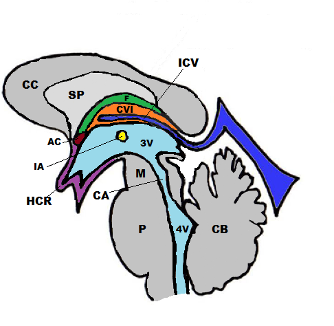
Sagittal Schematic View, Cavum Veli Interpositi. Corpus callosum (CC), septum pellucidum (SP), internal cerebral vein (ICV), fornix (FF), anterior commissure (AC), interthalamic adhesion (IA), hypothalamus, chiasmatic region (HCR), third ventricle (3V), cerebral aqueduct (CA), fourth ventricle (4V), Midbrain (M), Pons (P), Cerebellum (CB).
Contributed by A De Leucio, MD
(Click Image to Enlarge)
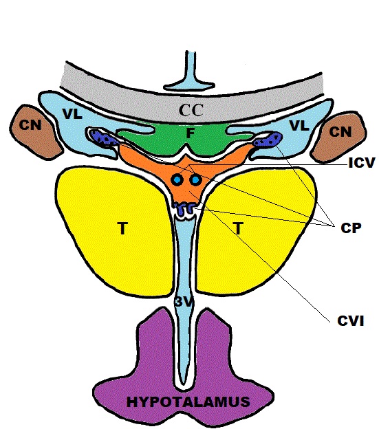
Coronal Schematic View, Cavum Veli Interpositi. Corpus callosum (CC), internal cerebral vein (ICV), fornix (F), lateral ventricle (VL), thalamus (T), caudate nucleus (CN), third ventricle (3V), choroid plexi (CP).
Contributed by A De Leucio, MD
(Click Image to Enlarge)
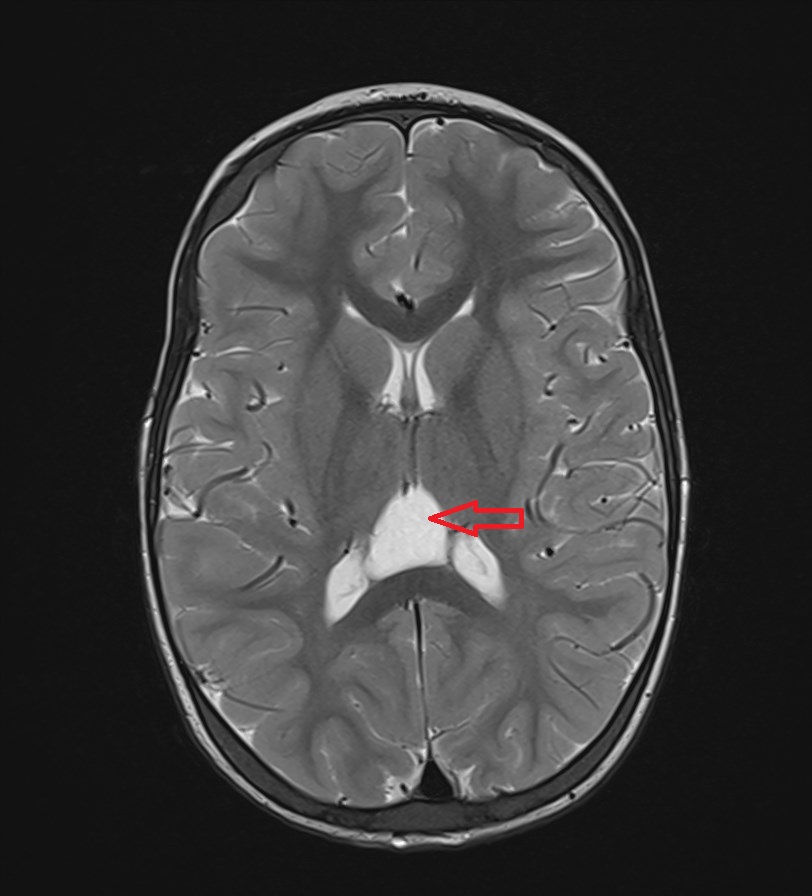
Axial T2: Cavum Veli Interpositi
Contributed by A De Leucio, MD
(Click Image to Enlarge)
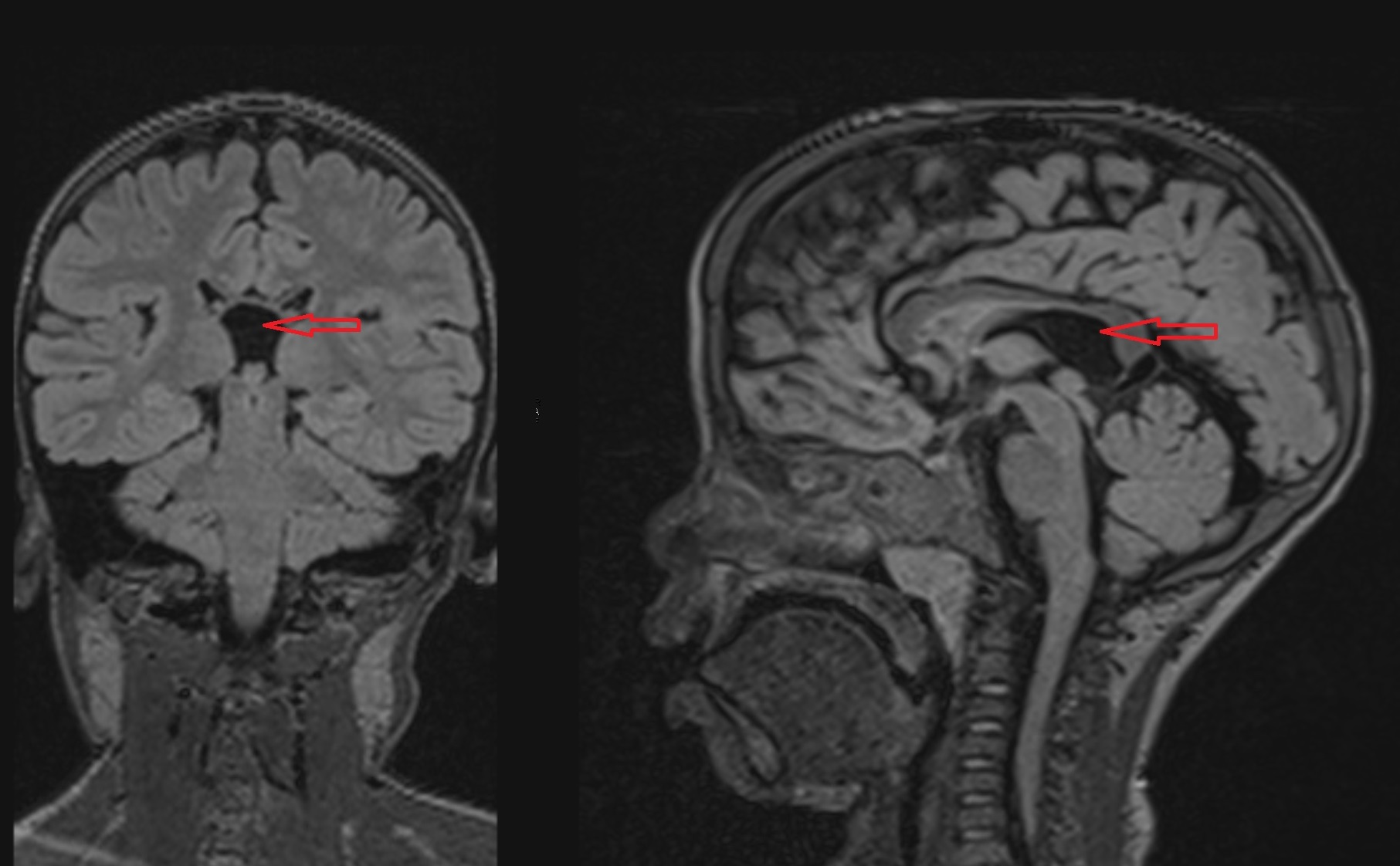
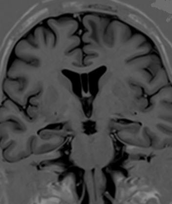
Coronal and Sagittal FLAIR: Cavum Veli Interpositi
Contributed by A De Leucio, MD
(Click Image to Enlarge)
Cavum Septum Pellucidum
Contributed by S Munakomi, MD
References
Macpherson P, Teasdale E. CT demonstration of a 5th ventricle--a finding to KO boxers? Neuroradiology. 1988:30(6):506-10 [PubMed PMID: 3265765]
Chen CY, Chen FH, Lee CC, Lee KW, Hsiao HS. Sonographic characteristics of the cavum velum interpositum. AJNR. American journal of neuroradiology. 1998 Oct:19(9):1631-5 [PubMed PMID: 9802483]
Kier LE. The evolutionary and embryologic basis for the development and anatomy of the cavum veli interpositi. AJNR. American journal of neuroradiology. 2000 Mar:21(3):612-4 [PubMed PMID: 10730668]
Level 3 (low-level) evidenceSaba L,Anzidei M,Raz E,Suri J,Piga M,Grassi R,Catalano C, MR and CT of brain's cava. Journal of neuroimaging : official journal of the American Society of Neuroimaging. 2013 Jul; [PubMed PMID: 23320830]
Aldur MM, Celik HH, Gürcan F, Sancak T. Frequency of cavum veli interpositi in non-psychotic population: a magnetic resonance imaging study. Journal of neuroradiology = Journal de neuroradiologie. 2001 Jun:28(2):92-6 [PubMed PMID: 11466492]
Kempe LG, Busch E. Clinical significance of the cisterna veli interpositi. Acta neurochirurgica. 1967:16(3):241-8 [PubMed PMID: 5299324]
Raimondi AJ, Gutierrez FA, Jones RR, Winston SR. Cystic cavum veli interpositi associated with normal or low pressure hydrocephalus. Child's brain. 1975:1(5):291-305 [PubMed PMID: 1175442]
Level 3 (low-level) evidenceGangemi M,Donati P,Maiuri F,Sigona L, Cyst of the velum interpositum treated by endoscopic fenestration. Surgical neurology. 1997 Feb; [PubMed PMID: 9040815]
Level 3 (low-level) evidenceTong CK, Singhal A, Cochrane DD. Endoscopic fenestration of cavum velum interpositum cysts: a case study of two symptomatic patients. Child's nervous system : ChNS : official journal of the International Society for Pediatric Neurosurgery. 2012 Aug:28(8):1261-4. doi: 10.1007/s00381-012-1770-4. Epub 2012 Apr 29 [PubMed PMID: 22543434]
Level 3 (low-level) evidenceKrejčí T, Vacek P, Krejčí O, Chlachula M, Szathmaryová S, Lipina R. Symptomatic cysts of the cavum septi pellucidi, cavum vergae and cavum veli interpositi: A retrospective duocentric study of 10 patients. Clinical neurology and neurosurgery. 2019 Oct:185():105494. doi: 10.1016/j.clineuro.2019.105494. Epub 2019 Aug 19 [PubMed PMID: 31472394]
Level 2 (mid-level) evidence