Introduction
Superior keratoconus is a rare corneal disorder classified as a progressive noninflammatory ectasia, most commonly bilateral and asymmetric, with few occurrences of unilateral presentation.[1] Controversy exists regarding the true presence of isolated superior keratoconus, but several cases have been confirmed and documented based on corneal topography measurements.[2][3][4] Superior keratoconus characteristically presents as a superior corneal protrusion with superior central and paracentral corneal thinning and steepening, resulting in visual disturbances.[5]
Ocular manifestations vary and can remain subclinical for years with incremental advancement. Clinical onset and diagnosis typically occur between ages 10 and 40.[6] Secondary causes of superior keratoconus commonly include mechanical disruption, such as in cases of blepharoptosis.[7] As for the symptoms, patients usually experience worsening and fluctuating visual acuity with associated glare, halos, and photophobia.[8]
Recent advancements in diagnostic imaging, such as corneal topography and tomography, have improved clinicians' ability to detect and monitor superior keratoconus. These imaging techniques allow for detailed corneal curvature, elevation, and thickness mapping, providing essential information for diagnosis and management.[9] Studies have shown that superior keratoconus can be identified by distinctive topographic patterns, including superior steepening and inferior flattening, critical for differentiating it from other corneal ectasias.
The management of superior keratoconus involves both nonsurgical and surgical approaches. Nonsurgical options include using rigid gas permeable (RGP) contact lenses or scleral lenses to improve vision by providing a more regular refractive surface. These lenses are particularly beneficial in cases where glasses are insufficient to correct vision due to irregular astigmatism. For progressive cases, corneal collagen cross-linking (CXL) is a well-established treatment aimed at halting the progression of ectasia. CXL works by strengthening the corneal stroma through the application of riboflavin (vitamin B2) and ultraviolet-A (UVA) light, which induces the formation of additional covalent bonds within the corneal collagen fibers. This procedure has been shown to stabilize the cornea and prevent further deterioration in many patients with superior keratoconus.[10]
Surgical interventions such as intracorneal ring segments (ICRS) or keratoplasty may be required in advanced stages of superior keratoconus, marked by corneal scarring or significant thinning. ICRS involves the insertion of semicircular plastic rings into the corneal stroma to flatten the cornea and improve vision. This procedure may be particularly useful in cases presenting with moderate ectasia and contact lens intolerance.
Penetrating keratoplasty (PKP) or deep anterior lamellar keratoplasty (DALK) may be considered in severe cases where other treatments are ineffective.[11] These procedures involve replacing the damaged corneal tissue with healthy donor tissue, offering the potential for significant visual improvement. However, PKP and DALK come with risks, such as graft rejection and the need for long-term immunosuppressive therapy.
Genetic factors have also been implicated in the development of superior keratoconus. Studies have identified several genetic markers associated with keratoconus, suggesting a hereditary component to the disease. Understanding these genetic factors could pave the way for future therapies targeting the underlying causes of corneal ectasia.[12]
Moreover, research into the biomechanical properties of the cornea has provided insights into the pathophysiology of superior keratoconus. Advanced technologies like Brillouin microscopy and optical coherence elastography are being explored to assess corneal stiffness and viscoelasticity, which may help in early diagnosis and personalized treatment strategies.
The prognosis of superior keratoconus is similar to that of general keratoconus.[13] Patients typically experience progressive corneal thinning and worsening visual acuity. Severe stages of this disease may cause acute corneal hydrops and subsequent corneal stromal scarring. Current management strategies are aimed at stabilizing the cornea and improving visual acuity. Options include contact lens fitting, corneal cross-linking (CXL) or transplants, and intrastromal ring insertions.[14]
Superior keratoconus is a complex corneal disorder requiring a multifaceted approach for effective management. Early detection and intervention are crucial in preventing significant visual impairment. Ongoing research and technological advancements continue to enhance our understanding and treatment of this challenging condition, offering hope for better patient outcomes in the future.
Etiology
Register For Free And Read The Full Article
 Search engine and full access to all medical articles
- 10 free questions in your specialty
- Free CME/CE Activities
- Free daily question in your email
- Save favorite articles to your dashboard
- Emails offering discounts
Learn more about a Subscription to StatPearls Point-of-Care
Etiology
The exact etiology of superior keratoconus is not well understood. However, several risk factors and associated conditions have been identified, including eye rubbing, blepharoptosis, and connective tissue disorders like Ehlers-Danlos, Down, and Marfan syndromes.[15] Superior keratoconus is also linked to atopy.[16] However, this association is highly controversial, as superior keratoconus in patients with atopy may be largely due to eye rubbing in response to allergic conjunctivitis. These risk factors are similar to those of general keratoconus, but their relationship with superior keratoconus remains undetermined due to the small number of confirmed cases.
Like other forms of keratoconus, the etiology of superior keratoconus is multifactorial, involving a combination of genetic, environmental, and biochemical factors. Understanding the causes of superior keratoconus is essential for early diagnosis, management, and potential prevention strategies.
Genetic Factors
Superior keratoconus shows evidence of a hereditary component, often appearing in individuals with a family history of the disease. Research has identified several genetic loci and specific genes that contribute to its development, highlighting a complex genetic basis.
Hereditary component
Keratoconus often runs in families, suggesting a genetic predisposition. Multiple studies have identified various genetic loci associated with the disease, indicating a polygenic inheritance pattern.[17] Specific genes such as VSX1, SOD1, and ZEB1 have been implicated in the development of keratoconus, although no single gene mutation has been identified as the primary cause.
Genetic syndromes
Superior keratoconus, like other forms, is more prevalent in individuals with certain genetic disorders such as Down, Marfan, and Ehlers-Danlos syndromes. These conditions are characterized by connective tissue abnormalities that may predispose individuals to corneal ectasia.
Environmental Factors
Environmental factors play a significant role in the development and progression of superior keratoconus. External influences, such as mechanical trauma and allergic conditions, can exacerbate corneal weakening and contribute to the disease.
Eye rubbing
Chronic eye rubbing is strongly associated with the development and progression of keratoconus. The mechanical trauma from repeated rubbing can lead to microtrauma and weakening of the corneal structure.[18]
Atopy and allergies
Individuals with atopic conditions such as asthma, eczema, and allergic conjunctivitis are at increased risk of developing keratoconus. Chronic inflammation and rubbing due to itching may contribute to corneal weakening.[19]
Biochemical Factors
Biochemical factors significantly influence the development of superior keratoconus through disruptions in the corneal structure. Enzymatic activity imbalances and increased oxidative stress can compromise corneal integrity and contribute to disease progression.
Enzymatic imbalance
An imbalance between proteolytic enzymes and their inhibitors in the corneal stroma has been proposed as a contributing factor. Increased activity of matrix metalloproteinases (MMPs) and decreased levels of tissue inhibitors of metalloproteinases (TIMPs) can lead to extracellular matrix degradation, resulting in corneal thinning and ectasia.[20]
Oxidative stress
Increased oxidative stress and decreased antioxidant defenses in the cornea are believed to play a role in the pathogenesis of keratoconus. Oxidative damage to corneal cells and extracellular matrix components can weaken the cornea and predispose it to deformation.[21]
Mechanical Factors: Biomechanical Properties
Abnormal corneal biomechanical properties, such as reduced corneal stiffness and altered collagen cross-linking, can contribute to the development of keratoconus. These changes make the cornea more susceptible to deformation under normal intraocular pressure.[22]
Hormonal Factors
Hormonal changes, particularly during puberty and pregnancy, may influence the progression of keratoconus. Hormonal fluctuations can affect the biomechanical properties of the cornea, potentially exacerbating ectasia.[23]
The etiology of superior keratoconus is complex and multifactorial, involving genetic predisposition, environmental influences, biochemical imbalances, mechanical factors, and possibly hormonal changes. Understanding these factors can help identify individuals at risk for developing superior keratoconus and initiate early intervention.
Epidemiology
The incidence of isolated superior keratoconus is largely unknown and has not been specifically reported due to its extreme rarity. However, the disease is thought to account for less than 1% of all confirmed keratoconus cases. The incidence rate of general keratoconus is thought to range from 1.5 to 25 per 100,000 persons per year, affecting all ethnicities and sexes. The current estimated prevalence rate is thought to be between 0.2 and 4,790 per 100,000 persons per year.[24] Most superior keratoconus cases are undiagnosed until the later stages due to their initially subclinical presentation.
Superior keratoconus is a rare variant of keratoconus characterized by corneal thinning and steepening, primarily in the superior cornea. The epidemiology of superior keratoconus resembles that of general keratoconus but includes some distinct aspects due to its unusual presentation.
General Epidemiology of Keratoconus
The prevalence of keratoconus varies widely across different populations. The condition's presentation and demographic patterns can offer insights into its broader epidemiological profile.
Prevalence
Studies estimate the prevalence of keratoconus to range from 50 to 230 per 100,000 in the general population.[25] The prevalence of keratoconus is approximately 54.5 per 100,000 individuals in the United States.[26] Higher prevalence rates have been reported in certain ethnic groups, such as people of Middle Eastern, South Asian, and African descent.[27]
Age distribution
Keratoconus typically presents during adolescence and early adulthood, with the age of onset commonly between 10 and 25 years. The condition progresses over 10 to 20 years before stabilizing.[28] The disease is often diagnosed in the 2nd and 3rd decades of life, corresponding to the period of rapid corneal changes.[29]
Sex distribution
Keratoconus has a slight male predominance. The male-to-female ratio varies in different studies but is generally around 1.5:1.
Specific Epidemiology of Superior Keratoconus
Superior keratoconus is a rarer form of keratoconus with limited prevalence data. This disorder's geographical, age, and sex distribution generally align with the broader patterns observed in keratoconus.
Frequency
Superior keratoconus is less common than the typical inferior form. Precise prevalence data for superior keratoconus are limited due to its rarity and the challenge of differentiating it from other corneal ectatic disorders.[30] Case series and reports indicate that superior keratoconus constitutes a small percentage of all keratoconus cases, with some estimates suggesting it accounts for less than 5% of all cases.
Geographical distribution
Like general keratoconus, superior keratoconus does not show a significant geographical preference. However, the higher prevalence of keratoconus in certain ethnic groups may also be reflected in the distribution of superior keratoconus within those populations.[31]
Age and sex distribution
The age of onset and progression pattern of superior keratoconus are similar to those of typical keratoconus, with onset in the 2nd to 3rd decades of life. The male predominance observed in general keratoconus is also seen in superior keratoconus, although specific data on sex distribution are scarce.[32]
Global Perspective
Studies from various parts of the world, including the Middle East, Asia, and Africa, have reported higher rates of keratoconus. These rates reflect genetic and environmental factors that may also influence the prevalence of superior keratoconus in these regions.[33]
By understanding the epidemiological characteristics of superior keratoconus, healthcare providers can better identify at-risk populations and implement early screening and intervention strategies to manage this rare corneal disorder effectively.
Pathophysiology
The proposed pathophysiological mechanisms of superior keratoconus are thought to be the same as general keratoconus, although not entirely understood. Many proteins, such as TGF-β1, collagen type IV, secreted protein acidic and cysteine-rich (SPARC), wingless-related integration site (Wnt), Hedgehog (Hh), and cellular adhesion molecules, are believed to be dysregulated in patients with superior keratoconus.[34] These abnormally expressed proteins may lead to structural changes in the cornea via keratocyte apoptosis and stromal necrosis.
Dysregulation of oxidative stress markers and antioxidants may also play a significant role in the pathogenesis of superior keratoconus. Corneal stromal thinning and degeneration may be caused by decreased concentrations of antioxidants such as aldehyde/NADPH dehydrogenase, lactoferrin, transferrin, and albumin and increased concentrations of reactive oxidative stress markers such as reactive oxygen species, nitrogen, and malondialdehyde in the stroma. Additionally, increased levels of lysosomal enzymes have been found in the stromal keratocytes of patients with keratoconus, potentially explaining damage seen in the anterior stromal membrane. Physical stresses, such as increased intraocular pressure and eye rubbing, may exacerbate this degradation.[35]
Histopathology
Histopathological findings of superior keratoconus may demonstrate both stromal and epithelial thinning, predominantly in the superior cornea. Breaks in the Bowman layer, Descemet membrane, or both may also be observed. Panstromal scarring and Descemet membrane rupture with subsequent hydrops may be seen in the later stages of superior keratoconus.[36]
Histopathological examination of corneal tissue in the superior keratoconus reveals a range of microscopic changes that reflect structural abnormalities and disease progression. These findings help understand the underlying pathophysiology and differentiate superior keratoconus from other ectatic corneal disorders.
Epithelial Changes
The corneal epithelium in keratoconus often shows thinning and irregularity. This disruption can lead to breaks in the Bowman layer, contributing to the characteristic corneal steepening. Irregularities in the epithelium's basal cell layer are frequently observed, indicating chronic stress and damage.[37]
Bowman Layer Breaks and Scarring
The Bowman layer often shows localized breaks and, in advanced cases, scarring. These breaks are significant as they contribute to corneal protrusion and the irregular astigmatism seen clinically.[38]
Stromal Changes
The stromal layer shows significant thinning, which is more pronounced in the superior part of the cornea. This thinning is a key feature that differentiates superior keratoconus from other forms of keratoconus.[39] The normally regular arrangement of collagen lamellae in the stroma becomes disrupted. Abnormal collagen fibrils appear interspersed, weakening the corneal structure.[40] Increased apoptosis of keratocytes, the cells responsible for maintaining the corneal stroma, is observed. This cell loss contributes to stromal thinning and weakening.[41]
Changes in the Descemet Membrane and Endothelium
Clinically visible vertical stress lines in the Descemet membrane called "Vogt striae" arise, correlating with breaks or folds seen histologically. The endothelium is generally spared in early keratoconus. However, advanced cases may show endothelial cell loss and pleomorphism, reflecting the overall corneal stress.[42]
Presence of Inflammatory Cells
Keratoconus is primarily a noninflammatory disorder. However, mild infiltration of inflammatory cells is sometimes observed, particularly in areas of significant tissue damage or scarring.[43]
Significance of Histopathological Findings
Histopathological examination provides crucial insights into the progression of superior keratoconus. The observed changes, such as stromal thinning, collagen disorganization, and keratocyte apoptosis, highlight the disease's degenerative nature. These findings support the clinical and imaging diagnoses and help differentiate superior keratoconus from other corneal pathologies like pellucid marginal degeneration or keratoglobus. Understanding the histopathology of superior keratoconus aids in developing targeted treatments aimed at strengthening the corneal structure, such as CXL, and provides the basis for ongoing research into the pathophysiology of corneal ectatic disorders.
History and Physical
Patient Presentation
Patients with superior keratoconus often present with progressively worsening and fluctuating visual acuity with associated glare, halos, and photophobia. A thorough medical history gathering may reveal atopy, eye rubbing, or a family history of keratoconus. External examination of the patient's eyes may reveal abnormal protrusion and an inverse Munson sign, characterized by a V-shaped indentation of the upper eyelid during upgaze (see Image. Keratoconus).
Onset and progression
Symptoms typically begin in the 2nd to 3rd decade of life, although they can start earlier in some cases. Superior keratoconus gradually worsens over time, with periods of relative stability interspersed with episodes of rapid deterioration.
Family history
A positive family history of keratoconus or other ectatic corneal disorders may be present, suggesting a genetic component.[44] This information is crucial during history-taking to identify individuals at higher risk and to guide further diagnostic evaluation.
Associated conditions
Frequent eye rubbing, often due to associated conditions such as allergies or atopy, may be a contributing factor. Conditions like Down syndrome and connective tissue disorders like Marfan and Ehlers-Danlos syndromes may be associated with keratoconus.[45]
Physical Examination Findings
Slit-lamp biomicroscopy may show normal findings in subclinical superior keratoconus.[46] Findings in the later stages of superior keratoconus may include the following:
- Superior central and paracentral corneal thinning and superior steepening
- Superior Vogt striae or vertical stress lines formed by the compression of the Descement membrane
- Partial breaks in the Bowman or Descemet membrane
- Complete or partial Fleischer rings or iron deposits in the basal corneal epithelial cells, visualized as a brown or yellow ring using a cobalt blue filter attached to the slit-lamp [47]
- Rizutti's sign, which is a bright reflection of the nasal limbal area when light shines on the temporal limbus
- Acute corneal hydrops with subsequent stromal scarring [48]
- Scissoring reflex on retinoscopy, indicating irregular astigmatism [49]
- Corneal topography reveals superior corneal steepening and asymmetry, which are key diagnostic features distinguishing superior keratoconus from other variants.[50]
- The posterior segment is typically normal unless complications develop from advanced disease.
By taking a thorough history and performing a detailed physical examination, including specialized diagnostic tests, clinicians can accurately diagnose superior keratoconus and differentiate it from other corneal conditions. This comprehensive approach ensures appropriate disease management and monitoring to prevent significant visual impairment.
Evaluation
The evaluation of superior keratoconus involves a combination of clinical examination, advanced imaging techniques, and other diagnostic tests. These methods are used to accurately assess disease extent, monitor progression, and guide treatment decisions.
Clinical Examination
The initial diagnosis of superior keratoconus involves measuring the patient's refraction. Additionally, standard visual acuity tests, including testing with and without corrective lenses, determine the level of vision impairment.
Slit Lamp Biomicroscopy
Slit-lamp biomicroscopy is used to assess for clinical signs of superior keratoconus, as described above. This modality allows for a detailed examination of the corneal structure and identification of key features.
Corneal Topography
Corneal topography is a diagnostic method used to screen and evaluate patients with superior keratoconus. Findings include superior central and paracentral corneal thinning with superior steepening. The S-I ratio, a value comparing the average power difference between the cornea's superior and inferior hemispheres, may be between 1.4 and 1.8 in patients with superior keratoconus.[51] Changes in the cornea's anterior elevation may be 20 µm or greater. Changes in the cornea's posterior elevation can be 50 µm or greater.[52] Corneal pachymetry at the thinnest point of the cornea may be 495 µm or less. Corneal topographical patterns may include an asymmetric bowtie with or without superior steening, skewed radial axes, and an inferior flat area of decreased steepening.
Anterior Segment Optical Coherence Tomography
Anterior segment optical coherence tomography (OCT) is useful as a pachymetry tool in revealing corneal thickness asymmetries that may be masked in patients with superior keratoconus due to epithelial remodeling. Fourier-domain OCT devices are also useful in assessing corneal epithelial thickness distribution changes.[53]
Retinoscopy
Retinoscopy is a highly sensitive and reliable test for detecting keratoconus, including the early stages of the disease. Findings include a scissoring reflex, characterized by 2 light bands moving toward and away from each other.[54]
Corneal Tomography
Corneal tomography provides a 3-dimensional image of the cornea, measuring both anterior and posterior surfaces as well as corneal thickness. Techniques like Scheimpflug imaging and optical coherence tomography (OCT) are commonly used. Tomography is essential for detecting early keratoconus and subclinical cases, and various ophthalmology societies recommend it for comprehensive assessment.[55]
Pachymetry
Pachymetry measures the cornea's thickness, which is crucial for assessing the extent of corneal thinning and determining eligibility for treatments like CXL. Regular pachymetric assessments are recommended to monitor disease progression and treatment response.[56]
Biomechanical Assessment
Devices such as the Ocular Response Analyzer (ORA) and CorVis ST measure corneal biomechanical properties, providing additional information about corneal rigidity and susceptibility to ectasia. These assessments are useful adjuncts to topography and tomography, aiding in early diagnosis and risk stratification.[57]
Refraction and Keratometry
Detailed refraction tests are used to measure the degree of myopia or astigmatism. Keratometry is used to measure the curvature of the cornea. These modalities are essential for documenting changes over time. Routine refraction and keratometry are part of the standard evaluation protocol for patients with keratoconus.[58]
Genetic Testing
Genetic testing can identify mutations associated with keratoconus, providing insights into familial patterns and risk factors. While not routine, genetic testing may be recommended in cases with a strong family history or atypical presentation.[59]
Other Diagnostics
Endothelial cell density and morphology may also be assessed. These attributes may be affected in advanced keratoconus or after corneal surgery. Specular microscopy is recommended for preoperative assessment and postoperative monitoring in patients with keratoconus undergoing surgical interventions.[60]
By following these evaluation protocols and guidelines, healthcare providers can accurately diagnose superior keratoconus, monitor its progression, and tailor treatment plans to patients' needs, improving outcomes and quality of life.
Treatment / Management
The treatment options for superior keratoconus are similar to those of general keratoconus. Management options depend on the disease stage and progression.[61] Management is also classified into nonsurgical and surgical techniques. (B3)
Nonsurgical Management
Nonsurgical management of superior keratoconus focuses on preserving and improving visual acuity through conservative corrective measures. Treatment strategies vary depending on the severity of the condition, ranging from glasses and contact lenses to specialized scleral lenses.
Mild superior keratoconus
Treatment focuses on maintaining adequate visual acuity for the patient, usually in the form of prescribing glasses. Hard contact lenses such as RGP or scleral lenses may be considered based on patient preference.[62]
Moderate superior keratoconus
Patients at this stage typically use contact lenses to correct their vision. Options may include RGP lenses, which correct corneal irregularities. However, patients may find these lenses intolerable, and they are usually not prescribed for individuals currently or previously with corneal scarring. Scleral lenses are being used now for patients with moderate keratoconus, as these lenses allow a tear vault to form in the space between the cornea and the lens, aiding in correcting irregular astigmatism and refractive errors. Scleral lenses also have a lower risk of dislocation on the cornea and are found to be more comfortable by patients.[63](A1)
Severe superior keratoconus
Patients with severe keratoconus are at high risk of corneal stromal scarring due to the likelihood of experiencing episodes of acute corneal hydrops. Scleral lenses are the preferred treatment option for these patients.
Surgical Management
Surgical management of superior keratoconus involves various procedures aimed at stabilizing the cornea and improving visual acuity. Treatment options range from cross-linking and corneal transplants to specialized techniques like DALK and intrastromal corneal ring segments (ICRSs).
Cross-linking
This procedure is indicated for progressive keratoconus and promotes the strengthening and stability of the cornea's collagen fibrils. Cross-linking involves removing a portion of the central epithelium and administering riboflavin solution drops. This step is followed by UV-A light exposure that activates riboflavin and generates reactive oxygen species to form covalent bonds between the collagen and proteoglycans in the cornea.[64] This procedure is often coupled with photorefractive keratectomy to aid in refractive correction.[65] Contraindications for this procedure include corneal thickness of less than 400 µm, prior or active herpetic eye infection, severe corneal scarring, certain autoimmune disorders, and severe ocular surface disease.
Penetrating keratoplasty
Corneal transplants are indicated when lenses are no longer effective in correcting visual acuity. Approximately 10% to 20% of patients with keratoconus will require a corneal transplant.[66] Penetrating keratoplasty (PKP) is a full-thickness transplant that involves removing all layers of the host cornea and replacing them with a healthy donor cornea. Patients typically achieve adequate visual acuity after surgery.
PKP may be indicated in patients with keratoconus and concurrent endothelial dysfunction or deep stromal scarring. Residual astigmatism after surgery may be corrected with contact lenses or other forms of refractive correction.[67] A known complication of PKP is graft rejection, so patients typically are treated with corticosteroids for a year or longer.[68] Contraindications include severe ocular surface disease, certain autoimmune disorders, very poor eyelid hygiene, severe corneal neovascularization, and various forms of ocular surface inflammation.
Deep anterior lamellar keratoplasty
DALK is a partial-thickness corneal transplant that replaces the host cornea's epithelium and anterior stroma. DALK offers faster visual recovery and a reduced risk of graft rejection and failure compared to PKP.[69] This procedure is preferred for patients without endothelial dysfunction. Contraindications are similar to those of PKP.[70](A1)
Intrastromal corneal ring segments
Small plastic rings are inserted into the stroma to flatten the cornea, improving visual acuity and reducing myopia. Notably, ICRS does not treat or slow the progression of keratoconus. The procedure is only useful for correcting visual acuity. Contraindications include a corneal thickness of less than 450 µm, corneal scarring, and the possibility of improving vision with contact lenses.
Other Interventions
Other nonsurgical approaches are available for managing superior keratoconus. These modalities include pharmacological treatments to control inflammation and visual rehabilitation techniques to enhance the quality of life for patients with significant visual impairment. Topical corticosteroids and nonsteroidal anti-inflammatory drugs may be used postoperatively to control inflammation and prevent complications.[71] For patients with significant visual impairment not correctable by surgery or contact lenses, low vision aids and rehabilitation services are essential to improving their quality of life.[72](A1)
Monitoring and Follow-Up
Regular follow-up is crucial for monitoring disease progression and treatment effectiveness. Advanced imaging techniques, such as corneal topography and OCT, assess corneal shape and thickness changes over time.[73]
Psychological Support
Living with a chronic condition like superior keratoconus can be challenging, and psychological support, including counseling and support groups, can help patients cope with the emotional and psychological impacts of the disease. By adhering to these guidelines and employing a multifaceted approach to treatment, healthcare providers can effectively manage superior keratoconus, improving visual outcomes and patients' quality of life.[74](A1)
Differential Diagnosis
Several differential diagnoses must be considered when evaluating a patient with corneal thinning, including the following:
- Corneal ectasia (iatrogenic)
- Terrien marginal degeneration
- Fuchs endothelial dystrophy
- Contact lens warpage
Superior keratoconus, a keratoconus variant, also shares many clinical features with other corneal ectatic disorders and conditions that affect corneal curvature and thickness. Accurate diagnosis is crucial, and several other conditions must be considered and ruled out. Below are some of the key differential diagnoses.
Pellucid Marginal Degeneration
Pellucid marginal degeneration (PMD) is a bilateral, noninflammatory corneal thinning disorder characterized by a peripheral band of thinning, typically in the inferior cornea but occasionally affecting the superior cornea. PMD presents with a classic "kissing doves" or "crab-claw" pattern on corneal topography, which differs from the asymmetric, superior steepening seen in superior keratoconus.[75]
Keratoglobus
Keratoglobus is a rare ectatic disorder marked by diffuse, generalized corneal thinning, producing a globular shape. Unlike superior keratoconus, which shows localized superior thinning and steepening, keratoglobus affects the entire cornea, resulting in a more uniform, globular protrusion.[76]
Terrien Marginal Degeneration
Terrien marginal degeneration is a rare, slowly progressive thinning disorder that typically begins superiorly and progresses circumferentially. This condition is usually characterized by vascularization and lipid deposition at the leading edge of the thinning area, features not seen in superior keratoconus.[77]
Posterior Keratoconus
Posterior keratoconus is characterized by localized posterior corneal surface thinning, leading to an irregular posterior corneal curvature. This disorder primarily affects the posterior cornea, unlike the superior keratoconus, which involves anterior corneal thinning and steepening.
Contact Lens-Induced Corneal Warpage
Long-term use of rigid contact lenses may cause corneal shape changes that mimic ectatic disorders. Corneal warpage typically resolves after discontinuing contact lens use, and the topographic changes are usually symmetric, unlike the progressive nature of superior keratoconus.[78]
Post-Laser-Assisted In Situ Keratomileusis Ectasia
Laser-assisted in situ keratomileusis (LASIK) reshapes the cornea with a laser to correct vision and reduce the need for corrective lenses. Post-LASIK ectasia is a complication characterized by progressive corneal thinning and bulging. The history of refractive surgery and the location of ectasia corresponding to the LASIK flap help differentiate this condition from superior keratoconus.[79]
Corneal Dystrophies
Several corneal dystrophies, such as lattice and granular dystrophies, can cause corneal structural changes. These dystrophies typically present with distinct opacities or deposits within the corneal layers, visible on slit-lamp examination and not seen in keratoconus.[80]
Accurate diagnosis involves a thorough clinical examination, corneal topography, and tomography. Advanced imaging techniques, such as OCT and Scheimpflug imaging, can provide detailed maps of the corneal surface and thickness, aiding in differentiation. By considering these differential diagnoses, clinicians can more accurately diagnose superior keratoconus and implement appropriate management strategies to improve patient outcomes.
Pertinent Studies and Ongoing Trials
Hersh et al conducted a study titled "Corneal collagen crosslinking for keratoconus and corneal ectasia: one-year results," which evaluated the efficacy of CXL in patients with keratoconus and corneal ectasia. The results suggested significant corneal stabilization, with reduced progression of ectasia over one year, supporting the use of CXL as an effective treatment to halt the progression of superior keratoconus.[81]
Wittig-Silva et al conducted a randomized controlled trial of CXL in progressive keratoconus. The 3-year follow-up data from this trial showed sustained stabilization and improvement in corneal curvature, providing robust evidence for the long-term benefits of CXL in managing keratoconus, including superior keratoconus.[82]
Reinhart et al compared the outcomes of DALK with PKP in patients with keratoconus. The group's research revealed that DALK was a viable alternative to PKP, with fewer complications and better postoperative recovery, making it a recommended option for advanced superior keratoconus.[83]
Ongoing trials at the National Eye Institute and other research institutions continue to evaluate the long-term outcomes of various treatment modalities for superior keratoconus, including newer surgical techniques and genetic therapies. These studies aim to enhance understanding of the disease, improve diagnostic techniques, and develop more effective treatments, ultimately improving patient outcomes and quality of life for those with superior keratoconus.
Treatment Planning
Given the disease presentation and progression variability among patients, treatment planning for superior keratoconus involves a detailed and personalized approach. Below are the key components of treatment planning for superior keratoconus.
Diagnosis and Initial Assessment
- Comprehensive eye examination: Includes visual acuity tests, slit-lamp examination, and retinoscopy to evaluate the degree of corneal thinning and protrusion
- Corneal imaging: Advanced imaging techniques such as corneal topography and tomography are essential for detailed corneal surface mapping and staging the disease [84]
Nonsurgical Management
- Contact lenses: Customized contact lenses, including RGP and scleral lenses, are often the first line of treatment to improve visual acuity by providing a more regular refractive surface.
- Monitoring: Regular follow-up visits to monitor disease progression using corneal imaging and visual acuity tests. The frequency of visits may vary based on the severity and progression of the disease.[85]
Corneal Cross-Linking
- Indications: Recommended for patients with progressive keratoconus to strengthen corneal tissue and halt progression
- Procedure: CXL involves riboflavin application and subsequent exposure to UVA light. The total dose and duration are tailored to the patient's corneal thickness and curvature.
- Follow-up: Post-CXL monitoring includes regular check-ups to assess corneal stability and detect any adverse effects.[84]
Surgical Interventions
- ICRS
- Indications: Suitable for patients with moderate keratoconus and intolerant to contact lenses or have significant visual impairment
- Procedure: Insertion of plastic rings into the corneal stroma to flatten the cornea and improve vision
- Follow-up: Regular monitoring for complications such as infection or displacement of the rings.[86]
- Optical Penetrating Keratoplasty (OPK) vs DALK
- Indications: Considered for advanced cases with severe corneal scarring or thinning
- Procedure: Replacement of the damaged corneal tissue with donor tissue. The choice between therapeutic keratoplasty (TPK) and DALK depends on the depth of corneal involvement.
- Follow-up: Long-term follow-up is critical to monitor for graft rejection and ensure graft stability.[87]
Postoperative and Rehabilitation Care
- Medication adherence: Postsurgical patients are prescribed antibiotics and anti-inflammatory eye drops to prevent infection and reduce inflammation.
- Visual rehabilitation: Use of corrective lenses or visual aids to maximize postoperative visual outcomes.
- Psychological support: Counseling and support groups to help patients cope with the psychological impact of chronic eye disease.[88]
Advanced Imaging and Technology
Advanced imaging modalities include topography and tomography. These techniques provide detailed views of corneal structure, aiding in precise diagnosis and monitoring of keratoconus progression.[89]
Individualized Treatment Plans
- Personalized approach: Each patient's treatment plan is tailored based on the severity of keratoconus, rate of progression, and individual needs.
- Interdisciplinary collaboration: Coordination among ophthalmologists, corneal specialists, contact lens specialists, and other healthcare providers ensures comprehensive care.[90]
Healthcare providers can effectively manage superior keratoconus and improve patient outcomes by adopting a comprehensive and individualized approach to treatment planning.
Toxicity and Adverse Effect Management
Managing toxicity and adverse effects is critical to treating superior keratoconus, particularly given the various treatments involved, including medications, contact lenses, and surgical interventions. Below are key considerations and strategies for managing these issues.
Contact Lens-Related Complications
- Toxicity: Extended use of contact lenses, especially RGP and scleral lenses, can lead to corneal hypoxia, epithelial breakdown, and microbial keratitis. Management involves ensuring proper fitting and periodic evaluation of the lenses. Patients should be educated on proper lens hygiene and the importance of follow-up visits.
- Adverse effects: Common issues include discomfort, dry eye symptoms, and lens intolerance. Management involves using lubricating eye drops to alleviate dryness, taking breaks from lens-wearing to allow the cornea to recover, and, if necessary, switching to a different type of lens.[91]
Medication-Related Toxicity
- Corticosteroids: Topical corticosteroids are often used postsurgically to reduce inflammation, but prolonged use can cause elevated intraocular pressure and cataract formation. Management consists of monitoring intraocular pressure regularly and adjusting the dosage as necessary. Consider steroid-sparing agents if long-term treatment is required.
- Antibiotics: Long-term use of these medications can lead to resistance or toxicity. Management comprises using the lowest effective dose for the shortest duration possible. The patient should be regularly evaluated for signs of toxicity or resistance, and the treatment regimen should be adjusted accordingly.[92]
Corneal Cross-Linking Complications
- Toxicity: CXL involves the use of riboflavin and ultraviolet-A (UVA) light, which can cause corneal haze and endothelial cell damage if not performed correctly. Management involves ensuring strict adherence to the protocol to minimize UVA exposure. The cornea must be monitored postprocedure for any signs of haze or damage. Adjust treatment parameters based on individual corneal thickness.
- Adverse Effects: Pain, photophobia, and transient visual disturbances are common after CXL. Management consists of providing adequate pain relief and anti-inflammatory medications postprocedure. Educate patients about the expected temporary nature of these side effects.
Surgical Complications
- OPK and DALK: These procedures carry a risk of graft rejection and failure. Management involves implementing strict postoperative monitoring and immunosuppressive therapy as needed. Patients should be educated on the signs of rejection and the importance of early intervention.
- Adverse Effects: Infection, suture-related complications, and irregular astigmatism may arise. Management comprises prophylactic antibiotics to prevent infection, regular follow-up to remove sutures, and glasses or contact lenses to correct postoperative refraction errors.
Psychological Impact
The chronic nature of superior keratoconus and its impact on vision can lead to significant psychological stress, anxiety, and depression. Management involves providing psychological support and counseling. Referral to a mental health professional may be necessary for patients exhibiting significant distress.
By addressing these potential toxicities and adverse effects through vigilant monitoring, patient education, and timely intervention, healthcare providers can optimize the management of superior keratoconus and improve patient outcomes.[93]
Staging
Amsler-Krumeich Classification
Like other forms of keratoconus, superior keratoconus may be staged to assess the severity of the disease and guide treatment decisions. The Amsler-Krumeich classification is one of the most widely used staging systems for keratoconus and may also be applied to superior keratoconus.
Stage 1
- Findings: Slight central or paracentral inferior steepening, mild myopia, or astigmatism less than 5.00 diopters (D)
- Corneal thickness: Minimum thickness greater than 500 microns
- Clinical presentation: Often asymptomatic or mild visual disturbances
- Management: Regular monitoring, corrective lenses (glasses or contact lenses) [94]
Stage 2
- Findings: More pronounced inferior steepening, myopia, or astigmatism between 5.00 and 8.00 D.
- Corneal thickness: Minimum thickness between 400 and 500 microns
- Clinical presentation: Increased visual disturbances and need for more frequent changes in corrective lenses
- Management: Contact lenses (RGP or scleral lenses), CXL
Stage 3
- Findings: Severe inferior steepening, myopia, or astigmatism between 8.00 and 10.00 D, presence of corneal thinning and ectasia
- Corneal thickness: Minimum thickness between 200 and 400 microns
- Clinical presentation: Significant visual impairment, frequent changes in lens prescription, and possible corneal scarring
- Management: Contact lenses, CXL, consideration of surgical options such as ICRS or TPK
Stage 4
- Findings: Advanced inferior steepening, myopia, or astigmatism greater than 10.00 D, marked corneal thinning, and central scarring
- Corneal thickness: Minimum thickness of less than 200 microns
- Clinical presentation: Severe visual impairment, intolerance to contact lenses, significant corneal scarring
- Management: Surgical intervention, primarily TPK or DALK, with a potential need for corneal transplantation
Additional Considerations for Superior Keratoconus
- Topographic and tomographic analysis: Advanced imaging techniques such as corneal topography and tomography are essential for accurately staging superior keratoconus. These tools help assess the extent of corneal steepening, thinning, and asymmetry, which are critical for staging and treatment planning.[95]
- Clinical course and progression: Superior keratoconus may have a different progression pattern compared to classic keratoconus, often presenting with asymmetric and more localized superior involvement. Regular monitoring and early intervention are crucial to prevent significant visual impairment.
- Individualized treatment plans: The staging of superior keratoconus should guide individual treatment plans, taking into account the specific characteristics of the superior corneal involvement and the patient's overall ocular health.[96]
By utilizing the Amsler-Krumeich classification and incorporating advanced diagnostic tools, clinicians can effectively stage superior keratoconus and implement appropriate management strategies to optimize patient outcomes.
Prognosis
The prognosis of superior keratoconus is similar to that of general keratoconus. The condition is characterized by progressive corneal thinning and worsening of visual acuity. Approximately 10% to 20% of patients will require some form of corneal transplant during the course of the disease.
The prognosis of superior keratoconus varies depending on the stage of the disease at diagnosis, the rate of progression, and the effectiveness of the management strategies employed. Like other forms of keratoconus, superior keratoconus is a progressive condition that can lead to significant visual impairment if not properly managed.
Factors Influencing Prognosis
- Early detection and intervention: Early diagnosis through regular screenings and the use of advanced imaging techniques such as corneal topography can significantly improve the prognosis. Early intervention with treatments such as CXL can halt the progression of the disease and preserve vision.
- Treatment effectiveness: Treatment success plays a critical role in determining the prognosis. Treatments such as RGP and scleral lenses, CXL, and, in severe cases, OPK or DALK are essential in managing the condition and improving visual outcomes.[97]
- Stage at diagnosis: The stage of superior keratoconus at the time of diagnosis is a key determinant of prognosis. Patients diagnosed at an earlier stage generally have a better prognosis due to the availability of more effective treatment options to halt disease progression.
- Compliance with treatment: Patient adherence to prescribed treatments and follow-up schedules is crucial for achieving optimal outcomes. Noncompliance can lead to disease progression and poorer visual prognosis.[98]
Long-Term Prognosis
- Stable vision with treatment: With appropriate and timely treatment, many patients with superior keratoconus can achieve stable vision and maintain a good quality of life. CXL, in particular, has been shown to be effective in stabilizing the cornea and preventing further ectasia.
- Potential for visual rehabilitation: Even in advanced stages, surgical interventions such as TPK and DALK can restore significant visual function. However, these procedures carry risks such as graft rejection and require long-term follow-up.
- Risk of complications: Patients with superior keratoconus are at risk of complications such as corneal scarring, acute hydrops, and graft failure post-keratoplasty. These complications can adversely affect the prognosis and necessitate additional treatments.
- Genetic factors: Genetic predisposition may influence the prognosis. Patients with a family history of keratoconus may experience a more rapid progression and require more aggressive management.[99]
Overall Prognosis
The overall prognosis for superior keratoconus is generally favorable with early detection and appropriate management. Advances in diagnostic technologies and treatments have significantly improved the ability to manage this condition effectively. Regular monitoring and individualized treatment plans are essential to optimize visual outcomes and enhance the quality of life for patients with superior keratoconus.
Complications
Complications of superior keratoconus are similar to those of general keratoconus and can significantly impact visual acuity and quality of life. The primary complications associated with superior keratoconus are described below.
Progressive Corneal Thinning and Ectasia
The hallmark of keratoconus is progressive corneal thinning and bulging, which is more pronounced in the superior portion of superior keratoconus. This thinning can lead to significant visual distortion and impairment.[100]
Corneal Scarring
Scarring can occur as the cornea thins and becomes more irregular, particularly if the condition progresses to advanced stages. Corneal scarring can severely affect vision and may necessitate surgical intervention.[101]
Acute Corneal Hydrops
This condition is a sudden and severe complication characterized by the rupture of the Descemet membrane, leading to the influx of aqueous humor into the cornea. Acute corneal hydrops results in corneal swelling, pain, and a sudden decrease in vision.[102]
Contact Lens Intolerance
Many patients with superior keratoconus rely on RGP or scleral lenses for vision correction. However, the corneal shape may become too irregular as the disease progresses, making it difficult or impossible to fit contact lenses comfortably.
Graft Failure Post-Keratoplasty
Patients undergoing OPK or DALK have an increased risk of graft rejection or failure. This complication can occur due to immune reactions, infection, or improper healing.
Visual Disturbances
Patients with superior keratoconus often experience significant visual disturbances, including ghosting, glare, halos, and double vision. These symptoms can impair daily activities and reduce the overall quality of life.[103]
Psychological Impact
The chronic and progressive nature of superior keratoconus can lead to psychological stress and anxiety. Patients may experience frustration and depression due to the condition's impact on their vision and the limitations it imposes on their lifestyle.
Recurrent Corneal Infections
Due to compromised corneal integrity, patients with superior keratoconus are at an increased risk of recurrent corneal infections. The resulting inflammation can further complicate the condition and lead to additional scarring and vision loss.[104]
Management of Complications
- Regular monitoring and early intervention: Early detection and management of complications are crucial. Regular follow-up visits and advanced imaging techniques can help monitor the progression and detect complications early.
- Customized treatment plans: Treatment plans should be tailored to the individual patient's needs, considering the severity of the disease and any associated complications. Measures may include the use of customized contact lenses, CXL, or surgical interventions.
- Psychological support: Providing psychological support and counseling can help patients cope with the emotional impact of the disease and improve their overall well-being.
Understanding and managing the complications associated with superior keratoconus can help healthcare providers improve patient outcomes and quality of life.
Postoperative and Rehabilitation Care
Postoperative Care
- Immediate postoperative management: Close monitoring is essential after surgical interventions, such as OPK or ICRS insertion, to detect and manage any complications early. Measures include regular follow-up visits to assess wound healing, suture integrity, and early signs of graft rejection or infection.
- Medication adherence: Patients should be instructed to adhere strictly to prescribed medications, including antibiotics and anti-inflammatory eye drops, to prevent infection and control inflammation. The regimen typically includes topical corticosteroids and antibiotics to support healing and reduce the risk of graft rejection.
- Monitoring for graft rejection: Vigilant monitoring for signs of graft rejection is crucial, especially in the first year postsurgery. Symptoms such as redness, pain, photophobia, and a sudden decrease in vision should prompt immediate consultation with an ophthalmologist.
Rehabilitation Care
- Visual rehabilitation: Postoperative visual rehabilitation may involve fitting the patient with specialized contact lenses, such as RGP or scleral lenses, to correct residual refractive errors and improve visual acuity. These lenses help create a more regular corneal surface, essential for optimal vision.
- Physical and occupational therapy: Patients with significant visual impairment may benefit from physical and occupational therapy to enhance their functional abilities and adapt to visual changes. Measures include training in the use of low-vision aids and strategies to perform daily activities safely and effectively.
- Patient education and support: Ongoing education about the nature of the disease, the importance of follow-up care, and the management of symptoms is essential. Support groups and counseling services can offer emotional support and practical advice, helping patients cope with the psychological impact of living with a chronic eye condition.
- Lifestyle modifications: Patients should be advised on lifestyle modifications that can support their recovery and overall eye health. Helpful measures include avoiding activities that may cause eye injury, protecting the eyes from UV exposure, and maintaining good overall health through proper nutrition and regular exercise.[105]
Long-Term Follow-Up
Regular long-term follow-up appointments are necessary to monitor the graft's stability and the cornea's overall health. The healthcare team can assess the effectiveness of the rehabilitation strategies and make necessary adjustments to the treatment plan during these visits. By implementing a comprehensive postoperative and rehabilitation care plan, healthcare providers can significantly improve visual outcomes and quality of life for patients with superior keratoconus.
Consultations
- Ophthalmologist consultation: Early referral to an ophthalmologist is essential for accurately diagnosing and managing superior keratoconus. Comprehensive eye examinations, including corneal topography and tomography, are critical for assessing the extent of corneal thinning and ectasia.
- Corneal specialist consultation: Patients with advanced or rapidly progressing superior keratoconus should be referred to a corneal specialist. These specialists can offer advanced treatment options such as CXL and OPK.
- Genetic counseling: Given the hereditary nature of keratoconus, genetic counseling may be beneficial for patients and their families. Genetic counselors can provide information about the risk of transmission to offspring and discuss potential genetic testing options.[106]
- Contact lens specialist consultation: Consultation with a contact lens specialist is important for fitting specialized lenses, such as RGP or scleral lenses, which can significantly improve visual acuity in patients with irregular astigmatism caused by superior keratoconus.[107]
- Low vision specialist: In cases where vision cannot be fully restored with surgical or nonsurgical treatments, a low vision specialist can provide aids and training to help patients maximize their remaining vision and improve their quality of life.[108]
- Psychological support: The progressive nature of superior keratoconus and its impact on vision can cause significant psychological stress. Consultation with a mental health professional can help patients cope with the emotional and psychological challenges of living with a chronic eye condition.
Deterrence and Patient Education
Patients should be routinely monitored with eye examinations to detect superior keratoconus as early as possible. Patients diagnosed with the condition should be advised to avoid eye rubbing as much as possible. Individuals with a family history of superior keratoconus should be screened promptly. Patients should be educated on the progressive nature of superior keratoconus and must be made aware of available treatment options upon diagnosis. Patients should also be informed of the 10% to 20% chance that they will likely require some form of corneal transplantation to improve their visual acuity. Providing emotional and psychological support in the form of support groups is highly beneficial for individuals with this chronic and progressive disease. Patients should also be encouraged to comply with routine follow-up appointments to monitor disease progression.
Deterrence
- Early screening: Regular eye examinations, especially in individuals with a family history of keratoconus or other corneal ectasias, can help in early detection and management. Corneal topography and tomography should be employed as part of routine screenings in at-risk populations.[109]
- Avoidance of eye rubbing: Patients should be educated about the harmful effects of vigorous eye rubbing, which can exacerbate corneal thinning and ectasia. Protective measures such as wearing glasses or using allergy medications to reduce eye irritation can be beneficial.
- Protective eyewear: Wearing protective eyewear during activities that pose a risk of eye injury can prevent mechanical trauma to the cornea, a known risk factor for developing superior keratoconus.[110]
Patient Education
- Understanding the condition: Patients should be informed about the nature of superior keratoconus, including its progressive nature, potential impact on vision, and the importance of regular monitoring. Visual aids and detailed explanations can help patients better understand their condition.[111]
- Treatment options: Educating patients about the available treatment options, including nonsurgical methods like contact lenses and surgical interventions such as CXL and OPK, can help them make informed decisions about their care.
- Follow-up and monitoring: Emphasizing the importance of regular follow-up appointments to monitor disease progression and adjust treatment plans accordingly is crucial. Patients should understand that early intervention can significantly improve outcomes.[112]
- Lifestyle modifications: Patients should be advised on lifestyle modifications that can help manage their condition. Measures include using prescribed eyewear, avoiding contact sports without proper eye protection, and managing underlying conditions such as allergies that might exacerbate eye rubbing.[113]
- Support resources: Providing information about support groups, counseling services, and patient advocacy organizations can help patients cope with the psychological and emotional aspects of living with a chronic eye condition.[114]
By implementing these deterrence strategies and educating patients effectively, the progression of superior keratoconus can be managed more optimally, leading to better visual outcomes and quality of life for affected individuals.
Pearls and Other Issues
Pearls
- Early detection and diagnosis: Superior keratoconus can remain subclinical for years, making early detection challenging. Regular screening and corneal topography in at-risk populations, such as those with a family history of keratoconus, can aid in early diagnosis and timely intervention.
- Differentiation from other ectasias: Superior keratoconus must be distinguished from other corneal ectasias, such as PMD and post-LASIK ectasia, using detailed corneal imaging techniques like topography and tomography.
- Customized contact lenses: RGP and scleral lenses are crucial in managing irregular astigmatism and improving visual acuity in patients with superior keratoconus. Each lens should be customized to the patient's unique corneal shape.
Disposition
Superior keratoconus is a chronic, progressive condition that requires ongoing management and monitoring. Patients often need long-term follow-up with regular assessments to monitor disease progression and adjust treatment plans accordingly.
Pitfalls
- Misdiagnosis: Superior keratoconus can be misdiagnosed or overlooked due to its rarity and often subtle presentation, leading to inappropriate treatment. Clinicians should maintain a high index of suspicion in patients with unexplained visual disturbances and corneal thinning.
- Treatment resistance: Some individuals may not respond adequately to conventional contact lenses or CXL treatments. Exploring advanced surgical options and adjunctive therapies becomes necessary in such cases.
- Recurrence after surgery: A significant risk of graft failure and recurrence of ectasia remains despite successful PKP. Close postoperative monitoring and early intervention in case of complications are essential.
Prevention
While the exact etiology of superior keratoconus is not fully understood, preventive measures focus on mitigating risk factors and early intervention. Patients with known risk factors, such as a family history of keratoconus or mechanical eye rubbing, should be educated about the importance of regular eye exams and avoiding eye trauma. Genetic counseling may also be beneficial for individuals with a strong family history of the disease.[115]
Additional Information
Recent advancements in corneal imaging, such as OCT and Scheimpflug imaging, have significantly enhanced our understanding of corneal biomechanics and the early detection of keratoconus. Research into the genetic basis of keratoconus is ongoing, and future therapies may involve targeted treatments based on genetic findings. Superior keratoconus, although rare, requires a comprehensive and individualized approach to management, incorporating both medical and surgical options to optimize visual outcomes and quality of life for patients.[116]
Enhancing Healthcare Team Outcomes
Superior keratoconus is a progressive eye disease that can lead to significant visual impairment. Early detection and intervention can slow its progression. Patients with this condition and at-risk individuals benefit from a collaborative healthcare approach, which ensures patient-centered care and minimizes overall morbidity. Primary care providers, ophthalmologists, optometrists, and other healthcare professionals involved in patient care must have the necessary clinical expertise to diagnose and manage superior keratoconus effectively, identifying at-risk individuals and understanding the various diagnostic methods available.
A strategic approach is vital for early screening of at-risk patients and regular monitoring of people diagnosed with the condition to prevent disease progression. Seamless interprofessional communication facilitates efficient information exchange and collaborative decision-making among team members. This coordination reduces delays, enhances patient safety, and ultimately improves outcomes. Patient-centered care prioritizes the overall well-being of individuals affected by superior keratoconus, emphasizing their needs and preferences throughout the treatment journey.
Interdisciplinary Approach
An effective management strategy for superior keratoconus involves a collaborative, interdisciplinary approach. This team should include ophthalmologists, corneal specialists, contact lens specialists, genetic counselors, low vision specialists, and mental health professionals. Each member brings specialized knowledge and skills, facilitating comprehensive care tailored to the patient's unique needs.[117]
Regular Training and Education
Continuous education and training for healthcare professionals on the latest diagnostic tools, treatment modalities, and research findings are essential. Workshops, seminars, and conferences focused on corneal ectasias can help keep the healthcare team updated on advancements in the field, thereby enhancing patient outcomes.[118]
Effective Communication
Clear and effective communication among team members ensures that all aspects of the patient's condition and treatment plan are understood and coordinated. Regular team meetings and case discussions can help review patient progress, address complications early, and make necessary treatment plan adjustments.[119]
Patient-Centered Care
Patient-centered care is crucial. Measures involve addressing the clinical aspects of superior keratoconus and considering the disease's psychological and social impacts. Providing patients with detailed information about their condition, treatment options, and potential outcomes can empower them to participate actively in their care.[120]
Utilization of Advanced Technologies
Incorporating advanced diagnostic and treatment technologies, such as corneal topography, tomography, and cross-linking procedures, can significantly improve diagnostic accuracy and treatment effectiveness. These technologies should be made accessible to all team members to ensure a standardized approach to patient care.[121]
Outcome Tracking and Research
Implementing a robust system for tracking patient outcomes and conducting ongoing research can provide valuable insights into the effectiveness of various treatment strategies. Data collected from patient outcomes can help in refining treatment protocols and identifying best practices for managing superior keratoconus.[122]
Support and Counseling Services
Integrating support and counseling services into the care plan can help patients cope with the emotional and psychological challenges associated with superior keratoconus. Mental health professionals can provide the necessary support to improve patients' overall well-being and adherence to treatment plans.
By fostering a collaborative, educated, and patient-centered healthcare team, outcomes for individuals with superior keratoconus can be significantly enhanced. This holistic approach ensures that all aspects of the patient's health and well-being are addressed, leading to better management of the condition and improved quality of life for patients.
Media
(Click Image to Enlarge)
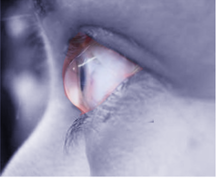
Keratoconus. This image shows corneal steepening that is characteristic of keratoconus.
Image courtesy S Bhimji MD
References
Rogers GJ, Attenborough M. Bilateral superior keratoconus: two case reports. Eye (London, England). 2014 Oct:28(10):1254-7. doi: 10.1038/eye.2014.146. Epub 2014 Jul 4 [PubMed PMID: 24993320]
Level 3 (low-level) evidenceWeed KH, McGhee CN, MacEwen CJ. Atypical unilateral superior keratoconus in young males. Contact lens & anterior eye : the journal of the British Contact Lens Association. 2005 Dec:28(4):177-9 [PubMed PMID: 16332502]
Prisant O, Legeais JM, Renard G. Superior keratoconus. Cornea. 1997 Nov:16(6):693-4 [PubMed PMID: 9395882]
Mounir A, Mostafa EM. Combined accelerated corneal collagen crosslinking and intrastromal Kerarings implantation for treatment of advanced superior keratoconus. GMS ophthalmology cases. 2020:10():Doc10. doi: 10.3205/oc000137. Epub 2020 Feb 27 [PubMed PMID: 32269908]
Level 3 (low-level) evidenceChiang CC, Lin JM, Tsai YY. Superior keratoconus with inferior paracentral corneal thinning and inferior peripheral pellucid marginal degeneration. Eye (London, England). 2007 Feb:21(2):266-8 [PubMed PMID: 16858442]
Godefrooij DA, de Wit GA, Uiterwaal CS, Imhof SM, Wisse RP. Age-specific Incidence and Prevalence of Keratoconus: A Nationwide Registration Study. American journal of ophthalmology. 2017 Mar:175():169-172. doi: 10.1016/j.ajo.2016.12.015. Epub 2016 Dec 28 [PubMed PMID: 28039037]
Kim T, Khosla-Gupta B, Debacker C. Blepharoptosis-induced superior keratoconus. American journal of ophthalmology. 2000 Aug:130(2):232-4 [PubMed PMID: 11004300]
Galperín G, Berra M. Bilateral superior keratoconus. Archivos de la Sociedad Espanola de Oftalmologia. 2021 Aug:96(8):438-441. doi: 10.1016/j.oftale.2020.09.003. Epub 2021 Jan 6 [PubMed PMID: 34340783]
Kanclerz P, Khoramnia R, Wang X. Current Developments in Corneal Topography and Tomography. Diagnostics (Basel, Switzerland). 2021 Aug 13:11(8):. doi: 10.3390/diagnostics11081466. Epub 2021 Aug 13 [PubMed PMID: 34441401]
Niazi S, Jiménez-García M, Findl O, Gatzioufas Z, Doroodgar F, Shahriari MH, Javadi MA. Keratoconus Diagnosis: From Fundamentals to Artificial Intelligence: A Systematic Narrative Review. Diagnostics (Basel, Switzerland). 2023 Aug 21:13(16):. doi: 10.3390/diagnostics13162715. Epub 2023 Aug 21 [PubMed PMID: 37627975]
Level 1 (high-level) evidenceSakellaris D, Balidis M, Gorou O, Szentmary N, Alexoudis A, Grieshaber MC, Sagri D, Scholl H, Gatzioufas Z. Intracorneal Ring Segment Implantation in the Management of Keratoconus: An Evidence-Based Approach. Ophthalmology and therapy. 2019 Oct:8(Suppl 1):5-14. doi: 10.1007/s40123-019-00211-2. Epub 2019 Oct 11 [PubMed PMID: 31605316]
Gurnani B, Czyz CN, Mahabadi N, Havens SJ. Corneal Graft Rejection. StatPearls. 2024 Jan:(): [PubMed PMID: 30085585]
Baptista PM, Ambrosio R, Oliveira L, Meneres P, Beirao JM. Corneal Biomechanical Assessment with Ultra-High-Speed Scheimpflug Imaging During Non-Contact Tonometry: A Prospective Review. Clinical ophthalmology (Auckland, N.Z.). 2021:15():1409-1423. doi: 10.2147/OPTH.S301179. Epub 2021 Apr 6 [PubMed PMID: 33854295]
Zadnik K, Money S, Lindsley K. Intrastromal corneal ring segments for treating keratoconus. The Cochrane database of systematic reviews. 2019 May 14:5(5):CD011150. doi: 10.1002/14651858.CD011150.pub2. Epub 2019 May 14 [PubMed PMID: 31087649]
Level 1 (high-level) evidenceTananuvat N, Leeungurasatien P, Wiriyaluppa C. Superior keratoconus with hydrops. International ophthalmology. 2009 Oct:29(5):419-21. doi: 10.1007/s10792-008-9244-0. Epub 2008 Jul 10 [PubMed PMID: 18615241]
Yang K, Li D, Xu L, Pang C, Zhao D, Ren S. Independent and interactive effects of eye rubbing and atopy on keratoconus. Frontiers in immunology. 2022:13():999435. doi: 10.3389/fimmu.2022.999435. Epub 2022 Sep 29 [PubMed PMID: 36248837]
Park S, Kim JY, Lee SH, Suh B, Keam B, Kim TM, Kim DW, Heo DS. KRAS G12C mutation as a poor prognostic marker of pemetrexed treatment in non-small cell lung cancer. The Korean journal of internal medicine. 2017 May:32(3):514-522. doi: 10.3904/kjim.2015.299. Epub 2017 Apr 14 [PubMed PMID: 28407465]
Najmi H, Mobarki Y, Mania K, Altowairqi B, Basehi M, Mahfouz MS, Elmahdy M. The correlation between keratoconus and eye rubbing: a review. International journal of ophthalmology. 2019:12(11):1775-1781. doi: 10.18240/ijo.2019.11.17. Epub 2019 Nov 18 [PubMed PMID: 31741868]
Level 2 (mid-level) evidenceAhuja P, Dadachanji Z, Shetty R, Nagarajan SA, Khamar P, Sethu S, D'Souza S. Relevance of IgE, allergy and eye rubbing in the pathogenesis and management of Keratoconus. Indian journal of ophthalmology. 2020 Oct:68(10):2067-2074. doi: 10.4103/ijo.IJO_1191_19. Epub [PubMed PMID: 32971611]
Caban M, Owczarek K, Lewandowska U. The Role of Metalloproteinases and Their Tissue Inhibitors on Ocular Diseases: Focusing on Potential Mechanisms. International journal of molecular sciences. 2022 Apr 12:23(8):. doi: 10.3390/ijms23084256. Epub 2022 Apr 12 [PubMed PMID: 35457074]
Wojcik KA, Kaminska A, Blasiak J, Szaflik J, Szaflik JP. Oxidative stress in the pathogenesis of keratoconus and Fuchs endothelial corneal dystrophy. International journal of molecular sciences. 2013 Sep 23:14(9):19294-308. doi: 10.3390/ijms140919294. Epub 2013 Sep 23 [PubMed PMID: 24065107]
Hamid A, Jahadi-Hosseini H, Khalili MR, Jahanbani-Ardakani H. Corneal Biomechanical Changes after Corneal Cross-Linking in Patients with Keratoconus. Journal of current ophthalmology. 2022 Oct-Dec:34(4):409-413. doi: 10.4103/joco.joco_177_21. Epub 2023 Apr 29 [PubMed PMID: 37180532]
Coco G, Kheirkhah A, Foulsham W, Dana R, Ciolino JB. Keratoconus progression associated with hormone replacement therapy. American journal of ophthalmology case reports. 2019 Sep:15():100519. doi: 10.1016/j.ajoc.2019.100519. Epub 2019 Jul 16 [PubMed PMID: 31372581]
Level 3 (low-level) evidenceSantodomingo-Rubido J, Carracedo G, Suzaki A, Villa-Collar C, Vincent SJ, Wolffsohn JS. Keratoconus: An updated review. Contact lens & anterior eye : the journal of the British Contact Lens Association. 2022 Jun:45(3):101559. doi: 10.1016/j.clae.2021.101559. Epub 2022 Jan 4 [PubMed PMID: 34991971]
Salman A, Darwish T, Ghabra M, Kailani O, Haddeh Y, Askar M, Ali A, Ali A, Alhassan S. Prevalence of Keratoconus in a Population-Based Study in Syria. Journal of ophthalmology. 2022:2022():6064533. doi: 10.1155/2022/6064533. Epub 2022 Jun 23 [PubMed PMID: 35783343]
Gordon-Shaag A, Millodot M, Shneor E, Liu Y. The genetic and environmental factors for keratoconus. BioMed research international. 2015:2015():795738. doi: 10.1155/2015/795738. Epub 2015 May 17 [PubMed PMID: 26075261]
Spanakis EK, Golden SH. Race/ethnic difference in diabetes and diabetic complications. Current diabetes reports. 2013 Dec:13(6):814-23. doi: 10.1007/s11892-013-0421-9. Epub [PubMed PMID: 24037313]
Olivo-Payne A, Abdala-Figuerola A, Hernandez-Bogantes E, Pedro-Aguilar L, Chan E, Godefrooij D. Optimal management of pediatric keratoconus: challenges and solutions. Clinical ophthalmology (Auckland, N.Z.). 2019:13():1183-1191. doi: 10.2147/OPTH.S183347. Epub 2019 Jul 10 [PubMed PMID: 31371915]
Bui AD, Truong A, Pasricha ND, Indaram M. Keratoconus Diagnosis and Treatment: Recent Advances and Future Directions. Clinical ophthalmology (Auckland, N.Z.). 2023:17():2705-2718. doi: 10.2147/OPTH.S392665. Epub 2023 Sep 16 [PubMed PMID: 37736107]
Level 3 (low-level) evidenceDeshmukh R, Ong ZZ, Rampat R, Alió Del Barrio JL, Barua A, Ang M, Mehta JS, Said DG, Dua HS, Ambrósio R Jr, Ting DSJ. Management of keratoconus: an updated review. Frontiers in medicine. 2023:10():1212314. doi: 10.3389/fmed.2023.1212314. Epub 2023 Jun 20 [PubMed PMID: 37409272]
Marx-Gross S, Fieß A, Münzel T, Wild PS, Beutel ME, Schmidtmann I, Lackner KJ, Pfeiffer N, Schuster AK. Much higher prevalence of keratoconus than announced results of the Gutenberg Health Study (GHS). Graefe's archive for clinical and experimental ophthalmology = Albrecht von Graefes Archiv fur klinische und experimentelle Ophthalmologie. 2023 Nov:261(11):3241-3247. doi: 10.1007/s00417-023-06132-y. Epub 2023 Jun 14 [PubMed PMID: 37314521]
Chung IK, Kim BS, Han KD, Yoo YS, Kim H, Jeong C. Ten-year incidence of keratoconus in relation to sex, age, and thyroid gland dysfunction: a nationwide population-based cohort study (2009-2018). Annals of translational medicine. 2024 Jun 10:12(3):45. doi: 10.21037/atm-23-1906. Epub 2024 May 20 [PubMed PMID: 38911561]
Sharif R, Bak-Nielsen S, Hjortdal J, Karamichos D. Pathogenesis of Keratoconus: The intriguing therapeutic potential of Prolactin-inducible protein. Progress in retinal and eye research. 2018 Nov:67():150-167. doi: 10.1016/j.preteyeres.2018.05.002. Epub 2018 Jul 13 [PubMed PMID: 29758268]
Loukovitis E, Kozeis N, Gatzioufas Z, Kozei A, Tsotridou E, Stoila M, Koronis S, Sfakianakis K, Tranos P, Balidis M, Zachariadis Z, Mikropoulos DG, Anogeianakis G, Katsanos A, Konstas AG. The Proteins of Keratoconus: a Literature Review Exploring Their Contribution to the Pathophysiology of the Disease. Advances in therapy. 2019 Sep:36(9):2205-2222. doi: 10.1007/s12325-019-01026-0. Epub 2019 Jul 30 [PubMed PMID: 31363996]
Level 3 (low-level) evidenceSherwin T, Brookes NH, Loh IP, Poole CA, Clover GM. Cellular incursion into Bowman's membrane in the peripheral cone of the keratoconic cornea. Experimental eye research. 2002 Apr:74(4):473-82 [PubMed PMID: 12076091]
Khaled ML, Helwa I, Drewry M, Seremwe M, Estes A, Liu Y. Molecular and Histopathological Changes Associated with Keratoconus. BioMed research international. 2017:2017():7803029. doi: 10.1155/2017/7803029. Epub 2017 Jan 30 [PubMed PMID: 28251158]
Fania L, Didona D, Morese R, Campana I, Coco V, Di Pietro FR, Ricci F, Pallotta S, Candi E, Abeni D, Dellambra E. Basal Cell Carcinoma: From Pathophysiology to Novel Therapeutic Approaches. Biomedicines. 2020 Oct 23:8(11):. doi: 10.3390/biomedicines8110449. Epub 2020 Oct 23 [PubMed PMID: 33113965]
Johns LK, Leon P, Cortes-Barrantes P, Hanbazazh M, Zurakowski D, Dryja TP, Ciolino JB. Recurrent keratoconus: an analysis of breaks in Bowman's layer in corneal grafts. BMJ open ophthalmology. 2023 Apr:8(1):. doi: 10.1136/bmjophth-2022-001225. Epub [PubMed PMID: 37278429]
Falgayrettes N, Patoor E, Cleymand F, Zevering Y, Perone JM. Biomechanics of keratoconus: Two numerical studies. PloS one. 2023:18(2):e0278455. doi: 10.1371/journal.pone.0278455. Epub 2023 Feb 2 [PubMed PMID: 36730305]
Pirbhai A, Sheidow T, Hooper P. Prospective evaluation of digital non-stereo color fundus photography as a screening tool in age-related macular degeneration. American journal of ophthalmology. 2005 Mar:139(3):455-61 [PubMed PMID: 15767053]
Dupps WJ Jr, Wilson SE. Biomechanics and wound healing in the cornea. Experimental eye research. 2006 Oct:83(4):709-20 [PubMed PMID: 16720023]
Güngör IU, Beden U, Sönmez B. Bilateral horizontal Vogt's striae in keratoconus. Clinical ophthalmology (Auckland, N.Z.). 2008 Sep:2(3):653-5 [PubMed PMID: 19668769]
Galvis V, Sherwin T, Tello A, Merayo J, Barrera R, Acera A. Keratoconus: an inflammatory disorder? Eye (London, England). 2015 Jul:29(7):843-59. doi: 10.1038/eye.2015.63. Epub 2015 May 1 [PubMed PMID: 25931166]
Loukovitis E, Sfakianakis K, Syrmakesi P, Tsotridou E, Orfanidou M, Bakaloudi DR, Stoila M, Kozei A, Koronis S, Zachariadis Z, Tranos P, Kozeis N, Balidis M, Gatzioufas Z, Fiska A, Anogeianakis G. Genetic Aspects of Keratoconus: A Literature Review Exploring Potential Genetic Contributions and Possible Genetic Relationships with Comorbidities. Ophthalmology and therapy. 2018 Dec:7(2):263-292. doi: 10.1007/s40123-018-0144-8. Epub 2018 Sep 6 [PubMed PMID: 30191404]
Unni P, Lee HJ. Systemic Associations with Keratoconus. Life (Basel, Switzerland). 2023 Jun 10:13(6):. doi: 10.3390/life13061363. Epub 2023 Jun 10 [PubMed PMID: 37374145]
Henriquez MA, Hadid M, Izquierdo L Jr. A Systematic Review of Subclinical Keratoconus and Forme Fruste Keratoconus. Journal of refractive surgery (Thorofare, N.J. : 1995). 2020 Apr 1:36(4):270-279. doi: 10.3928/1081597X-20200212-03. Epub [PubMed PMID: 32267959]
Level 1 (high-level) evidenceKriszt Á, Losonczy G, Berta A, Takács L. Presence of Fleischer ring and prominent corneal nerves in keratoconus relatives and normal controls. International journal of ophthalmology. 2015:8(5):922-7. doi: 10.3980/j.issn.2222-3959.2015.05.12. Epub 2015 Oct 18 [PubMed PMID: 26558202]
Fan Gaskin JC, Patel DV, McGhee CN. Acute corneal hydrops in keratoconus - new perspectives. American journal of ophthalmology. 2014 May:157(5):921-8. doi: 10.1016/j.ajo.2014.01.017. Epub 2014 Jan 31 [PubMed PMID: 24491416]
Level 3 (low-level) evidenceVazirani J, Basu S. Keratoconus: current perspectives. Clinical ophthalmology (Auckland, N.Z.). 2013:7():2019-30. doi: 10.2147/OPTH.S50119. Epub 2013 Oct 14 [PubMed PMID: 24143069]
Level 3 (low-level) evidenceDoctor K, Vunnava KP, Shroff R, Kaweri L, Lalgudi VG, Gupta K, Kundu G. Simplifying and understanding various topographic indices for keratoconus using Scheimpflug based topographers. Indian journal of ophthalmology. 2020 Dec:68(12):2732-2743. doi: 10.4103/ijo.IJO_2111_20. Epub [PubMed PMID: 33229649]
Level 3 (low-level) evidenceCavas-Martínez F, De la Cruz Sánchez E, Nieto Martínez J, Fernández Cañavate FJ, Fernández-Pacheco DG. Corneal topography in keratoconus: state of the art. Eye and vision (London, England). 2016:3():5. doi: 10.1186/s40662-016-0036-8. Epub 2016 Feb 22 [PubMed PMID: 26904709]
Jafarinasab MR, Shirzadeh E, Feizi S, Karimian F, Akaberi A, Hasanpour H. Sensitivity and specificity of posterior and anterior corneal elevation measured by orbscan in diagnosis of clinical and subclinical keratoconus. Journal of ophthalmic & vision research. 2015 Jan-Mar:10(1):10-5. doi: 10.4103/2008-322X.156085. Epub [PubMed PMID: 26005546]
Kanellopoulos AJ, Asimellis G. OCT-derived comparison of corneal thickness distribution and asymmetry differences between normal and keratoconic eyes. Cornea. 2014 Dec:33(12):1274-81. doi: 10.1097/ICO.0000000000000275. Epub [PubMed PMID: 25321940]
Al-Mahrouqi H, Oraba SB, Al-Habsi S, Mundemkattil N, Babu J, Panchatcharam SM, Al-Saidi R, Al-Raisi A. Retinoscopy as a Screening Tool for Keratoconus. Cornea. 2019 Apr:38(4):442-445. doi: 10.1097/ICO.0000000000001843. Epub [PubMed PMID: 30640248]
Level 2 (mid-level) evidenceBelin MW, Ambrósio R. Scheimpflug imaging for keratoconus and ectatic disease. Indian journal of ophthalmology. 2013 Aug:61(8):401-6. doi: 10.4103/0301-4738.116059. Epub [PubMed PMID: 23925323]
Camburu G, Zemba M, Tătaru CP, Purcărea VL. The measurement of Central Corneal Thickness. Romanian journal of ophthalmology. 2023 Apr-Jun:67(2):168-174. doi: 10.22336/rjo.2023.29. Epub [PubMed PMID: 37522018]
Ramm L, Herber R, Lorenz G, Jasper CS, Pillunat LE, Pillunat KR. Evaluation of corneal biomechanical properties using the ocular response analyzer and the dynamic Scheimpflug-Analyzer Corvis ST in high pressure and normal pressure open-angle glaucoma patients. PloS one. 2023:18(1):e0281017. doi: 10.1371/journal.pone.0281017. Epub 2023 Jan 26 [PubMed PMID: 36701409]
Llorens-Quintana C, Lee DJ, Pavlatos E, Chamberlain W, Huang D, Li Y. Measuring corneal astigmatism using OCT in keratoconus. Journal of cataract and refractive surgery. 2022 Nov 1:48(11):1285-1291. doi: 10.1097/j.jcrs.0000000000000977. Epub [PubMed PMID: 35616503]
Chen S, Li XY, Jin JJ, Shen RJ, Mao JY, Cheng FF, Chen ZJ, Linardaki E, Voulgaraki S, Aslanides IM, Jin ZB. Genetic Screening Revealed Latent Keratoconus in Asymptomatic Individuals. Frontiers in cell and developmental biology. 2021:9():650344. doi: 10.3389/fcell.2021.650344. Epub 2021 May 31 [PubMed PMID: 34136477]
Kaur K, Gurnani B. Specular Microscopy. StatPearls. 2024 Jan:(): [PubMed PMID: 36256774]
Espandar L, Meyer J. Keratoconus: overview and update on treatment. Middle East African journal of ophthalmology. 2010 Jan:17(1):15-20. doi: 10.4103/0974-9233.61212. Epub [PubMed PMID: 20543932]
Level 3 (low-level) evidenceMohammadpour M, Heidari Z, Hashemi H. Updates on Managements for Keratoconus. Journal of current ophthalmology. 2018 Jun:30(2):110-124. doi: 10.1016/j.joco.2017.11.002. Epub 2017 Dec 6 [PubMed PMID: 29988906]
Levit A, Benwell M, Evans BJW. Randomised controlled trial of corneal vs. scleral rigid gas permeable contact lenses for keratoconus and other ectatic corneal disorders. Contact lens & anterior eye : the journal of the British Contact Lens Association. 2020 Dec:43(6):543-552. doi: 10.1016/j.clae.2019.12.007. Epub 2020 Jan 8 [PubMed PMID: 31924530]
Level 1 (high-level) evidenceSaad S, Saad R, Jouve L, Kallel S, Trinh L, Goemaere I, Borderie V, Bouheraoua N. Corneal crosslinking in keratoconus management. Journal francais d'ophtalmologie. 2020 Dec:43(10):1078-1095. doi: 10.1016/j.jfo.2020.07.002. Epub 2020 Sep 21 [PubMed PMID: 32972759]
De Rosa G, Rossi S, Santamaria C, Boccia R, De Rosa L, D'Alterio FM, Simonelli F. Combined photorefractive keratectomy and corneal collagen cross-linking for treatment of keratoconus: a 2-year follow-up study. Therapeutic advances in ophthalmology. 2022 Jan-Dec:14():25158414221083362. doi: 10.1177/25158414221083362. Epub 2022 Mar 23 [PubMed PMID: 35340750]
Arnalich-Montiel F, Alió Del Barrio JL, Alió JL. Corneal surgery in keratoconus: which type, which technique, which outcomes? Eye and vision (London, England). 2016:3():2. doi: 10.1186/s40662-016-0033-y. Epub 2016 Jan 18 [PubMed PMID: 26783544]
Al-Mohaimeed MM. Penetrating keratoplasty for keratoconus: visual and graft survival outcomes. International journal of health sciences. 2013 Jan:7(1):67-74 [PubMed PMID: 23559907]
Alkharashi M, AlAbdulhadi HA, Otaif W, Alahmadi AS, Alanazi B, Al Habash A, Aldayel A, Aljindan M, Almulhim A, Bin Helayel H. Incidence, Pathophysiology, Complications, and Management of Positive Vitreous Pressure During Penetrating Keratoplasty: A Literature Review. Clinical ophthalmology (Auckland, N.Z.). 2023:17():583-590. doi: 10.2147/OPTH.S382502. Epub 2023 Feb 14 [PubMed PMID: 36820300]
Liu H, Chen Y, Wang P, Li B, Wang W, Su Y, Sheng M. Efficacy and safety of deep anterior lamellar keratoplasty vs. penetrating keratoplasty for keratoconus: a meta-analysis. PloS one. 2015:10(1):e0113332. doi: 10.1371/journal.pone.0113332. Epub 2015 Jan 29 [PubMed PMID: 25633311]
Level 1 (high-level) evidenceNassiri N, Djalilian AR. Keratoplasty: moving to the front. Journal of ophthalmic & vision research. 2009 Jan:4(1):5-7 [PubMed PMID: 23056666]
Bindu S, Mazumder S, Bandyopadhyay U. Non-steroidal anti-inflammatory drugs (NSAIDs) and organ damage: A current perspective. Biochemical pharmacology. 2020 Oct:180():114147. doi: 10.1016/j.bcp.2020.114147. Epub 2020 Jul 10 [PubMed PMID: 32653589]
Level 3 (low-level) evidencevan Nispen RM, Virgili G, Hoeben M, Langelaan M, Klevering J, Keunen JE, van Rens GH. Low vision rehabilitation for better quality of life in visually impaired adults. The Cochrane database of systematic reviews. 2020 Jan 27:1(1):CD006543. doi: 10.1002/14651858.CD006543.pub2. Epub 2020 Jan 27 [PubMed PMID: 31985055]
Level 1 (high-level) evidenceMoshirfar M,Duong A,Ronquillo Y, Corneal Imaging StatPearls. 2022 Jan; [PubMed PMID: 32965828]
Durakovic E, Kandel H, Watson SL. Mental Health Impact of Keratoconus: A Systematic Review. Cornea. 2023 Sep 1:42(9):1187-1197. doi: 10.1097/ICO.0000000000003263. Epub 2023 May 12 [PubMed PMID: 37195289]
Level 1 (high-level) evidenceSahu J, Raizada K. Pellucid Marginal Corneal Degeneration. StatPearls. 2024 Jan:(): [PubMed PMID: 32965985]
Rathi VM, Murthy SI, Bagga B, Taneja M, Chaurasia S, Sangwan VS. Keratoglobus: An experience at a tertiary eye care center in India. Indian journal of ophthalmology. 2015 Mar:63(3):233-8. doi: 10.4103/0301-4738.156927. Epub [PubMed PMID: 25971168]
Chan AT, Ulate R, Goldich Y, Rootman DS, Chan CC. Terrien Marginal Degeneration: Clinical Characteristics and Outcomes. American journal of ophthalmology. 2015 Nov:160(5):867-872.e1. doi: 10.1016/j.ajo.2015.07.031. Epub 2015 Jul 23 [PubMed PMID: 26210866]
Christy J, Gurnani B, Kaur K, Moutappa F. Contact lens warpage: Lost but found. Indian journal of ophthalmology. 2020 Aug:68(8):1662. doi: 10.4103/ijo.IJO_2034_19. Epub [PubMed PMID: 32709811]
Wolle MA, Randleman JB, Woodward MA. Complications of Refractive Surgery: Ectasia After Refractive Surgery. International ophthalmology clinics. 2016 Spring:56(2):127-39. doi: 10.1097/IIO.0000000000000102. Epub [PubMed PMID: 26938343]
Moshirfar M, Bennett P, Ronquillo Y. Corneal Dystrophy. StatPearls. 2024 Jan:(): [PubMed PMID: 32491788]
Hersh PS, Greenstein SA, Fry KL. Corneal collagen crosslinking for keratoconus and corneal ectasia: One-year results. Journal of cataract and refractive surgery. 2011 Jan:37(1):149-60. doi: 10.1016/j.jcrs.2010.07.030. Epub [PubMed PMID: 21183110]
Wittig-Silva C, Whiting M, Lamoureux E, Lindsay RG, Sullivan LJ, Snibson GR. A randomized controlled trial of corneal collagen cross-linking in progressive keratoconus: preliminary results. Journal of refractive surgery (Thorofare, N.J. : 1995). 2008 Sep:24(7):S720-5. doi: 10.3928/1081597X-20080901-15. Epub [PubMed PMID: 18811118]
Level 1 (high-level) evidenceReinhart WJ, Musch DC, Jacobs DS, Lee WB, Kaufman SC, Shtein RM. Deep anterior lamellar keratoplasty as an alternative to penetrating keratoplasty a report by the american academy of ophthalmology. Ophthalmology. 2011 Jan:118(1):209-18. doi: 10.1016/j.ophtha.2010.11.002. Epub [PubMed PMID: 21199711]
Vohra V, Tuteja S, Gurnani B, Chawla H. Collagen Cross Linking for Keratoconus. StatPearls. 2024 Jan:(): [PubMed PMID: 32965942]
Gurnani B, Kaur K. Quality of life in patients with keratoconus. Indian journal of ophthalmology. 2022 Aug:70(8):3155. doi: 10.4103/ijo.IJO_927_22. Epub [PubMed PMID: 35919000]
Level 2 (mid-level) evidenceGurnani B, Kaur K. Evolving concepts in etiopathogenesis of keratoconus: Is it quasi-inflammatory or inflammatory? Indian journal of ophthalmology. 2023 Jun:71(6):2609-2610. doi: 10.4103/ijo.IJO_783_22. Epub [PubMed PMID: 37322699]
Gurnani B, Kaur K. Penetrating Keratoplasty. StatPearls. 2025 Jan:(): [PubMed PMID: 37276324]
Mahabadi N, Gurnani B, Czyz CN. Bacterial Endophthalmitis. StatPearls. 2025 Jan:(): [PubMed PMID: 31424768]
Gurnani B, Kaur K, Narayana S. Corneal ectasia, cerulean (blue dot) cataract with acute hydrops in a child with Down's syndrome and hypothyroidism - a rare presentation. GMS ophthalmology cases. 2023:13():Doc07. doi: 10.3205/oc000215. Epub 2023 Mar 1 [PubMed PMID: 37034411]
Level 3 (low-level) evidenceGurnani B, Kaur K, Chaudhary S, Kaur RP, Nayak S, Mishra D, Balakrishnan H, Parkash RO, Morya AK, Porwal A. Pediatric corneal transplantation: techniques, challenges, and outcomes. Therapeutic advances in ophthalmology. 2024 Jan-Dec:16():25158414241237906. doi: 10.1177/25158414241237906. Epub 2024 Mar 25 [PubMed PMID: 38533487]
Level 3 (low-level) evidenceGurnani B, Kaur K. Contact Lenses. StatPearls. 2025 Jan:(): [PubMed PMID: 35593861]
Pleyer U, Ursell PG, Rama P. Intraocular pressure effects of common topical steroids for post-cataract inflammation: are they all the same? Ophthalmology and therapy. 2013 Dec:2(2):55-72. doi: 10.1007/s40123-013-0020-5. Epub 2013 Sep 17 [PubMed PMID: 25135807]
Moschos MM, Gouliopoulos NS, Kalogeropoulos C, Androudi S, Kitsos G, Ladas D, Tsatsos M, Chatziralli I. Psychological Aspects and Depression in Patients with Symptomatic Keratoconus. Journal of ophthalmology. 2018:2018():7314308. doi: 10.1155/2018/7314308. Epub 2018 May 29 [PubMed PMID: 30002924]
Naderan M, Shoar S, Kamaleddin MA, Rajabi MT, Naderan M, Khodadadi M. Keratoconus Clinical Findings According to Different Classifications. Cornea. 2015 Sep:34(9):1005-11. doi: 10.1097/ICO.0000000000000537. Epub [PubMed PMID: 26203749]
Motlagh MN, Moshirfar M, Murri MS, Skanchy DF, Momeni-Moghaddam H, Ronquillo YC, Hoopes PC. Pentacam® Corneal Tomography for Screening of Refractive Surgery Candidates: A Review of the Literature, Part I. Medical hypothesis, discovery & innovation ophthalmology journal. 2019 Fall:8(3):177-203 [PubMed PMID: 31598520]
Asimellis G, Kaufman EJ. Keratoconus. StatPearls. 2025 Jan:(): [PubMed PMID: 29262160]
Gurnani B, Kaur K. Contact Lens–Related Complications. StatPearls. 2024 Jan:(): [PubMed PMID: 36512659]
Jimmy B, Jose J. Patient medication adherence: measures in daily practice. Oman medical journal. 2011 May:26(3):155-9. doi: 10.5001/omj.2011.38. Epub [PubMed PMID: 22043406]
Gurnani B, Kaur K. Therapeutic Keratoplasty. StatPearls. 2025 Jan:(): [PubMed PMID: 37276297]
Esporcatte LPG, Salomão MQ, Neto ABDC, Machado AP, Lopes BT, Ambrósio R Jr. Enhanced Diagnostics for Corneal Ectatic Diseases: The Whats, the Whys, and the Hows. Diagnostics (Basel, Switzerland). 2022 Dec 2:12(12):. doi: 10.3390/diagnostics12123027. Epub 2022 Dec 2 [PubMed PMID: 36553038]
Fuest M, Jhanji V, Yam GH. Molecular and Cellular Mechanisms of Corneal Scarring and Advances in Therapy. International journal of molecular sciences. 2023 Apr 24:24(9):. doi: 10.3390/ijms24097777. Epub 2023 Apr 24 [PubMed PMID: 37175482]
Level 3 (low-level) evidenceTuft SJ, Gregory WM, Buckley RJ. Acute corneal hydrops in keratoconus. Ophthalmology. 1994 Oct:101(10):1738-44 [PubMed PMID: 7936572]
Al Zabadi H, Shehadeh M, Amro L, Ghattass N, Taha I. Vision-related quality of life among patients with keratoconus: a cross sectional study. Frontiers in medicine. 2023:10():1208911. doi: 10.3389/fmed.2023.1208911. Epub 2023 Aug 10 [PubMed PMID: 37636581]
Level 2 (mid-level) evidenceLjubimov AV. Diabetic complications in the cornea. Vision research. 2017 Oct:139():138-152. doi: 10.1016/j.visres.2017.03.002. Epub 2017 Apr 28 [PubMed PMID: 28404521]
Lim L, Lim EWL. Current perspectives in the management of keratoconus with contact lenses. Eye (London, England). 2020 Dec:34(12):2175-2196. doi: 10.1038/s41433-020-1065-z. Epub 2020 Jul 8 [PubMed PMID: 32641797]
Level 3 (low-level) evidenceBiesecker B. Genetic Counseling and the Central Tenets of Practice. Cold Spring Harbor perspectives in medicine. 2020 Mar 2:10(3):. doi: 10.1101/cshperspect.a038968. Epub 2020 Mar 2 [PubMed PMID: 31570379]
Level 3 (low-level) evidenceNoufal KH, Babu SP. Short-term visual outcome with sclerocorneal contact lens on irregular cornea. Saudi journal of ophthalmology : official journal of the Saudi Ophthalmological Society. 2023 Jan-Mar:37(1):43-47. doi: 10.4103/sjopt.sjopt_268_21. Epub 2023 Mar 9 [PubMed PMID: 36968765]
Kaur K, Gurnani B. Low Vision Aids. StatPearls. 2024 Jan:(): [PubMed PMID: 36256771]
Zhang X, Munir SZ, Sami Karim SA, Munir WM. A review of imaging modalities for detecting early keratoconus. Eye (London, England). 2021 Jan:35(1):173-187. doi: 10.1038/s41433-020-1039-1. Epub 2020 Jul 16 [PubMed PMID: 32678352]
Mahan M, Purt B. Ocular Trauma Prevention Strategies and Patient Counseling. StatPearls. 2025 Jan:(): [PubMed PMID: 35593844]
Fournié P, Acquadro M, Touboul D, Cochener B, Chiambaretta F, Muraine M, Borderie V, Bourges JL, Benmedjahed K, Tugaut B, Bernheim D, Bourcier T, Burillon C, David T, Delbosc B, Gain P, Hoffart L, Labetoulle M, Laroche L, Malet F, Orignac I, Robert PY, Thuret G, Vabres B, Malecaze F, Arnould B. Keratoconus and the Impact of Treatment on Patients' Quality of Life: A Qualitative Study. Ophthalmology and therapy. 2023 Aug:12(4):1939-1956. doi: 10.1007/s40123-023-00717-w. Epub 2023 May 8 [PubMed PMID: 37157013]
Level 2 (mid-level) evidenceKaram M, Chouinard MC, Poitras ME, Couturier Y, Vedel I, Grgurevic N, Hudon C. Nursing Care Coordination for Patients with Complex Needs in Primary Healthcare: A Scoping Review. International journal of integrated care. 2021 Mar 19:21(1):16. doi: 10.5334/ijic.5518. Epub 2021 Mar 19 [PubMed PMID: 33776605]
Level 2 (mid-level) evidenceSadiq IZ. Lifestyle medicine as a modality for prevention and management of chronic diseases. Journal of Taibah University Medical Sciences. 2023 Oct:18(5):1115-1117. doi: 10.1016/j.jtumed.2023.04.001. Epub 2023 Apr 15 [PubMed PMID: 37187803]
Hughes RG, Reinhard SC, Given B, Petlick NH, Bemis A. Supporting Family Caregivers in Providing Care. Patient Safety and Quality: An Evidence-Based Handbook for Nurses. 2008 Apr:(): [PubMed PMID: 21328765]
Level 2 (mid-level) evidenceDebourdeau E, Planells G, Chamard C, Touboul D, Villain M, Demoly P, Babeau F, Fournie P, Daien V. New Keratoconus Risk Factors: A Cross-Sectional Case-Control Study. Journal of ophthalmology. 2022:2022():6605771. doi: 10.1155/2022/6605771. Epub 2022 Sep 28 [PubMed PMID: 36211596]
Level 2 (mid-level) evidenceChong YJ, Azzopardi M, Hussain G, Recchioni A, Gandhewar J, Loizou C, Giachos I, Barua A, Ting DSJ. Clinical Applications of Anterior Segment Optical Coherence Tomography: An Updated Review. Diagnostics (Basel, Switzerland). 2024 Jan 5:14(2):. doi: 10.3390/diagnostics14020122. Epub 2024 Jan 5 [PubMed PMID: 38248000]
Song M, Chen T, Moktar A, Chan E, Chong EW, Daniell M, Sahebjada S. Diagnosis and Management of Keratoconus-A Narrative Review of Clinicians' Perspectives. Children (Basel, Switzerland). 2022 Dec 15:9(12):. doi: 10.3390/children9121973. Epub 2022 Dec 15 [PubMed PMID: 36553416]
Level 3 (low-level) evidenceForsetlund L, O'Brien MA, Forsén L, Reinar LM, Okwen MP, Horsley T, Rose CJ. Continuing education meetings and workshops: effects on professional practice and healthcare outcomes. The Cochrane database of systematic reviews. 2021 Sep 15:9(9):CD003030. doi: 10.1002/14651858.CD003030.pub3. Epub 2021 Sep 15 [PubMed PMID: 34523128]
Level 1 (high-level) evidenceHughes RG, O’Daniel M, Rosenstein AH. Professional Communication and Team Collaboration. Patient Safety and Quality: An Evidence-Based Handbook for Nurses. 2008 Apr:(): [PubMed PMID: 21328739]
Level 2 (mid-level) evidenceEdgman-Levitan S, Schoenbaum SC. Patient-centered care: achieving higher quality by designing care through the patient's eyes. Israel journal of health policy research. 2021 Mar 5:10(1):21. doi: 10.1186/s13584-021-00459-9. Epub 2021 Mar 5 [PubMed PMID: 33673875]
Level 2 (mid-level) evidenceAtalay E, Özalp O, Yıldırım N. Advances in the diagnosis and treatment of keratoconus. Therapeutic advances in ophthalmology. 2021 Jan-Dec:13():25158414211012796. doi: 10.1177/25158414211012796. Epub 2021 Jun 24 [PubMed PMID: 34263132]
Level 3 (low-level) evidenceBhati D, Deogade MS, Kanyal D. Improving Patient Outcomes Through Effective Hospital Administration: A Comprehensive Review. Cureus. 2023 Oct:15(10):e47731. doi: 10.7759/cureus.47731. Epub 2023 Oct 26 [PubMed PMID: 38021686]