Introduction
Many military and law enforcement agencies now deploy dogs in high-risk and high-fatality operations, including explosive detection, patrol, tracking, and search and rescue.[1] These canine officers, including military working dogs (MWDs) and other canine officers (COs), are highly skilled and invaluable team members who put their lives on the line to ensure the safety and security of their colleagues and the public. In situations where veterinarians are seldom available immediately after an injury to these canine officers, it becomes crucial for dog handlers, medics, and other first responders to possess the knowledge and skills required for providing emergency care.
Anatomy and Physiology
Register For Free And Read The Full Article
Search engine and full access to all medical articles
10 free questions in your specialty
Free CME/CE Activities
Free daily question in your email
Save favorite articles to your dashboard
Emails offering discounts
Learn more about a Subscription to StatPearls Point-of-Care
Anatomy and Physiology
Anyone treating COs should understand the anatomical and physiologic differences between canines and humans. Below are the normal vital signs for canines.[2]
| Canine vital signs | At rest | Exercise |
| Temperature (°F) | 100.5-101.5 | 101.0-104.0 |
| Heart rate (bpm or beats per minute) | 60-75 | 75-130 |
| Respiratory rate (breaths/min) | 10-20 | 30-panting |
| Mucous membrane color | Pink | Bright pink |
| Capillary refill time (sec) | 1-2 | 1 |
Indications
The most common preventable causes of death in canine trauma-related incidents are hemorrhage from extremity wounds, tension pneumothorax, and airway obstruction. These 3 factors contribute significantly to prehospital fatalities in both civilian trauma and military combat casualties. It is estimated that 20% to 25% of these preventable deaths occur due to delays in implementing early and appropriate basic first-aid techniques.[3]
Extremity Wounds
To address life-threatening extremity hemorrhaging in canines effectively, it's important to avoid using commercially available limb tourniquets, which are typically too large and prone to slipping.[4] Instead, consider these steps:
- Utilize direct or circumferential pressure dressings in combination with hemostatic agents.
- Apply direct pressure to the wound or use wide, elastic bandages to wrap around the affected area as pressure dressings.
- Consider using commercially available hemostatic dressings or tranexamic acid (TXA) directly to the wound.
- Pack the wound with hemostatic gauze if the injury is noncompressible.[5] If the bleeding vessel is visible, employ a hemostat to clamp it.
The above measures can be more effective in managing extremity hemorrhaging in canines while avoiding the issues associated with tourniquets.
When canine blood is available for transfusion, the recommended volume is a 10 to 20 mL/kg bolus. Gradually administer over 4 hours, beginning with a quarter dose for the first 15 minutes, doubling the flow every 15 minutes until the desired rate is reached. For simplicity, canine Tactical Combat Casualty Care recommends a 500 mL initial bolus as most MWD weight exceeds 25 kg.[4] In emergencies where the dog is actively losing blood, the entire blood unit may be infused rapidly. However, this approach risks a transfusion reaction, with symptoms such as fever, rapid breathing, and facial swelling.
Blood transfusions should be given intravenously (IV) or intraosseously with a filter. It is crucial to use the same IV catheter exclusively for administering 0.9% saline and avoid introducing any other medications or drugs through it. A second IV catheter may be necessary if the animal requires additional drugs, such as antibiotics or analgesics.
If canine whole blood is unavailable, a 1:1 ratio of canine plasma to canine-packed red blood cells is an alternative option.[4][6] In veterinary medicine, blood products typically come in single (125 mL) or double (250 mL) units. Therefore, each component in this combination should be equivalent to 1 single or 1 double unit.
Under no circumstances should human blood products be transfused into an MWD, as there is a high chance of a hemolytic reaction occurring.[4]
TXA is readily available in most ambulances. In cases of significant blood loss, give 10 mg/kg of TXA as a slow IV push or dissolved in 100 mL normal saline or lactated Ringer's solution.[4] TXA is administered immediately if the injury occurred within the past 3 hours; it has been shown to reduce mortality from bleeding if given within this timeframe.[7][4][8] TXA is contraindicated after 3 hours from injury because delayed administration is associated with worse outcomes.[9]
Antibiotics are recommended for all open combat wounds.[4]
Chest Wounds
Penetrating chest injuries from ballistics and explosions are high-incidence injuries for military and police canines.[10][11]
Breath sounds should be auscultated first. Inspection for an open chest wound may require shaving hair for visualization.[12] If active bleeding occurs, apply direct pressure or a gloved hand directly over the wound. The sticky occlusive dressing designed for human skin may not work well on canine hair. If the canine is stable, the wound should be disinfected, and a chest seal should be placed over the wound.[12] The hair around the wound may need to be trimmed before the commercially available occlusive dressing can be applied.[4]
Ultrasound can be used to check for lung sliding or absence of pneumothorax. Lung ultrasound has higher sensitivity than x-ray for detection of mild pneumothorax.[13] A chest radiograph may be used to check for hemothorax or pneumothorax.[12] Without a radiograph, absent lung sounds, increased respiratory effort, a flail chest, and palpable displaced rib fractures suggest the thoracic cavity has been compromised.
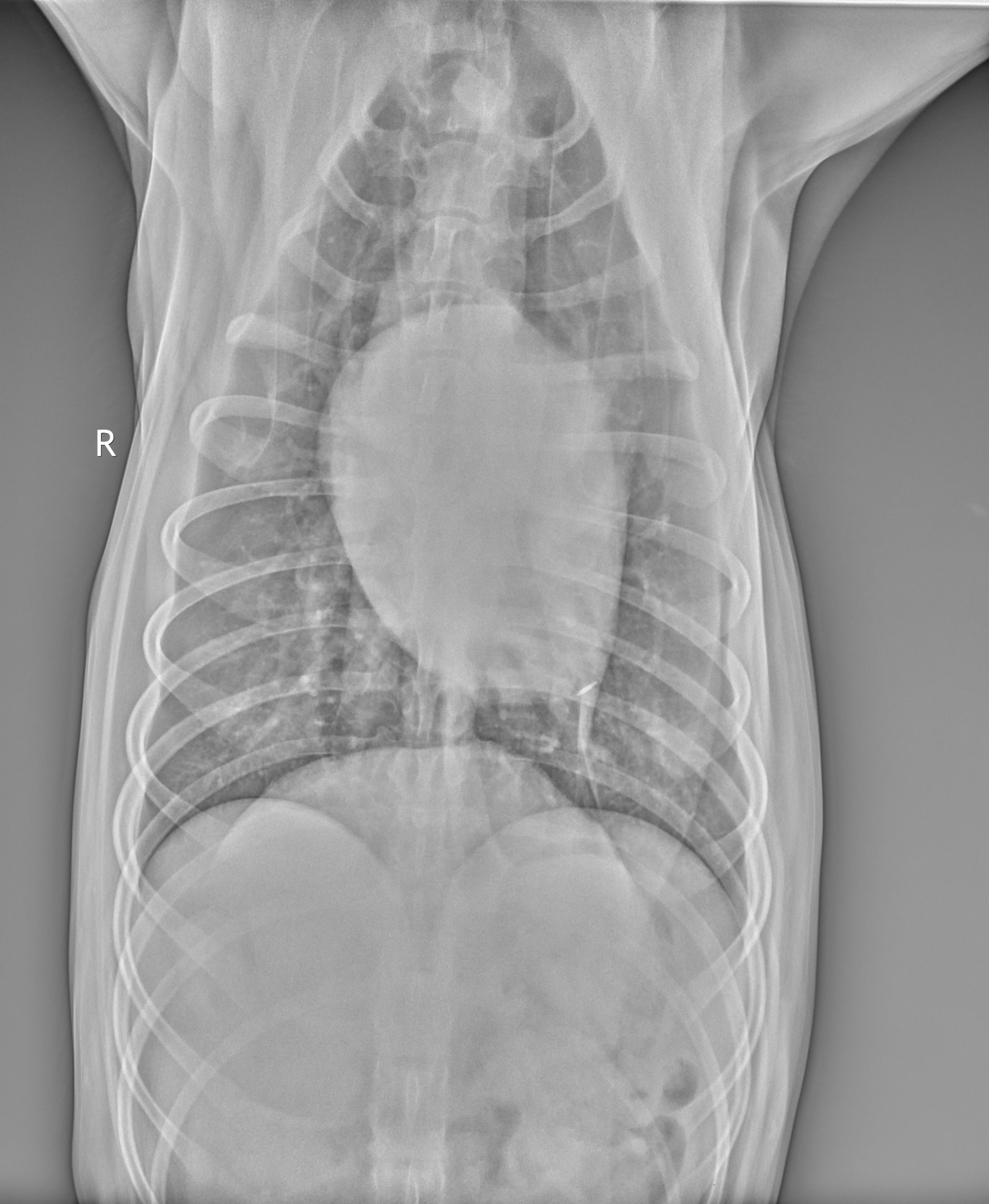
Diagnosis of tension pneumothorax may be confirmed with needle decompression in a canine who sustained a chest trauma with worsening respiratory distress, diminished or absent lung sounds, or deteriorating blood pressure. Placing a 14-gauge needle or IV catheter into the seventh to ninth rib space on the dorsal aspect of the thorax is appropriate (see Image. CXR Dorsal Lungs).[12]
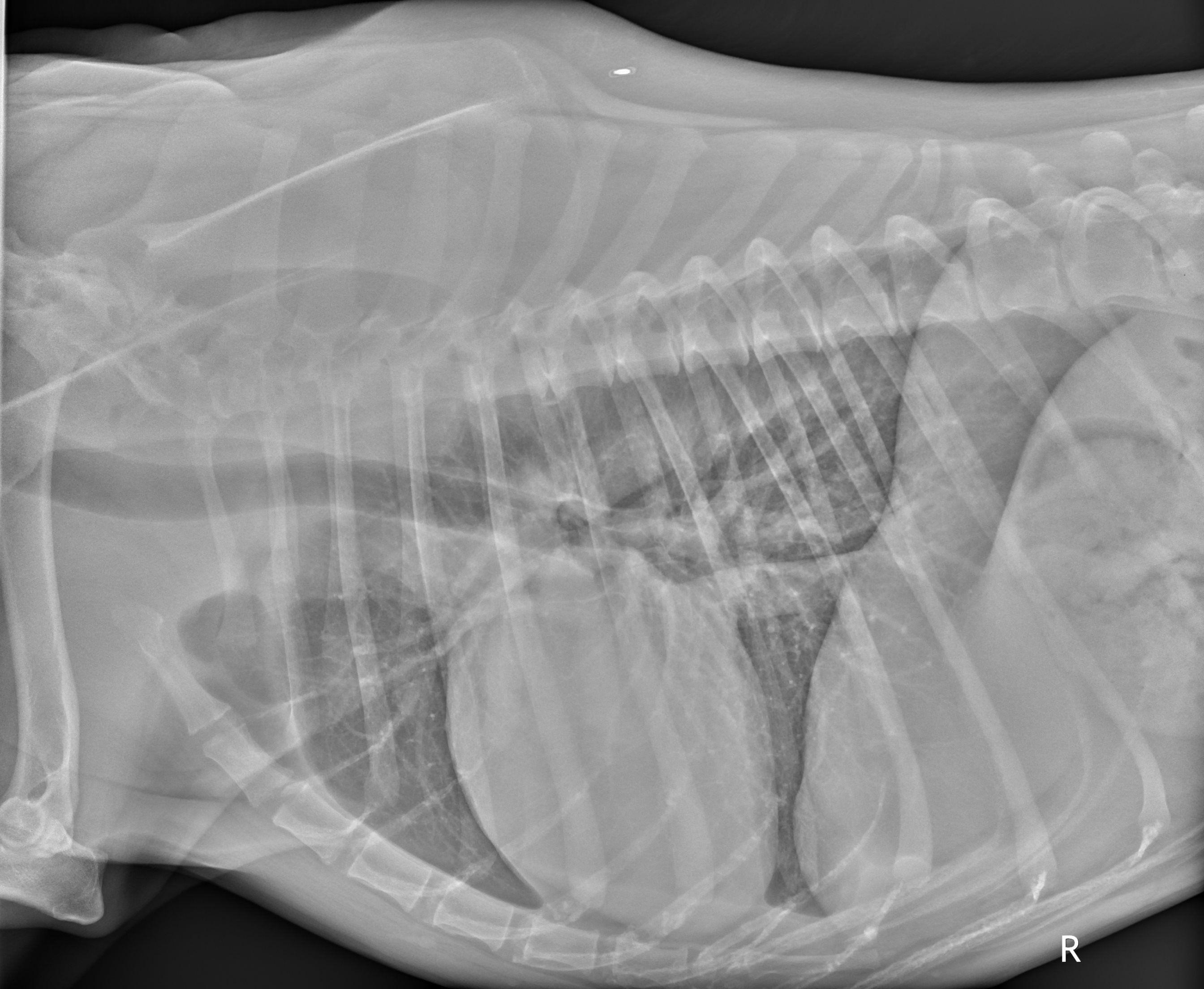
In cases where pleural effusion is a concern (ie, hemothorax, pyothorax), auscultation may reveal muffled heart and lung sounds. A chest radiograph can help make this diagnosis. A pulmonary contusion may take 24 to 72 hours to become visible on radiographs. In pulmonary contusion, IV fluids should be given judiciously. Most canines will require IV fluids after trauma. Regardless of fluid status, the primary treatment includes supplemental oxygen (see Image. CXR of Canine Costophrenic Margins).[12]
If respiratory distress persists despite oxygen supplementation, the canine may require intubation.
To prevent hypothermia, apply a ready-heat blanket and warm the IV fluids or blood products.
Abdominal Wounds
The presence of abdominal wounds or bruising suggests potential associated internal injuries.[12] If the bowel is eviscerated, it should be irrigated, replaced, and bandaged in the field.[12] Open thoracic or abdominal wounds require exploration and definitive treatment by a veterinarian as soon as possible.[12]
Preparation
An injured or frightened MWD may act unpredictably during medical care.[4] Gloves and appropriate personal protective equipment should be worn. A muzzle to protect the medical providers is appropriate unless respiratory distress precludes its use.
Sedation or pain medication should be considered.[4] Human medications should not be given to dogs without checking the safety profile. Many NSAIDs can kill a canine, with meloxicam being an exception. However, most working dogs are German shepherds or Belgian Malinois, typically weighing over 30 kg. Medication doses should be calculated for their weight.
Technique or Treatment
Airway
A dog that can bark, growl, or whine has a patent airway.[4] Signs of airway obstruction in canines include gagging, noisy breathing, rapid or labored breathing, excessive drooling, stridor, tripod position, cyanosis, and pawing at the mouth or neck.[4]
To treat airway obstruction, consider sedating the MWD to visualize the airway. Avoid placing hands directly into the mouth. A shoelace can be looped around the jaw and lifted to keep the mouth open. Other methods include using a leash, rope, or roll gauze between the canine teeth to open the mouth.[4] Dogs have significantly larger tracheal openings, which allows for the direct removal of foreign objects or their removal through suction.[4]
In respiratory failure, the following are recommended for MWD >25 kg when endotracheal intubation is necessary.[4]
- Laryngoscope (Miller blade #4 or #5)
- An endotracheal tube (ETT) with an internal diameter ranging from 8.0 to 10.0 mm, equipped with a cuff
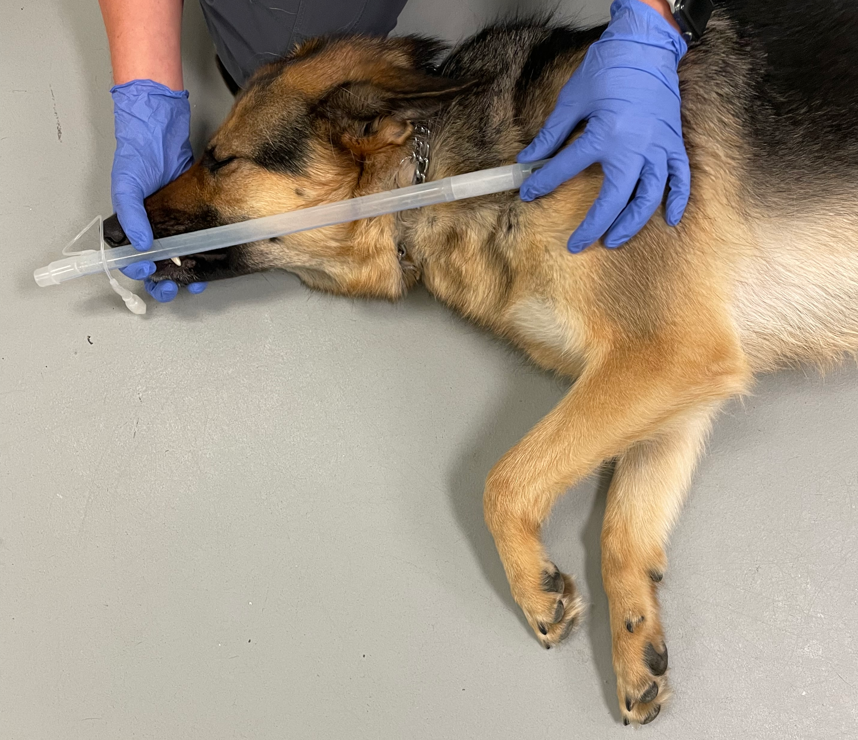
ETT depth can be measured by measuring the distance between the thoracic inlet or in-between the shoulder (most proximal front legs) and the central incisor. Before intubation, the ETT depth should be marked by tying a shoelace over the length of the ETT. After intubation, the shoelace can be secured by tying it around the mouth (see Image. Canine ETT Depth).
The following are medication tables for canines.
Table. Hemorrhage
| TXA | Canine whole blood | Canine plasma and canine PRBC | Reconstructed freeze-dried canine plasma | |
| Initial dosing | 10 mg/kg as a slow IV push or in a 100 mL NS/LR | 10-20 mL/kg bolus or 500 mL unit bolus[4] | 1:1 ratio of 250 mL of plasma plus 250 mL unit pRBC bolus[4] | 250 mL bolus[4] |
IV, intravenous; LR, lactated Ringer solution; NS, normal saline; PRBC, packed red blood cells; TXA, tranexamic acid.
Table. Analgesia
| Intranasal | Oral | IM | IV |
| Fentanyl: 4 μg/kg[4] | Meloxicam: 0.1 mg/kg orally once daily[4] | Morphine: 0.25-0.5 mg/kg[4] | Fentanyl: 2-5 μg/kg IV/IO (every 20-30 min)[4] |
| Hydromorphone: 0.1 mg/kg IV/IO/IM[4] | Ketamine: 2-5 mg/kg slow push over 1 min IV/IO/IM/IN[4] | ||
| Fentanyl:10 μg/kg (every 20-30 min)[4] |
IM, intramuscular; IN, intranasal; IO, intraosseous; IV, intravenous.
Table. Antibiotics*
| Ceftriaxone Sodium | Cefotaxime Sodium | Ertapenem | Moxifloxacin | Cefazolin |
| 25 mg/kg IV/IM every 12 h [4] | 25 mg/kg IV/IM every 8 h [4] | 15-30 mg/kg IV or SC every 8 h [4] | 400 mg orally once daily[4] | 15-35 mg/kg IV/IM/SC every 6-8 h |
IM, intramuscular; IV, intravenous; SC, subcutaneous.
*Antibiotics are recommended for all combat wounds.[4]
Complications
No trauma registry or formal collaborative system for collecting and reporting prehospital canine care exists.
Clinical Significance
The 3 most common, preventable, trauma-related deaths on the battlefield include hemorrhage from extremity wounds, tension pneumothorax, and airway obstruction.[14] Veterinarians may not be available for hours to days in a military combat setting. During Operation Iraqi Freedom, fewer than 30 veterinary personnel were available to care for over 600 deployed operational canines.[3]
One study revealed that of the 11 MWD casualties identified, most MWDs received at least 1 clinical intervention, and 50% survived their traumatic injuries.[11]
Enhancing Healthcare Team Outcomes
The Tactical Combat Casualty Care concept and guidelines have standardized point-of-injury care for human combat casualties and remain among the most significant interventions for reducing combat-related mortality from preventable deaths.[4][15]
The K9 Tactical Combat Casualty Care Initiative was designed to provide evidence-based, standardized guidelines for MWD to reduce preventable deaths.[4]
The MARCHE algorithm was slightly modified to MWD M3ARCH2E:
- M1: Muzzle
- M2: Massive hemorrhage
- M3: Medication
- A: Airway
- R: Respiration
- C: Circulation
- H1: Head injury
- H2: Hypothermia
- E: Everything else
Nursing, Allied Health, and Interprofessional Team Interventions
IV access for canines can be established on the cephalic vein on the lateral surface of the front legs or the saphenous veins on the lateral surface of the hind legs. The external jugular vein may be accessible on either side of the neck at the jugular furrow. The leg area should be clipped of fur and prepped with surgical disinfectant for sterility and bandage securement.[2]
Nursing, Allied Health, and Interprofessional Team Monitoring
A pulse oximeter can be applied to the lip or tongue. Other options include the toe web, ear, prepuce, or vulva.[12]
Media
(Click Image to Enlarge)

CXR of Canine Costophrenic Margins. Normal x-ray showing pulmonary vascular markings to bilateral periphery (costophrenic margins). Heart present in the midline, with the apex against the sternum, which is normal in “deep-chested” dogs, such as German shepherds and Labrador retrievers.
Contributed by Cecilia Winthrop, DVM
(Click Image to Enlarge)
(Click Image to Enlarge)
References
Miller L, Pacheco GJ, Janak JC, Grimm RC, Dierschke NA, Baker J, Orman JA. Causes of Death in Military Working Dogs During Operation Iraqi Freedom and Operation Enduring Freedom, 2001-2013. Military medicine. 2018 Sep 1:183(9-10):e467-e474. doi: 10.1093/milmed/usx235. Epub [PubMed PMID: 29547926]
Level 2 (mid-level) evidenceTaylor WM. Canine tactical field care. Part two--Massive hemorrhage control and physiologic stabilization of the volume depleted, shock-affected, or heatstroke-affected canine. Journal of special operations medicine : a peer reviewed journal for SOF medical professionals. 2009 Spring:9(2):13-21. doi: 10.55460/V7B3-973P. Epub [PubMed PMID: 19813515]
Level 3 (low-level) evidencePalmer LE, Maricle R, Brenner JA. The Operational Canine and K9 Tactical Emergency Casualty Care Initiative. Journal of special operations medicine : a peer reviewed journal for SOF medical professionals. 2015 Fall:15(3):32-38. doi: 10.55460/RMVA-7381. Epub [PubMed PMID: 26360351]
Edwards TH, Palmer LE, Baxter RL, Sager TC, Coisman JG, Brown JC, George C, McGraw AC. Canine Tactical Combat Casualty Care (K9TCCC) Guidelines. Journal of special operations medicine : a peer reviewed journal for SOF medical professionals. 2020 Spring:20(1):101-111. doi: 10.55460/YUMR-DBOP. Epub [PubMed PMID: 32203614]
Edwards TH, Dubick MA, Palmer L, Pusateri AE. Lessons Learned From the Battlefield and Applicability to Veterinary Medicine-Part 1: Hemorrhage Control. Frontiers in veterinary science. 2020:7():571368. doi: 10.3389/fvets.2020.571368. Epub 2021 Jan 14 [PubMed PMID: 33521075]
Edwards TH, Pusateri AE, Mays EL, Bynum JA, Cap AP. Lessons Learned From the Battlefield and Applicability to Veterinary Medicine - Part 2: Transfusion Advances. Frontiers in veterinary science. 2021:8():571370. doi: 10.3389/fvets.2021.571370. Epub 2021 May 7 [PubMed PMID: 34026881]
Level 3 (low-level) evidenceCRASH-3 trial collaborators. Effects of tranexamic acid on death, disability, vascular occlusive events and other morbidities in patients with acute traumatic brain injury (CRASH-3): a randomised, placebo-controlled trial. Lancet (London, England). 2019 Nov 9:394(10210):1713-1723. doi: 10.1016/S0140-6736(19)32233-0. Epub 2019 Oct 14 [PubMed PMID: 31623894]
Level 1 (high-level) evidenceStansfield R, Morris D, Jesulola E. The Use of Tranexamic Acid (TXA) for the Management of Hemorrhage in Trauma Patients in the Prehospital Environment: Literature Review and Descriptive Analysis of Principal Themes. Shock (Augusta, Ga.). 2020 Mar:53(3):277-283. doi: 10.1097/SHK.0000000000001389. Epub [PubMed PMID: 32044848]
Nishida T, Kinoshita T, Yamakawa K. Tranexamic acid and trauma-induced coagulopathy. Journal of intensive care. 2017:5():5. doi: 10.1186/s40560-016-0201-0. Epub 2017 Jan 20 [PubMed PMID: 28729903]
Johnson SA, Carr C, Reeves LK, Bean K, Schauer SG. Injuries and Interventions on Transported Military Working Dogs Within the US Central Command. Journal of special operations medicine : a peer reviewed journal for SOF medical professionals. 2022 Spring:22(1):97-101. doi: 10.55460/VTBK-XU21. Epub [PubMed PMID: 35278322]
Reeves LK, Mora AG, Field A, Redman TT. Interventions Performed on Multipurpose Military Working Dogs in the Prehospital Combat Setting: A Comprehensive Case Series Report. Journal of special operations medicine : a peer reviewed journal for SOF medical professionals. 2019 Fall:19(3):90-93. doi: 10.55460/LE5D-P32Y. Epub [PubMed PMID: 31539440]
Level 2 (mid-level) evidenceTaylor WM. Canine tactical field care part three - thoracic and abdominal trauma. Journal of special operations medicine : a peer reviewed journal for SOF medical professionals. 2010 Winter:10(1):50-8 [PubMed PMID: 20306416]
Level 3 (low-level) evidenceHwang TS, Yoon YM, Jung DI, Yeon SC, Lee HC. Usefulness of transthoracic lung ultrasound for the diagnosis of mild pneumothorax. Journal of veterinary science. 2018 Sep 30:19(5):660-666. doi: 10.4142/jvs.2018.19.5.660. Epub [PubMed PMID: 30041286]
Holcomb JB, Stansbury LG, Champion HR, Wade C, Bellamy RF. Understanding combat casualty care statistics. The Journal of trauma. 2006 Feb:60(2):397-401 [PubMed PMID: 16508502]
Level 3 (low-level) evidenceButler FK Jr, Blackbourne LH. Battlefield trauma care then and now: a decade of Tactical Combat Casualty Care. The journal of trauma and acute care surgery. 2012 Dec:73(6 Suppl 5):S395-402. doi: 10.1097/TA.0b013e3182754850. Epub [PubMed PMID: 23192061]

