Introduction
After 1960, the Fresnel principle, which existed for more than 150 years, was used for the first time to manage strabismus. In 1970, the optical scientific group from San Rafael of California designed a combination of a series of Fresnel press on prisms.[1] These prisms are made up of plastic membranes with powers ranging from 0.5-30 prism dioptres. The Fresnel prism is derived from the concept of hand-ground lenses prepared by the physics and French specialist Augustine Fresnel in 1921.[2]
Initially, they were intended for use in lighthouse beacons. Prisms have been used extensively in the field of ophthalmology, especially related to neuro-ophthalmology and strabismus.[3] Prisms are used extensively to increase the binocular field, relieve diplopia, or expand the field of vision. Fresnel prisms are thin transparent plastic membranes with multiple tiny prisms attached from base to base. Augustin Fresnel articulated the optics of Fresnel prisms.[4]
The angle of refraction of a prism depends upon the angle of the surface and the refractive index of the material of which the prism is made.[5] The angle of refraction is independent of the thickness of the prism. These offer the advantage of being lightweight and easy to handle. The most common use is to temporarily relieve the diplopia in cases like nerve palsies secondary to trauma, blowout fractures, decompensated phoria, divergence insufficiency, or convergence insufficiency.[6]
Fresnel prisms are most commonly used as stuck-on prisms. Traditionally, prisms have also been used in patients with stroke and homonymous hemianopia. The aim is to shift the peripheral image towards the central retinal meridian.[7]
Anatomy and Physiology
Register For Free And Read The Full Article
 Search engine and full access to all medical articles
- 10 free questions in your specialty
- Free CME/CE Activities
- Free daily question in your email
- Save favorite articles to your dashboard
- Emails offering discounts
Learn more about a Subscription to StatPearls Point-of-Care
Anatomy and Physiology
Fresnel prisms are made from a thin transparent plastic sheet consisting of multiple angular grooves on one side. These prisms are much lighter than the regularly used prisms of the same power.[8] It can be thought of as a series of tiny plastic prisms placed on a plastic platform that is thin and adjacent to each other in a format that the base of one is attached to the apex of the next prism. The magnification effect seen with conventional prisms is minimal with Fresnel prisms.[9]
Indications
Fresnel prisms have been used widely to treat squints, amblyopia, nerve palsies, and nystagmus.
- Phorias – Fresnel prisms are used to disrupt the fusion, which causes the eye to deviate. Hence promotes fusion and reduces the deviation.[10]
- Early onset strabismus – Prism adaptation in the preoperative period can help by enhancing surgical outcomes or, in some cases, by establishing fusion non-surgically.[11]
- Amblyopia – Fresnel prisms can be applied as a weak patch over the dominant eye in mild cases of amblyopia.[12]
- Nystagmus – Fresnel prisms can be used in large abnormal head posture cases. Prisms are placed with the base away from the direction of gaze preference; this allows eyes to rotate into position without a significant head turn. This help by redirecting the visual gaze towards the null point of nystagmus towards the field of minimal tremor.[13]
- Nerve palsies – Fresnel prisms can be advised as a temporary measure in patients with isolated nerve pathologies to avoid diplopia or to increase the field of gaze. This allows the maintenance of binocularity during the recovery period. The power of these might be changed as the muscle function recovers.[14]
- Incomitant strabismus – Fresnel prisms can be applied over part of a lens to allow correction in specific gaze positions. Occasionally different prism powers might be needed in various fields of gaze for which Fresnel prisms can be easily cut and placed over spectacles as needed.[15]
- Hemianopsia/ One-eyed patient– Fresnel prisms placed several degrees off the central gaze with the base towards the non-seeing eye can help by increasing the visual field.[16]
- Bedridden patients – Base-down prisms can be advised for patients to read or watch television. These prisms change the image's angle and allow reading without needing head elevation.[17]
- Rehabilitation – Fresnel prisms can be used with the base placed towards the patient's neglected side. This can help by making the patient more aware of the neglected side. In stroke patients who tend to lean towards one side, prisms can be placed with the base directed toward inappropriate posture. This can help in alleviating the postural deficits related to vestibular movements.[7]
- Multiplex prism- In a one-eyed patient's field, expansion cannot be achieved by a conventional Fresnel prism. Hence, multiplex prisms are new devices for patients with apical scotoma limiting monocular field expansion.[9]
Contraindications
There are no clear contraindications in prescribing Fresnel prisms. But there are a few precautions that should be kept in mind before prescribing Fresnel prisms.
- Prism Adaptation – This determines the deviation before planning surgical correction. In a few patients, prism adaptation can be harmful. For example, if prism adaptation occurs with the Fresnel prism and is not diagnosed by the clinician, this may increase underlying deviation.[18]
- Incomitant strabismus – In incomitant strabismus, the deviation varies in different gazes; thus, prescribing Fresnel prisms helps correct deviation only in one gaze. Fresnel prisms might not help correct diplopia in other directions of gaze or even worsen the underlying diplopia.[19]
- Dragged fovea – In this, the fovea of the patient is displaced secondary to an epiretinal membrane or an underlying maculopathy. Fresnel prisms can help in temporarily reducing central double vision. But, as the peripheral fusion overcomes the central fusion, the diplopia is again appreciated in a few seconds. If the orthoptists or ophthalmologists keep adding prism, this may lead to an actual ocular deviation because of prism and muscle length adaptation.[20]
Equipment
The Fresnel prism trial sets are an indispensable tool for evaluating ocular motility. These prisms allow for the accurate evaluation of large angle deviations. The Fresnel prisms are available as rigid and loose stick-on prisms.[21] The flexible prism membrane is made from optical-grade polyvinyl chloride (PVC). Any patient presenting with a squint or double vision needs to be thoroughly evaluated. The ophthalmic instruments required would include a loose Fresnel prism, binocular single vision charts, diplopia charting, Hess charting, and Goldman perimetry for testing the binocular field.[8]
Personnel
The Fresnel prisms can be prescribed permanently or temporarily based on the underlying indications. The most common personnel involved are either orthoptists or ophthalmologists.[21] Optometrists can also prescribe Fresnel prisms after due consultation with an ophthalmologist. The formula for prescribing Fresnel prisms is essential while prescribing these prisms. Prism dioptres of Fresnel prism = 2/3 (phoria) – 1/3 (compensating fusional vergence). If the patient has an exophoria of 9D and a base out prism of 6D is needed to blur, the prism power required would be 2/3(9) – 1/3(6). Thus, this patient would need four D dioptres base in prism to avoid diplopia in exophoria patients.[8]
Preparation
Preparing Fresnel prisms is done by placing the Fresnel prism lens in the desired direction on the carrier lens. The Fresnel prism is cut nearest to the size of the carrier lens. Final cuttings are done to match the Fresnel lens to the carrier lens in size and shape.[22]
It is essential to check the optical center of the Fresnel lens with that of the carrier lens. The smooth surface of the Fresnel prism is placed facing the inside surface of the spectacles. These are available from powers ranging from 0.5 prism dioptres to 30 prism dioptres.[23]
The flexible prism sheet is thinner than 1mm in thickness. The prism is cut in the spectacle glass's shape and stuck to it.[24] A prism adaptation test should be done before prescribing Fresnel prism. The prism is cut such that it is smaller than the spectacle lens, which is about 1mm inside the spectacle edge. It is essential to take care that the prism membrane should not overlap the spectacle lens edge or the frame, as this may allow air bubbles to be trapped between the prism and the spectacle lens.[25]
The prism is stuck to the spectacle lens with the help of water. If any air bubble gets entrapped, the air bubble enters, and the prism separates from the spectacle lens. Another entity is the hemianopic Fresnel prism which is used to treat patients with hemianopic visual field defects by putting a 30-diopter Fresnel plastic prism which is pasted on the glasses.[2]
Technique or Treatment
The technique of applying Fresnel prisms varies based on whether the spectacle lens is in or out of the frame.
Spectacle lens out of frame – The Fresnel prism is cut with the scissors as close as possible to the size of the carrier lens. The optical flush is trimmed along the beveled edge of the spectacle lens with the help of a razor blade. This is then inserted into the spectacle frame.[2]
Spectacle lens in the frame – The inside rim of the spectacle frame is traced over the Fresnel prism. This is then cut with the help of scissors and can be applied over the spectacle lens.[26]
Attachment of Fresnel prism sheet onto the spectacle lens. First, the spectacles and the Fresnel prisms are washed with a gentle liquid detergent to clean grease or soil. Then submerge the spectacles and Fresnel prism in a large cup full of lukewarm water. Clean small air bubbles clinging to the surface. Position the Fresnel prism with a smooth side towards the inner layer of the spectacle lens with water. The final adjustments can be made by pushing the Fresnel prism and pressing it dry. The edges of the Fresnel prisms should be inspected to rule out any overlapping over the spectacle frames. Remove and reapply if any dust particles or air bubbles are noticed.[27]
Complications
- Cosmetic blemish
- Harder to clean
- Difficult to reapply
- Discolours with age
- Chromatic aberrations[28]
Deterioration of visual acuity due to reflections is common with prisms greater than ten prism dioptres. The vision deterioration with Fresnel prisms is more than with conventional prisms. A 30-prism dioptre conventional prism might reduce visual acuity from 20/20 to 20/30. This is lesser than the Fresnel prism, which reduces visual acuity from 20/20 to 20/100.[8]
Clinical Significance
Fresnel prisms are used to relieve diplopia. These can be used in fourth and sixth nerve palsies, restrictive motility secondary to thyroid-related orbitopathy, and convergence insufficiency. Only some patients better accept these in comparison to conventional prisms. These are also used in patients temporarily before surgery. This helps confirm the deviation in prism dioptres and better surgical planning. After surgery, Fresnel prisms play an essential role in avoiding postoperative diplopia.[29]
Flanders et al. shared their clinical experience among 141 patients prescribed Fresnel prisms. They reported that 90% of 127 patients received Fresnel prisms over the non-dominant eye. Of the patients, 80% had a successful outcome, with relief of double vision. There was a dropout of 6%; eight patients discontinued prism because of decreased vision with prisms, persistent double vision, torsion, or optical aberrations.[29]
Enhancing Healthcare Team Outcomes
Patients who complain of double vision need a detailed squint evaluation. Clear history regarding onset, duration, progression, diurnal variation, and prior episode of trauma/ fever/ association with underlying systemic disease needs to be explored. An interprofessional collaborative approach involving orthoptists, optometrists, ophthalmologists, and physicians is required. A strabismologist or neuro-ophthalmologist opinion should be sought whenever needed.[29]
Some of these patients might have underlying uncontrolled diabetes or hypertension, which needs urgent intervention and be life-threatening to the patient. Interprofessional coordination between physicians, orthoptists, nurses, and ophthalmologists, including squint or neuro-ophthalmologists, ensures better management and helps the patient make better decisions. Once underlying causes have been ruled out, dispensing Fresnel prisms requires coordination between strabismologist and orthoptists.[21]
Nursing, Allied Health, and Interprofessional Team Interventions
The nursing, allied health staff, and interprofessional team help in recruitment, evaluation, prism prescription, regular counseling, and follow-up of patients requiring Fresnel prisms.[30]
Nursing, Allied Health, and Interprofessional Team Monitoring
The nursing, allied health staff, and interprofessional team help monitor these patients to determine whether they are improving with Fresnel prisms.[31]
Media
(Click Image to Enlarge)
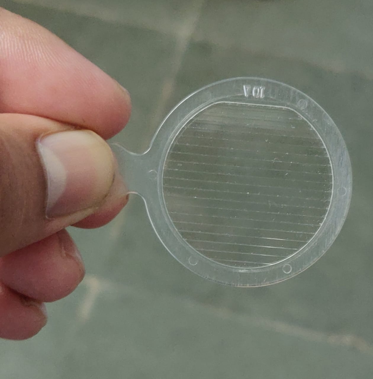
Digital image depicting a Fresnel prism Contributed by Kirandeep Kaur, MD
(Click Image to Enlarge)
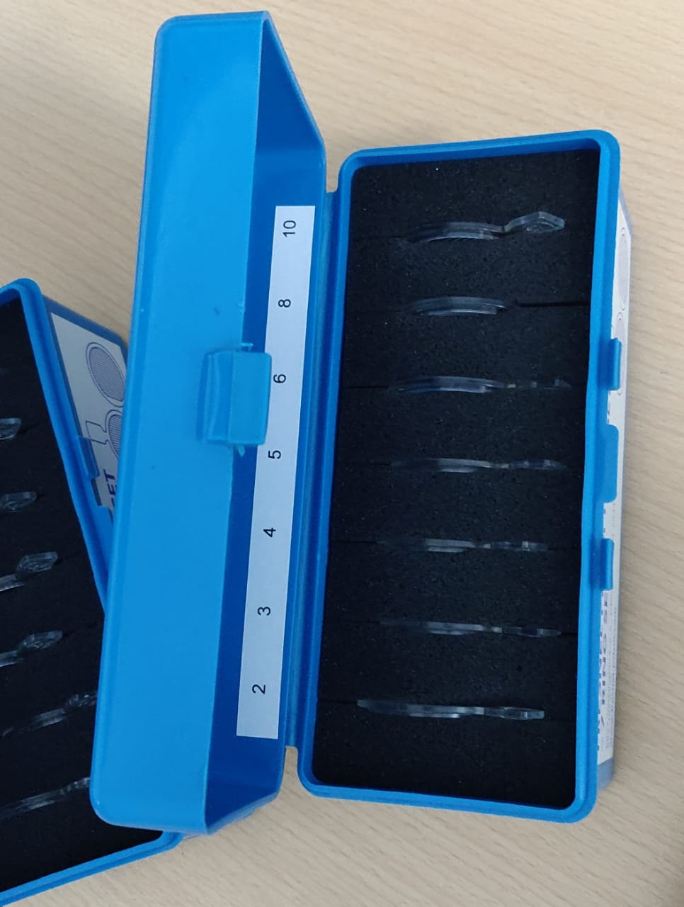
Digital image depicting a Fresnel prism box Contributed by Kirandeep Kaur, MD
References
Véronneau-Troutman S, Fresnel prism membrane in the treatment of strabismus. Canadian journal of ophthalmology. Journal canadien d [PubMed PMID: 5125648]
Smith JL,Weiner IG,Lucero AJ, Hemianopic Fresnel prisms. Journal of clinical neuro-ophthalmology. 1982 Mar; [PubMed PMID: 6226681]
Level 3 (low-level) evidenceKedar S,Ghate D,Corbett JJ, Visual fields in neuro-ophthalmology. Indian journal of ophthalmology. 2011 Mar-Apr; [PubMed PMID: 21350279]
Kaur K,Gurnani B, Prisms. StatPearls. 2022 Jan; [PubMed PMID: 35593813]
Hagen N,Tkaczyk TS, Compound prism design principles, I. Applied optics. 2011 Sep 1; [PubMed PMID: 22423145]
Gray LS, The prescribing of prisms in clinical practice. Graefe [PubMed PMID: 18379815]
Rossi PW,Kheyfets S,Reding MJ, Fresnel prisms improve visual perception in stroke patients with homonymous hemianopia or unilateral visual neglect. Neurology. 1990 Oct; [PubMed PMID: 2215953]
Level 1 (high-level) evidenceVéronneau-Troutman S, Fresnel prisms and their effects on visual acuity and binocularity. Transactions of the American Ophthalmological Society. 1978; [PubMed PMID: 754384]
Level 3 (low-level) evidencePeli E,Jung JH, Multiplexing Prisms for Field Expansion. Optometry and vision science : official publication of the American Academy of Optometry. 2017 Aug; [PubMed PMID: 28727615]
Peli E,Bowers AR,Keeney K,Jung JH, High-Power Prismatic Devices for Oblique Peripheral Prisms. Optometry and vision science : official publication of the American Academy of Optometry. 2016 May; [PubMed PMID: 26866438]
Brunnett SM,Munson MT,Kirschen DG, Fresnel vs. conventional prisms: their effects on the apparent fronto-parallel plane horopter. American journal of optometry and physiological optics. 1988 Jul; [PubMed PMID: 3207155]
Chen AM,Cotter SA, The Amblyopia Treatment Studies: Implications for Clinical Practice. Advances in ophthalmology and optometry. 2016 Aug; [PubMed PMID: 28435934]
Level 3 (low-level) evidenceTeodorescu L. ANOMALOUS HEAD POSTURES IN STRABISMUS AND NYSTAGMUS DIAGNOSIS AND MANAGEMENT. Romanian journal of ophthalmology. 2015 Jul-Sep:59(3):137-40 [PubMed PMID: 26978880]
Tamhankar MA,Ying GS,Volpe NJ, Success of prisms in the management of diplopia due to fourth nerve palsy. Journal of neuro-ophthalmology : the official journal of the North American Neuro-Ophthalmology Society. 2011 Sep; [PubMed PMID: 21378578]
Level 2 (mid-level) evidenceHaller T,Furr BA, Fresnel prism use among orthoptists. The American orthoptic journal. 2014; [PubMed PMID: 25313114]
Peli E, 2017 Charles F. Prentice Award Lecture: Peripheral Prisms for Visual Field Expansion: A Translational Journey. Optometry and vision science : official publication of the American Academy of Optometry. 2020 Oct; [PubMed PMID: 33055514]
Irsch K, Optical Issues in Measuring Strabismus. Middle East African journal of ophthalmology. 2015 Jul-Sep; [PubMed PMID: 26180462]
Gietzelt C,Fricke J,Neugebauer A,Hedergott A, Prism adaptation test before strabismus surgery in patients with decompensated esophoria and decompensated microesotropia. International ophthalmology. 2022 Jul; [PubMed PMID: 35038124]
Oystreck DT,Lyons CJ, Comitant strabismus: Perspectives, present and future. Saudi journal of ophthalmology : official journal of the Saudi Ophthalmological Society. 2012 Jul; [PubMed PMID: 23961004]
Level 3 (low-level) evidenceHatt SR,Leske DA,Klaehn LD,Kramer AM,Iezzi R Jr,Holmes JM, Treatment for Central-Peripheral Rivalry-Type Diplopia ("Dragged-Fovea Diplopia Syndrome"). American journal of ophthalmology. 2019 Dec; [PubMed PMID: 31323203]
Flanders M,Sarkis N, Fresnel membrane prisms: clinical experience. Canadian journal of ophthalmology. Journal canadien d [PubMed PMID: 10604055]
Level 3 (low-level) evidencePeli E,Vargas-Martin F,Kurukuti NM,Jung JH, Multi-periscopic prism device for field expansion. Biomedical optics express. 2020 Sep 1; [PubMed PMID: 33014587]
Hoppe E,Perlin RR, The effectivity of Fresnel prisms for visual field enhancement. Journal of the American Optometric Association. 1993 Jan; [PubMed PMID: 8454828]
Cheng C,Parreno J,Nowak RB,Biswas SK,Wang K,Hoshino M,Uesugi K,Yagi N,Moncaster JA,Lo WK,Pierscionek B,Fowler VM, Age-related changes in eye lens biomechanics, morphology, refractive index and transparency. Aging. 2019 Dec 16; [PubMed PMID: 31844034]
Koh KM,Samuel Kim U, Fresnel prism on hess screen test. Case reports in ophthalmological medicine. 2013; [PubMed PMID: 23710395]
Level 3 (low-level) evidenceShen J,Peli E,Bowers AR, Peripheral prism glasses: effects of moving and stationary backgrounds. Optometry and vision science : official publication of the American Academy of Optometry. 2015 Apr; [PubMed PMID: 25785533]
Sharma A,Tarbox L,Kurc T,Bona J,Smith K,Kathiravelu P,Bremer E,Saltz JH,Prior F, PRISM: A Platform for Imaging in Precision Medicine. JCO clinical cancer informatics. 2020 Jun; [PubMed PMID: 32479186]
Taylor SC, Cosmetic problems in skin of color. Skin pharmacology and applied skin physiology. 1999 May-Jun; [PubMed PMID: 10393522]
Anilkumar SE,Narendran K, Prisms in the treatment of diplopia with strabismus of various etiologies. Indian journal of ophthalmology. 2022 Feb; [PubMed PMID: 35086246]
Seaton J,Jones A,Johnston C,Francis K, Allied health professionals [PubMed PMID: 32297811]
Jones K,Adaji A,Schattner P, Involvement of practice nurses and allied health professionals in the development and management of care planning processes for patients with chronic disease - A pilot study. Malaysian family physician : the official journal of the Academy of Family Physicians of Malaysia. 2014; [PubMed PMID: 25606291]
Level 3 (low-level) evidence