Introduction
A patient with refractive error needs correction in the form of spectacle or contact lenses to see clearly.[1] The technique of finding out correct and suitable lenses for a particular patient is known as subjective refraction.[2]
Subjective refraction may be performed after objective refraction or can be done without that. However, it should be carried out after the objective refraction to be more precise.[3] This will help save time and energy in detecting the refractive error with accuracy. However, subjective refraction comes in handy in cases with corneal edema, dense lenticular opacity, or hazy media, where it is difficult to get a reliable retinoscopy.[4]
When the patient has been subjected to cycloplegic retinoscopy, it is better to perform subjective refraction once the drug effect wears off. It will be challenging to perform subjective refraction in very young children, mentally challenged patients, and uncooperative patients. Hence the prescription is given based on objective refraction in such cases.[5]
This activity will describe the instruments required, procedure, indications, interfering factors, and clinical significance of retinoscopy.
Specimen Collection
Register For Free And Read The Full Article
Search engine and full access to all medical articles
10 free questions in your specialty
Free CME/CE Activities
Free daily question in your email
Save favorite articles to your dashboard
Emails offering discounts
Learn more about a Subscription to StatPearls Point-of-Care
Specimen Collection
Instruments required
- Manual refraction unit or phoropter
- Trial frame
- Trial lenses and confirmation set
- Snellen's chart
- Pinhole
- Occluder
- Jackson cross cylinder- They combine two cylindrical lenses with equal power but the opposite sign (+/-), having an axis perpendicular to each other - they are used to diagnose astigmatism
- Duochrome test- To check the spherical power[6]
Procedures
Subjective Refraction Steps
- Subjective refraction monocularly
- Binocular balancing
- Near vision correlation[2]
Monocular Subjective Refraction
The primary goal of monocular refraction is to find the spherical power, cylindrical power, and axis.
Steps
- Baseline starting point selection and verification
- Cylindrical lens power and axis refinement and finalization
- Spherical lens refinement and finalization
Baseline Starting Point Selection and Verification
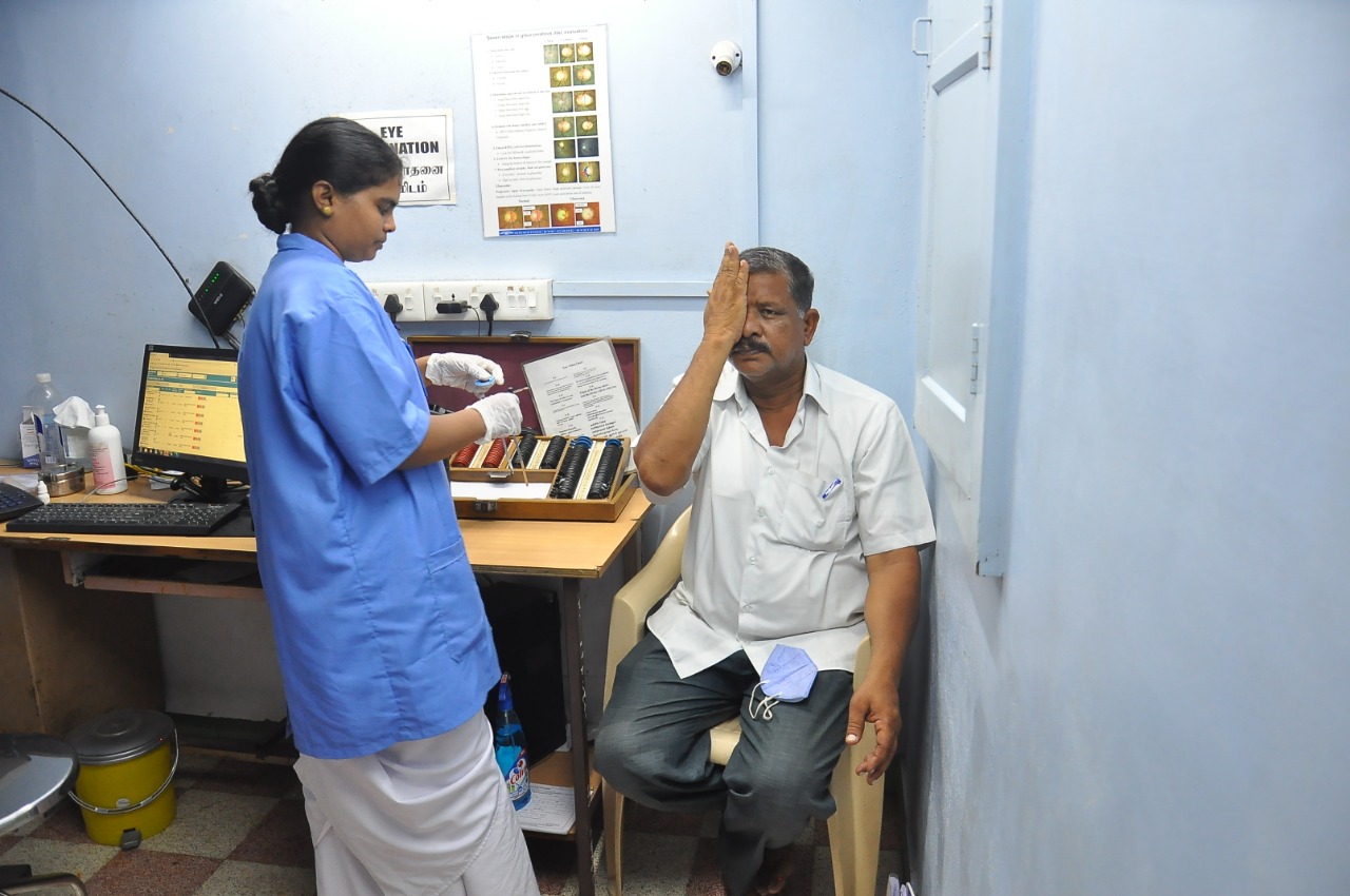
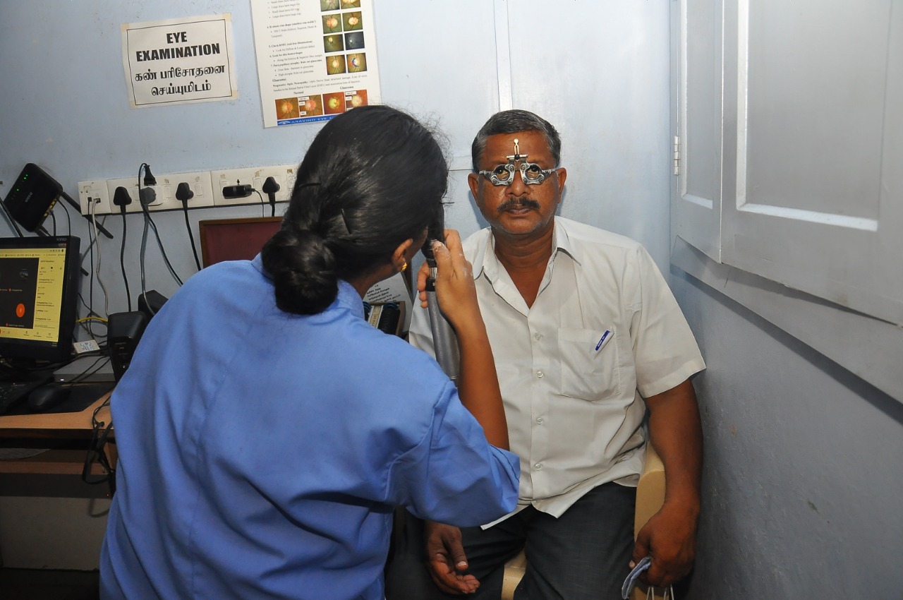
The subject is seated at 6 m. A trial frame is placed over the eye, and the visual acuity is tested for each eye separately. Although subjective refraction can be easily obtained directly, it is always recommended to get objective refraction done first.[7]
The objective refraction is done using retinoscopy, old glass prescription, and autorefractometry. While starting to perform the subjective refraction, the uncorrected visual acuity will give an idea about the amount of ametropia.[8] The verification of cylinder and sphere is usually performed by trial and error.[9]
Trial and Error Method
Spherical Lenses
This should be verified first. In the case of myopia, the weakest concave lens, and in hypermetropia, the strongest convex lens forms the best vision sphere.[10] To find the best vision sphere, a series of low power spheres such as +0.25, +0.5, -0.25, and -0.5 are quickly interchanged over the front trial lenses. The myopic patients are asked which correction makes the letter appear clearer and darker.[11]
Cylindrical Lenses
The cylindrical lenses need axis and power verification. Usually, the axis is checked first. 28030877
Axis Verification- This is done by rotating the cylinder's axis in increments of 5 and 10 degrees in either direction and confirming from the patient whether the visual acuity improved or not.[12] It is challenging to appreciate the change in smaller cylindrical powers at which axis the acuity is better. Under such cases, a more substantial cylindrical power is tried to verify the axis.[13]
Power Verification- is done after axis verification by changing the cylindrical power.[14]
Cylindrical Axis and Power Finalization and Refinement
Usually, the spherical component is refined and corrected after cylindrical astigmatism has been taken care of to obtain a sharper and clearer image. Hence, it is always mandatory to correct and refine the cylindrical astigmatic error before the spherical component. There are various methods of purifying the cylindrical correction.[15]
These include
- Astigmatic fan and block technique
- Jackson cross-cylinder technique
- Astigmatic clock dial and fogging technique
Astigmatic Fan and Block Technique
In some cases, the patient cannot respond well to cross-cylinder testing. In these cases, the Maddox V test comes in handy, an old fan and block technique method.[16] This consists of a series of radiating lines spaced at an interval of 10 degrees and arranged in the form of sun rays having a V and two sets of mutually perpendicular lines called blocks. The V and block can be simultaneously rotated at 180 degrees.[16]
Steps of Astigmatic Block and Fan Technique
Best Vision Sphere - The first step should be to obtain the visual acuity using a sphere only. It is thought that the best sphere gives the circle of least diffusion on the retina.[17]
Positive Sphere Addition - The positive sphere, half the amount of estimated astigmatism, should be added to bring the eye to a state of simple myopic astigmatism.[18]
Fan Chart- The patient should be seated at the fan chart and asked which line or group appears dark and which appears clearer. This gives a fair idea of the direction of astigmatic error. After this, a simple check must be performed by adding a +0.5 D sphere to confirm that the eye is in a state of simple myopic astigmatism. The black line should appear blurred, but if not more positive sphere should be added till it seems to be confused.[12]
In some cases, the clear line change through 90 degrees, indicating a state of simple hypermetropic astigmatism with an anterior focal line on the retina. Hence, furthermore, the sphere must be added till it appears blurred.[19]
Maddox Arrow - The focus should be directed to the Maddox arrow, and rotation should be done from the black limb until both limbs appear blurred. This helps in knowing the axis of astigmatism.
Blocks - Then direct the focus towards blocks and add a negative cylinder at the required axis until the second one becomes apparent as the first one. Suppose this doesn't happen, first under-correct and then overcorrect the astigmatic error.[20]
Second Check - A double-check must be performed by adding a +0.5 D sphere or a +0.25 D sphere with keen observation. Both blocks must blur equally, but astigmatism has been corrected if the black lines interchange. If the darker block becomes black again, the probably initial sphere correction was wrong and must be reassessed.[8]
Jackson Cross Cylinder Technique
This is a combination of two cylinders of equal strength but having an opposite sign, first described by Edward Jackson. The cylinders are mounted on a handle, and their axis is at the right angle. The commonly employed ones are +/- 0.25 and +/- 0.50. These are more useful than any other lenses in clinical refraction. These are usually employed to refine the cylinder's axis but can be used to assess complete astigmatic refraction.[21]
Steps
Sphere Adjustment
The sphere should be adjusted to most plus or least minus correction, giving the best-corrected visual acuity. This is achieved by performing fogging ( adding a positive sphere before the eye) while checking the visual acuity on the chart and then reducing the fog till a visual acuity is achieved. The primary aim is to place the circle of least diffusion on the retina, thus resulting in mixed astigmatism.[22]
Finding Astigmatism
If the cylindrical correction is not known initially, the Jackson cross-cylinder can still be used arbitrarily at 90 and 180 degrees to assess astigmatism. If the flip position is found, a cylindrical correction can be added with the axis parallel to the plus or minus axis of the cross-cylinder until the flip choices are equal. If the astigmatism is not discoverable at 90 and 180 degrees, then 45 and 135 degrees should be checked before assuming that no astigmatism is present.[12]
Axis Refinement
This should be attempted first. This is because a correct axis can be determined in the presence of an incorrect power, but full cylindrical power cannot be determined in the presence of an incorrect axis. For axis refinement, a cross-cylinder of +/- 0.5 D can be placed in front of the eye with the handle parallel to the axis in the trial frame, and then the patient is asked to tell any changes in the visual acuity.
If the patient discerns no difference between the two positions, the cylinder's axis is correct. If the visual acuity improves in a particular position, a plus cylinder should be rotated in the direction of the plus cylindrical component of the cross-cylinder. The test must be repeated several times till a neutral point is achieved.[12]
Cylindrical Power Refinement
To assess the cylinder's power, a cross-cylinder of +/- 0.25 D is placed in front of the eye, with its axis parallel to the cylinder's axis, first with the same sign and then with the opposite sign. In the first position, the correction is improved by 0.25 D, and in the second, it is diminished by the same power. If there is no improvement in any positions, then the cylinder's power is correct. However, if there is an improvement, the power should be verified again and improved.[2]
Astigmatic Clock Dial and Fogging
Steps
First, the visual acuity is obtained by placing a sphere with the other eye being occluded.[23]
Second, make the eye in compound myopic astigmatism but put a plus sphere before the eye to focus all the meridians before the eye. When the eye is fogged, the accommodation makes the image more blurred; hence the patient tends to relax accommodation and stabilize the refractive error of the eye.[24]
Third, the patient is asked to focus on the astigmatic dial and determine the darkest and sharpest lines.[25]
Fourth, minus cylindrical lenses of increasing power are used with an axis perpendicular to the black and sharp lines till the lines appear equal. A rotating cross dial is used and aligned with the principal meridian to compare the two meridians. The rule of 30 is used to calculate the axis of the minus cylinder. The lower number of the astigmatic dial is multiple with 30 to get the axis. For example, if the black line is seen at the 6 to 12 o'clock position, the lower number is multiplied by 30 to get the axis of the minus cylinder, i.e., 30 x 6= 180.[26]
Once all the lines in the astigmatic dial appear black equally, but still not incorrect focus because the eye is fogged. Now use the distance vision chart and reduce the plus sphere till the patient achieves a clear vision. [15]
Spherical Lens Refinement and Stabilization
After the cylindrical axis and power have been determined, the final step is sphere refinement by using fogging technique with Snellen's visual acuity chart, duo-chrome test, and pinhole visual acuity test.[27]
Fogging
A perfect way to determine spherical power is to fog the eye and then start unfogging by reducing the sphere by a +0.25 D sphere until a clear image is obtained. While termination of the test, it would be difficult to ascertain the endpoint as small changes in spherical power may not further refine the axis, and the examiner may land in a state of confusion. Hence, the Duochrome test is used to get the endpoint correction at this stage.[28]
Duochrome Test
This test is based on the chromatic aberration principle. The yellow light is focused on the retina in a normal human eye. The red light is focused at 0.24 D behind the retina, and the green light is concentrated at 0.20 D in front of the retina. In this test, the patient is told to read letters graded from 6/18 to 6/5 with green and red backgrounds.
In emmetropia, both colors give an equally sharp image. In myopia, the red appears clearer, and in hypermetropia, the green one. The lenses then should be adjusted to make the image appear clear. The only limitation with this test is that it doesn't relax the accommodation. Hence the test should be performed when the eye is slightly fogged. The letters on the red should appear clear, and minus power should be added till the red and green are equally clearer.[29]
Pinhole Testing
Finally, a pinhole test should be done to know that the correction in the trial frame is correct. Any improvement in the pinhole acuity indicates that the correction is incorrect.[30]
Binocular Balancing
This is the final step of subjective refraction, also known as vision equalization or equalization of accommodative effort. This process helps in the simultaneous focus of retinal images. A difference in appearance will lead to asthenopic symptoms and an imbalance in accommodation.[2]
The various proposed methods of binocular balancing are
- Fogging and alternate occlusion method
- Duochrome test with fogging
- Prism dissociation method
- Polaroid filters
- Turville infinity balance technique
Near Vision Correction
Correction of near vision is often necessary after the age of 40 years. Once the distant vision is corrected, the near vision should be corrected using near-vision charts such as Jaeger's chart, Snellen's chart, or number test types standardized by the faculty of Ophthalmologists N5 to N48.[31]
If the near vision is defective, further testing should be done as follows:
- Near the point of accommodation and amplitude of accommodation
- Determination of near point of convergence
- Dynamic retinoscopy
- Determination of near add[32]
Methodology to Assess Subjective Refraction
- History
- Subjective Refraction
- Assessing the fixation target
- Instruction to patients
- Visual acuity assessment
- Starting point
- Localizing principal meridian
- Astigmatism procedure
- Near vision assessment + Near correction
- Measurement of interpupillary distance
- Advice and prescription[2]
Indications
- Glass prescription
- Contact lens prescription
- Refractive error
- Keratoconus
- Early cataract
- Visual acuity not correlating with clinical findings[33]
Potential Diagnosis
The final diagnosis is made as follows:
Documenting the visual acuity with Snellen's chart and pinhole acuity. For example
- Uncorrected distant visual acuity (UDCVA) or visual acuity without correction – 20/40
- Best-corrected distant visual acuity (BCVA) or corrected distant visual acuity (CDVA)- 20/20
- Spherical correction- -2D sphere
- Cylindrical correction- -1D cylinder[34]
|
Eye |
UCDVA |
Sphere |
Cylinder |
Axis |
BCDVA/ CDVA |
Pinhole (Ph) |
|
Right eye (OD) |
20/40 |
-2D |
-1D |
60 degree |
20/20 |
20/20 |
|
Left eye (OS) |
20/40 |
-2D |
-1D |
150 degree |
20/20 |
20/20 |
The prescription is written as follows:
OD- -2 D/ -1D x 60 – 20/20
OS- -2 D/ -1D x 150 – 20/20
Duochrome test
R=G, where R= red and G= green
Interfering Factors
- Subjective refraction is dependent on patient cooperation
- It depends on the patient's ability to understand and respond to commands accurately
- Difficult in patients with the language barrier
- Difficult to perform in patients who are deaf and mute
- Pediatric patients
- Lack of skill of the optometrists/ophthalmologists
Patient Safety and Education
It is vital to educate the patient regarding the importance and technique of subjective refraction. The patient should receive education on the need for refractive error correction and the importance of preventing amblyopia in pediatric patients. The need for regular spectacle wear should be emphasized, and then if needed, patients should understand the need for laser refractive correction in the future.
The patient should also be educated regarding timely follow-up and subjective refraction evaluation to detect changes in refractive error. Moreover, they should also be told they need Schiempflug imaging in cases of high cylindrical power and retinal assessment in subjects with high myopia.[35]
Clinical Significance
Any patient presenting for routine ophthalmic examination may have an underlying refractive error. It is the duty of the examining ophthalmologist or the optometrist to determine the underlying refractive error and treat them correctly. The patient may be advised to use contact lenses or spectacles based on the need. Correction of refractive error in the pediatric age group is critical to prevent amblyopia.[36]
Media
(Click Image to Enlarge)
(Click Image to Enlarge)
References
Heus P, Verbeek JH, Tikka C. Optical correction of refractive error for preventing and treating eye symptoms in computer users. The Cochrane database of systematic reviews. 2018 Apr 10:4(4):CD009877. doi: 10.1002/14651858.CD009877.pub2. Epub 2018 Apr 10 [PubMed PMID: 29633784]
Level 1 (high-level) evidenceHervella L, Villegas EA, Prieto PM, Artal P. Assessment of subjective refraction with a clinical adaptive optics visual simulator. Journal of cataract and refractive surgery. 2019 Jan:45(1):87-93. doi: 10.1016/j.jcrs.2018.08.022. Epub 2018 Oct 8 [PubMed PMID: 30309774]
Hastings GD, Marsack JD, Nguyen LC, Cheng H, Applegate RA. Is an objective refraction optimised using the visual Strehl ratio better than a subjective refraction? Ophthalmic & physiological optics : the journal of the British College of Ophthalmic Opticians (Optometrists). 2017 May:37(3):317-325. doi: 10.1111/opo.12363. Epub 2017 Mar 30 [PubMed PMID: 28370389]
Ilechie AA, Addo NA, Abraham CH, Owusu-Ansah A, Annan-Prah A. Accuracy of Noncycloplegic Refraction for Detecting Refractive Errors in School-aged African Children. Optometry and vision science : official publication of the American Academy of Optometry. 2021 Aug 1:98(8):920-928. doi: 10.1097/OPX.0000000000001742. Epub [PubMed PMID: 34460453]
Hashemi H, Khabazkhoob M, Asharlous A, Soroush S, Yekta A, Dadbin N, Fotouhi A. Cycloplegic autorefraction versus subjective refraction: the Tehran Eye Study. The British journal of ophthalmology. 2016 Aug:100(8):1122-7. doi: 10.1136/bjophthalmol-2015-307871. Epub 2015 Nov 5 [PubMed PMID: 26541436]
Wesemann W, Rassow B. Modern instruments for subjective refraction. A comparative study. Ophthalmology. 1986 Sep:93(9 Suppl):52-60 [PubMed PMID: 3808640]
Level 2 (mid-level) evidenceRossato M, Nart A, Messina G, Favro F, Rossato V, Rrutja E, Biancalana V. The Refraction Assessment and the Electronic Trial Frame Measurement during Standing or Sitting Position Can Affect Postural Stability. International journal of environmental research and public health. 2022 Jan 29:19(3):. doi: 10.3390/ijerph19031558. Epub 2022 Jan 29 [PubMed PMID: 35162580]
Gantz L, Schrader S, Ruben R, Zivotofsky AZ. Can the red-green duochrome test be used prior to correcting the refractive cylinder component? PloS one. 2015:10(3):e0118874. doi: 10.1371/journal.pone.0118874. Epub 2015 Mar 16 [PubMed PMID: 25775478]
DeCarlo DK, McGwin G Jr, Searcey K, Gao L, Snow M, Waterbor J, Owsley C. Trial frame refraction versus autorefraction among new patients in a low-vision clinic. Investigative ophthalmology & visual science. 2013 Jan 2:54(1):19-24. doi: 10.1167/iovs.12-10508. Epub 2013 Jan 2 [PubMed PMID: 23188726]
Level 2 (mid-level) evidenceNorton TT, Siegwart JT Jr, Amedo AO. Effectiveness of hyperopic defocus, minimal defocus, or myopic defocus in competition with a myopiagenic stimulus in tree shrew eyes. Investigative ophthalmology & visual science. 2006 Nov:47(11):4687-99 [PubMed PMID: 17065475]
Level 3 (low-level) evidenceMaría Revert A, Conversa MA, Albarrán Diego C, Micó V. An alternative clinical routine for subjective refraction based on power vectors with trial frames. Ophthalmic & physiological optics : the journal of the British College of Ophthalmic Opticians (Optometrists). 2017 Jan:37(1):24-32. doi: 10.1111/opo.12343. Epub [PubMed PMID: 28030877]
Sha J, Fedtke C, Tilia D, Yeotikar N, Jong M, Diec J, Thomas V, Bakaraju RC. Effect of cylinder power and axis changes on vision in astigmatic participants. Clinical optometry. 2019:11():27-38. doi: 10.2147/OPTO.S190120. Epub 2019 Mar 19 [PubMed PMID: 30936760]
Arnold RW, Beveridge JS, Martin SJ, Beveridge NR, Metzger EJ, Smith KA. Grading Sphero-Cylinder Spectacle Similarity. Clinical optometry. 2021:13():23-32. doi: 10.2147/OPTO.S289770. Epub 2021 Jan 20 [PubMed PMID: 33505178]
Abelman H, Abelman S. Tolerance and nature of residual refraction in symmetric power space as principal lens powers and meridians change. Computational and mathematical methods in medicine. 2014:2014():492383. doi: 10.1155/2014/492383. Epub 2014 Nov 12 [PubMed PMID: 25478004]
Waddell K. Spherical refraction for general eye workers. Community eye health. 2000:13(33):6-7 [PubMed PMID: 17491944]
Murphy PJ, Beck AJ, Coll EP. An assessment of the orthogonal astigmatism test for the subjective measurement of astigmatism. Ophthalmic & physiological optics : the journal of the British College of Ophthalmic Opticians (Optometrists). 2002 May:22(3):194-200 [PubMed PMID: 12090633]
Level 1 (high-level) evidenceCholewiak SA, Love GD, Banks MS. Creating correct blur and its effect on accommodation. Journal of vision. 2018 Sep 4:18(9):1. doi: 10.1167/18.9.1. Epub [PubMed PMID: 30193343]
Althomali TA. Relative Proportion Of Different Types Of Refractive Errors In Subjects Seeking Laser Vision Correction. The open ophthalmology journal. 2018:12():53-62. doi: 10.2174/1874364101812010053. Epub 2018 Apr 30 [PubMed PMID: 29872484]
Archer SM. Monocular diplopia due to spherocylindrical refractive errors (an American Ophthalmological Society thesis). Transactions of the American Ophthalmological Society. 2007:105():252-71 [PubMed PMID: 18427616]
Level 3 (low-level) evidenceZareei A, Abdolahian M, Bamdad S. Cycloplegic Effects on the Cylindrical Components of the Refraction. Journal of ophthalmology. 2021:2021():8810782. doi: 10.1155/2021/8810782. Epub 2021 Apr 2 [PubMed PMID: 33884203]
Del Priore LV, Guyton DL. The Jackson cross cylinder. A reappraisal. Ophthalmology. 1986 Nov:93(11):1461-5 [PubMed PMID: 3808608]
Nakata S, Ikeda T, Nakatani H, Sakamoto M, Higashidutsumi M, Honda T, Kawayoshi A, Iwamura Y. Evaluation of an automatic fogging disinfection unit. Environmental health and preventive medicine. 2001 Oct:6(3):160-4. doi: 10.1007/BF02897964. Epub [PubMed PMID: 21432255]
Li T, Qureshi R, Taylor K. Conventional occlusion versus pharmacologic penalization for amblyopia. The Cochrane database of systematic reviews. 2019 Aug 28:8(8):CD006460. doi: 10.1002/14651858.CD006460.pub3. Epub 2019 Aug 28 [PubMed PMID: 31461545]
Level 1 (high-level) evidenceRemón L, Monsoriu JA, Furlan WD. Influence of different types of astigmatism on visual acuity. Journal of optometry. 2017 Jul-Sep:10(3):141-148. doi: 10.1016/j.optom.2016.07.003. Epub 2016 Sep 14 [PubMed PMID: 27639497]
Perches S, Collados MV, Ares J. Retinal Image Simulation of Subjective Refraction Techniques. PloS one. 2016:11(3):e0150204. doi: 10.1371/journal.pone.0150204. Epub 2016 Mar 3 [PubMed PMID: 26938648]
Elliott DB, Black AA, Wood JM. Subjective Verticality Is Disrupted by Astigmatic Visual Distortion in Older People. Investigative ophthalmology & visual science. 2020 Apr 9:61(4):12. doi: 10.1167/iovs.61.4.12. Epub [PubMed PMID: 32293665]
Venkataraman AP, Sirak D, Brautaset R, Dominguez-Vicent A. Evaluation of the Performance of Algorithm-Based Methods for Subjective Refraction. Journal of clinical medicine. 2020 Sep 29:9(10):. doi: 10.3390/jcm9103144. Epub 2020 Sep 29 [PubMed PMID: 33003297]
O'BRIEN JM, BANNON RE. The fogging method of refraction; a comparative analysis. American journal of ophthalmology. 1948 Nov:31(11):1453-9 [PubMed PMID: 18894717]
Level 2 (mid-level) evidenceBRINKBO B. Duochrome test as an aid in determinations of refraction. Acta ophthalmologica. 1954:32(5):585-8 [PubMed PMID: 14387639]
Loewenstein JI, Palmberg PF, Connett JE, Wentworth DN. Effectiveness of a pinhole method for visual acuity screening. Archives of ophthalmology (Chicago, Ill. : 1960). 1985 Feb:103(2):222-3 [PubMed PMID: 3977693]
Radner W. Near vision examination in presbyopia patients: Do we need good homologated near vision charts? Eye and vision (London, England). 2016:3():29 [PubMed PMID: 27844022]
Yazdani N, Khorasani AA, Moghadam HM, Yekta AA, Ostadimoghaddam H, Shandiz JH. Evaluating Three Different Methods of Determining Addition in Presbyopia. Journal of ophthalmic & vision research. 2016 Jul-Sep:11(3):277-81. doi: 10.4103/2008-322X.188387. Epub [PubMed PMID: 27621785]
Lee S, Jung G, Lee HK. Comparison of Contact Lens Corrected Quality of Vision and Life of Keratoconus and Myopic Patients. Korean journal of ophthalmology : KJO. 2017 Dec:31(6):489-496. doi: 10.3341/kjo.2016.0107. Epub 2017 Oct 20 [PubMed PMID: 29022291]
Level 2 (mid-level) evidenceLeube A, Kraft C, Ohlendorf A, Wahl S. Self-assessment of refractive errors using a simple optical approach. Clinical & experimental optometry. 2018 May:101(3):386-391. doi: 10.1111/cxo.12650. Epub 2018 Jan 21 [PubMed PMID: 29356102]
Donahue SP. The relationship between anisometropia, patient age, and the development of amblyopia. Transactions of the American Ophthalmological Society. 2005:103():313-36 [PubMed PMID: 17057809]
Medical Advisory Secretariat. Routine eye examinations for persons 20-64 years of age: an evidence-based analysis. Ontario health technology assessment series. 2006:6(15):1-81 [PubMed PMID: 23074485]

