Introduction
Lensometry measures the strength and prescription of eyeglasses or contact lenses using a lensmeter, an instrument sometimes referred to as a focimeter, vertometer, or Lensometer™. The two common indications for lensometry are the measurement of the power of spectacles during an ophthalmic examination and the verification of spectacle prescriptions created in an optical laboratory.[1]
Lensometry is based on the concept of lens neutralization, which states that the resultant power of a lens system is equal to the power required to neutralize its properties towards a net value of zero. The inbuilt lenses of a lensmeter are an adjustable collimator.[2] Lensmeters may be manual, semi-automated, or fully automated.[3][4] The lensmeter makes it possible to measure the dioptric vertex power, optical center, cylindrical axis, and prism of a lens; some lensmeters can also measure the amounts of tint, light transmission, and ultraviolet transmission of a lens.
This activity reviews the indications, contraindications, necessary preparation, technique, and clinical significance of lensometry while highlighting the role of the interprofessional eye care team in caring for patients who use lenses to correct refractive errors.
Indications
Register For Free And Read The Full Article
 Search engine and full access to all medical articles
- 10 free questions in your specialty
- Free CME/CE Activities
- Free daily question in your email
- Save favorite articles to your dashboard
- Emails offering discounts
Learn more about a Subscription to StatPearls Point-of-Care
Indications
The indications for lensometry include measuring the power, optic center, prismatic power and direction of a lens, and marking the optical axis and lens alignment before fitting.
Contraindications
There are no contraindications to lensometry.
Equipment
Several manual lensmeters are commercially available (see Image. Manual Lensmeter). A typical lensmeter consists of the following parts:
- Power switch: switches the equipment on and off.
- Illumination system: may be battery-powered or connected to AC/DC output mains. Plug-in lensmeters incorporate a transformer.
- Focusable eyepiece: focusable aperture through which the mires are seen, enabling the user to adjust the equipment for individual refractive errors before use.
- Power wheel: a rotary wheel calibrated in diopters. Equipment may have the powers engraved or displayed on the wheel or have the powers located inside the equipment.
- Axis scale: a fixed or rotary semi-circle calibrated from 0 to 180°.
- Reticle: a black target inside the equipment that helps the user center the lens and read cylindrical axes.
- Instrument table or lens stop aperture: where the user reads the lens if it is placed with its back surface away from the user. The lens must make adequate and proper contact with the table or aperture to avoid reading errors.[5]
- Lens holder: a spring-loaded device to hold the lens against or release it from the lens stop aperture or instrument table.
- Lens height adjustment knob or lever: used to raise or lower the instrument table to easily switch between measuring near and distant portions of a lens sample.
- Lens ink well or marker: pins set in temporary ink wells that can be lifted and manipulated onto the lens to imprint 3 reference points; helpful in marking the optical center after neutralization.
- Prism control system: this optional built-in accessory comprises a prism control knob, axis scale, and power scale.
Automated or semi-automated lensmeters are different but comparable to manual lensmeters (see Image. Semi-Automated Lensmeter). Automated or semi-automated lensmeters typically consist of the following components:
- Power switch: switches the equipment on and off.
- LCD screen: displays lens parameters.
- Lens-holding device: to hold the lens in place.
- Lens frame table: to rest the lens.
- Printer: may be built-in or a separate accessory.
- Lens marker lever and pins: to mark the lens.
- Nosepiece holder
Personnel
Any personnel trained and found to be proficient may perform lensometry.
Preparation
Before using the lensmeter, the measured lens must be clean and ready. Before measuring a lens, it is possible to determine whether it is single-vision or multifocal and if any tints or photochromic coatings are present. Lensometry can be used to measure powers in both naturally occurring lens materials, like CR-39 (Columbia resin, #39), and synthetic lenses, like polycarbonates.[6][7]
The lensmeter must be calibrated and prepared before measuring a lens to ensure accurate reading. Calibration and preparation require the following steps:
- To calibrate the lensmeter and adjust the instrument for the refractive error of the user, the user must look into the eyepiece towards the reticle. The reticle is a black target inside the equipment that includes a protractor for determining the axes of the cylinder. The dials must be adjusted to 0 and the light source properly aligned. Rotate the eyepiece counterclockwise until the reticle goes out of focus, then rotate it clockwise until the reticle comes into its sharpest view. Placing a white piece of paper on the instrument table may facilitate visualizing the reticle.
- Switch the equipment on and rotate the power wheel to the 0 value. The illuminated mires should be sharpest at this point when viewed inside the lensometer. If the mires are not sharp, adjust the power slowly in both directions until the mires are sharpest. This new nonzero power is recorded as the instrument error. The user may either send this information and the equipment for calibration or choose to work with the instrument error. Working with the instrument error requires subtracting the value from the spherical power of the final prescription of every lens measured using that particular piece of equipment.
Technique or Treatment
Measurement of Spherical Power
Measuring the spherical power of a lens or lenses with a manual lensmeter requires execution of the following steps after the equipment has been calibrated and prepared:
- Place the lens on the stage of the lensmeter with the convex side facing upward.
- Align the lens by positioning the target in front of the lens and centering the target in the viewfinder.
- Measure the lens by adjusting the 2 dials corresponding to the sphere and cylinder to focus on the target and measuring the refractive power of the lens in diopters.
- If the patient has a prescription for two lenses, repeat the above steps for the other lens.
- Verify the lensometry results against the known prescription, making adjustments to ensure the accuracy of the corrective lenses.
- Compensate for any spherical and cylindrical instrument error to arrive at the final back vertex power of the analyzed lens.
Measurement of Cylindrical Power
Cylindrical lenses contain 2 distinct powers, and only half of the mires will become sharp at each major meridian during routine lensometry.[8] It is customary to neutralize one meridian first, turn the measurement axis 90°, and measure the other meridian. This is a cross-cylinder prescription, and combining the two measured meridians is transposition. The first step of transposition is to take the maximum plus-powered spherical power; this will be the sphere. The second step is to calculate the dioptric difference between the two powers obtained and denote it as the minus cylindrical value. The axis of the resultant prescription is the power not chosen initially as the sphere. For example,
- To transpose a finding of + 1.00 DC x 90 and +1.50 DC x 180, select the maximum plus-powered cylinder (+1.50 DS) as the sphere. Take the dioptric difference as the minus cylinder (+1.50 - (+1.00) DC = -0.50 DC). The axis of the cylinder is the axis of the +1.00 DS not chosen as the sphere (90). The final prescription is +1.50 DS - 0.50 DC x 90.
To compensate for a cylindrical error, note the cylindrical value that is the null point of the equipment. If the null values of the equipment are 15º and 195º, the lower value must be subtracted from any eventual axis read. For example,
- If the lowest null value is 15º, and the measurement yields a finding of -0.75 DS - 0.50 DC x 145, the final prescription will be -0.75 DS - 0.50 DC x 130.
Measurements of Multifocal or Varifocal Lenses
A multifocal lens prescription consists of a distance correction and at least one reading or intermediate addition ground into the same lens.[9] The equivalent near correction is the dioptric addition of the distance correction and the reading addition. When measuring a multifocal lens, first measure the powers in the distance portion and then the powers of the near portion. The reading addition is obtained by subtracting the spherical power in the distance portion from the spherical power in the near portion. The equivalent near and distance powers can be estimated from partial prescriptions. The reading add is almost always a spherical power added to the lower portion of the lens. The amount of reading power is permanently engraved into the lens material at the point of production.
Varifocal lenses have markings that may be permanently engraved into the lenses or ink-based and temporary.[10] If these guide markings are not present, lensometry remains useful and can still be performed.[11] Horizontal alignment markers remain even when temporary markings have been removed, and permit proper alignment of the lenses during glazing. The equivalent distance power will be located precisely in the middle of the horizontal alignment markers in each varifocal lens. The horizontal markers may be connected using temporary ink to find the midpoint and measure the distance power.
Prismatic Effects
Errors in measurement, such as decentration, can induce prismatic effects.[4] While prismatic effects may be desirable when managing binocular vision anomalies, they may be a nuisance and can be a source of significant asthenopia.[12] The total amount of induced prism is obtained from the formula:
P = D x C,
where P = prismatic value, D = dioptric power of the lens, and C = the amount of decentration in centimeters.
Complications
There are no known complications of lensometry.
Clinical Significance
The recent advent of tunable lenses has fueled research into dynamic focimetry to measure changes in powers in real-time with little or no human input.[13][14] Garcia-Domene et al also described a novel focimeter for measuring the power of an intra-ocular lens in situ.[15]
Enhancing Healthcare Team Outcomes
The lensmeter measures the power and characteristics of ophthalmic lenses used to correct refractive errors of binocular vision. Spectacle and contact lens corrections remain commonplace despite the availability of corrective refractive surgical procedures such as laser-assisted in situ keratomileusis (LASIK), laser-assisted subepithelial keratectomy (LASEK), and photorefractive keratectomy (PRK).[16][17] Refractive errors are the most common reason patients seek ocular care.[18]
Lensometry confirms prescription powers before dispensing and neutralizes the powers of unknown lenses.[19] All ocular care providers must have a lensmeter, and staff must be appropriately trained in its use. The lensmeter is used in the assembly and confirmation of lens prescriptions to ensure the best-corrected vision for the patient.
Media
(Click Image to Enlarge)
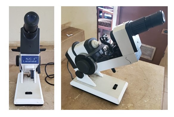
Manual Lensmeter
Contributed by Mutali Musa, OD, MSc, FNCO
(Click Image to Enlarge)
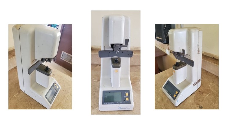
Semi-Automated Lensmeter
Contributed by Mutali Musa, OD, MSc, FNCO
References
Heidarian A, Mason D. Health information technology adoption in New Zealand optometric practices. Clinical & experimental optometry. 2013 Nov:96(6):557-65 [PubMed PMID: 24730034]
Cordero-Dávila A, Cruz-Ponce S, González-García J. Lensometer with autocollimation and a square Ronchi grid. Applied optics. 2020 Feb 20:59(6):1726-1731. doi: 10.1364/AO.377172. Epub [PubMed PMID: 32225677]
Papas E, Dahms A, Carnt N, Tahhan N, Ehrmann K. Power profiles and short-term visual performance of soft contact lenses. Optometry and vision science : official publication of the American Academy of Optometry. 2009 Apr:86(4):318-23. doi: 10.1097/OPX.0b013e318198959e. Epub [PubMed PMID: 19225434]
West CE, Hunter DG. Displacement of optical centers in over-the-counter readers: a potential cause of diplopia. Journal of AAPOS : the official publication of the American Association for Pediatric Ophthalmology and Strabismus. 2014 Jun:18(3):293-4. doi: 10.1016/j.jaapos.2014.01.008. Epub 2014 Apr 24 [PubMed PMID: 24767829]
Jin T, Gao X. Overcoming the error of optical power measurement caused by the curvature radius. Optics express. 2022 May 9:30(10):17115-17129. doi: 10.1364/OE.455280. Epub [PubMed PMID: 36221541]
Karaaslan H, Engin B. ESR dosimetric properties of gamma irradiated different origin eyeglass samples. Applied radiation and isotopes : including data, instrumentation and methods for use in agriculture, industry and medicine. 2021 Dec:178():109987. doi: 10.1016/j.apradiso.2021.109987. Epub 2021 Oct 16 [PubMed PMID: 34688023]
Pościk A, Jachowicz M. Mechanical properties of protective spectacles fitted with corrective lenses. International journal of occupational safety and ergonomics : JOSE. 2017 Sep:23(3):440-446. doi: 10.1080/10803548.2016.1234131. Epub 2016 Oct 13 [PubMed PMID: 27649581]
Aluyi-Osa G, Musa MJ, Zeppieri M. Jackson Cross Cylinder. StatPearls. 2023 Jan:(): [PubMed PMID: 36508527]
Chukwuyem EC, Musa MJ, Zeppieri M. Prescribing Glasses for Presbyopia. StatPearls. 2023 Jan:(): [PubMed PMID: 36943962]
Ferrer-Altabás S, Picazo-Bueno JÁ, Granero-Montagud L, Micó V. Shadowfocimetry: adapting the holographic principle to a manual focimeter for visualization/marking of permanent engravings in progressive addition lenses. Optics letters. 2022 May 1:47(9):2298-2301. doi: 10.1364/OL.454962. Epub [PubMed PMID: 35486785]
Level 3 (low-level) evidenceRadhakrishnan H, Lam CSY, Charman WN. Multiple segment spectacle lenses for myopia control. Part 1: Optics. Ophthalmic & physiological optics : the journal of the British College of Ophthalmic Opticians (Optometrists). 2023 Sep:43(5):1125-1136. doi: 10.1111/opo.13191. Epub 2023 Jun 28 [PubMed PMID: 37378657]
Ellison AC, Campbell AJ, Robertson MC, Sanderson GF. Prismatic displacement effect of progressive multifocal glasses on reaction time and accuracy in elderly people. Clinical ophthalmology (Auckland, N.Z.). 2014:8():891-902. doi: 10.2147/OPTH.S58193. Epub 2014 May 9 [PubMed PMID: 24872674]
Akondi V, Sawides L, Marrakchi Y, Gambra E, Marcos S, Dorronsoro C. Experimental validations of a tunable-lens-based visual demonstrator of multifocal corrections. Biomedical optics express. 2018 Dec 1:9(12):6302-6317. doi: 10.1364/BOE.9.006302. Epub 2018 Nov 15 [PubMed PMID: 31065430]
Level 1 (high-level) evidenceDorronsoro C, Barcala X, Gambra E, Akondi V, Sawides L, Marrakchi Y, Rodriguez-Lopez V, Benedi-Garcia C, Vinas M, Lage E, Marcos S. Tunable lenses: dynamic characterization and fine-tuned control for high-speed applications. Optics express. 2019 Feb 4:27(3):2085-2100. doi: 10.1364/OE.27.002085. Epub [PubMed PMID: 30732252]
García-Domene MC, Díez-Ajenjo MA, Peris-Martínez C, Navea A, Artigas JM. A rapid method for measuring intraocular lens power in vitro with a focimeter. Experimental eye research. 2015 Nov:140():190-192. doi: 10.1016/j.exer.2015.09.009. Epub 2015 Sep 18 [PubMed PMID: 26386149]
Abdelzaher HA, Sidky MK, Awadein A, Hosny M. Aniseikonia and visual functions with optical correction and after refractive surgery in axial anisometropia. International ophthalmology. 2022 Jun:42(6):1669-1677. doi: 10.1007/s10792-021-02161-w. Epub 2022 Jan 30 [PubMed PMID: 35094222]
Moshirfar M, Thomson AC, Thomson RJ, Martheswaran T, McCabe SE. Use of presbyopia-correcting intraocular lenses in patients with prior corneal refractive surgery. Current opinion in ophthalmology. 2021 Jan:32(1):45-53. doi: 10.1097/ICU.0000000000000722. Epub [PubMed PMID: 33122489]
Level 3 (low-level) evidenceMusa MJ, Zeppieri M. Spectacle Correction of Ametropias. StatPearls. 2023 Jan:(): [PubMed PMID: 36251812]
Yuen GS, Chou BR, Ngo TP, Cheng BB, Dain SJ. Prescription compliance in ophthalmic lenses. Clinical & experimental optometry. 2011 Jul:94(4):341-7. doi: 10.1111/j.1444-0938.2010.00566.x. Epub 2011 Jan 24 [PubMed PMID: 21255076]