Introduction
Refractive errors are an important common cause of visual disturbance worldwide.[1] The prevalence of types and degree of refractive errors may vary from region to region.[2] Vitale et al., in their analysis of the United States, found astigmatism as the most common refractive error with a prevalence of 36.2%, followed by myopia with a prevalence of 33.1%, and least being hyperopia with a prevalence of 3.6%.[3]
Further, myopia was more prevalent in females (39.9%) than males (32.6%). Sethu et al. found the prevalence of refractive error of at least 0.50 diopter (D) spherical, equivalent to 53.1%. Myopia and hypermetropia were found to be the most common refractive errors accounting for 27.7% and 22.9%, respectively.[4]
Symptoms related to refractive errors are quite disturbing and may even disrupt the normal lifestyle of individuals. Refractive error patients form the majority of outpatient patients visiting an optometrist or ophthalmology clinic. A study by Schiefer et al. found refractive errors accounted for 21.1% of the patients presenting to an ophthalmologist.[5]
The evaluation of these patients can be occasionally aided by topical agents called cycloplegic drugs.[6] Thus refraction can be broadly divided into cycloplegic and non-cycloplegic refraction. Cycloplegic drugs are often used to evaluate patients for underlying refractive errors. Cycloplegics cause temporary paralysis of ciliary muscles allowing the determination of total refractive errors.[7] Cycloplegic retinoscopy is also known as wet retinoscopy. Cycloplegics have been used since the 19 century to assess refractive errors by relaxing the accommodation.[8]
Noncycloplegic refraction is performed without any drug administration. It doesn't affect the accommodation and pupil dilatation.[9] The various listed methods of non-cycloplegic refraction are retinoscopy, autorefraction, and objective and subjective refraction. A detailed description of how to perform retinoscopy and refraction is beyond the scope of this chapter and is discussed in separate chapters.
Difference between Cycloplegic and Noncycloplegic Refraction
|
S. No |
Characteristic |
Cycloplegic |
Noncycloplegic |
|
1 |
Pupil dilatation |
Dilated |
Undilated |
|
2 |
Accommodation |
Lost |
Intact |
|
3 |
Time |
Need more time |
Need less time |
|
4 |
Drugs |
Atropine, Cyclopentolate, Tropicamide, Homatropine |
Not needed |
|
5 |
Types |
- |
Retinoscopy, Autorefractor, Objective and Subjective retinoscopy |
|
6 |
Cost |
Costly |
Less costly |
|
7 |
Patient convenience |
Less |
More |
Function
Register For Free And Read The Full Article
 Search engine and full access to all medical articles
- 10 free questions in your specialty
- Free CME/CE Activities
- Free daily question in your email
- Save favorite articles to your dashboard
- Emails offering discounts
Learn more about a Subscription to StatPearls Point-of-Care
Function
Cycloplegics are drugs that paralyze the ciliary muscles and cause relaxation of accommodation. Once the ciliary muscles are relaxed, the anterior zonules stretch, and posterior zonules lose tension leading to thinning of the lens. This allows the eye to be relaxed and helps in focusing on distance.[10]
Cycloplegic drugs are the anticholinergic agents that block acetylcholine's muscarinic action in ciliary muscle receptors. This inhibits the cholinergic stimulation of the iris sphincter and ciliary muscles, allowing relaxation of accommodation and inhibiting the accommodative power of the eye.[11]
The various cycloplegic agents are atropine sulfate, homatropine hydrobromide, cyclopentolate hydrochloride, and tropicamide.
Atropine Sulphate
It is an anticholinergic agent that acts directly on muscarinic receptors of structure innervated by post-ganglionic parasympathetic fibers. It is available in 0.01%, 0.5%, 1% and 3% topical ophthalmic drops and ointment. For refraction, 1% atropine ointment should be applied as a morning dose for three days. Atropine is one of the strongest drugs available for cycloplegia. It paralyzes the sphincter pupillae and ciliary muscles, thus causing mydriasis and cycloplegia. Atropine is a competitive inhibitor of the muscarinic action of acetylcholine.[12]
Apart from its use as a cycloplegic agent, atropine is also indicated for pupillary dilatation in inflammatory conditions to prevent pain and release synechiae. It is also used in ciliary block glaucoma, for penalization as part of amblyopia management, to treat accommodation spasms, to prevent undue vagal response in Tensilon test, and pre and post-operatively in some intraocular surgeries. Low dose atropine in the form of 0.01% topical application is widely accepted to control myopia progression. Atropine is contraindicated in patients with primary angle glaucoma or narrow anterior chambers. It is also contraindicated among those with hypersensitivity to any preparation components.[8]
Homatropine Hydrobromide
It is an anticholinergic agent used as a 2% sterile ophthalmic solution. It acts in the same manner as atropine by blocking the muscarinic action of acetylcholine, thus causing mydriasis and cycloplegia. It is also used for penalization therapy in amblyopia and to treat ocular inflammatory conditions like iritis. This is used as one drop repeated every 10 minutes six times for performing cycloplegic refraction. Retinoscopy can be performed after about 90 minutes of installing the first drop. The effect lasts for 48 to 72 hours. A drug correction of 0.5D is done from the retinoscopy value as tonus allowance.[12]
Cyclopentolate Hydrochloride
It is also an anti-cholinergic agent used as a 1% sterile ophthalmic solution as a cycloplegic action. It is also available as 0.5% and 2% formulations. Cyclopentolate has a faster onset of action and a shorter duration of effect. Cycloplegia action occurs in 30 to 45 minutes of instillation. It is applied as one drop and repeated after 5 minutes. The effect of the drug lasts 6 to 18 hours. A drug correction of 0.75D is done from the retinoscopy value as tonus allowance.[8]
Tropicamide
This is a commonly used anti-cholinergic mydriatic drug. The drug has a powerful mydriatic effect, fast onset of action, and low side effect profile. Tropicamide is available at a dose of 0.5% and 1%. It works by blocking the muscarinic acetylcholine receptors. The 1 % preparation paralyzes the accommodation. The 0.5% strength produces mydriasis with slight cycloplegia. Tropicamide is implicated in mydriasis and cycloplegia, diagnosis, and short-acting mydriatic for pre and post-operative stages. The pupillary dilatation is less dependent on iris pigmentation. For premature infants combination of 2.5% phenylephrine and 5% is recommended due to less dilatation alone.[13]
Characteristic Features of Cycloplegics and Mydriatics
|
S. No |
Drug |
Age Group |
Dose |
Peak effect |
Timing of Retinoscopy |
Duration of action |
Cycloplegic effect |
Tonus allowance |
|
1 |
Atropine Sulfate (1% ointment) |
Below 5 years |
Three times x 3 days |
2-3 days |
4 day |
10-20 days |
After 3 weeks of retinoscopy |
1 D |
|
2 |
Homatropine hydrobromide (2% drops) |
5-8 years |
One drop for every 10 mins ( 6 times) |
1-1.5 hours |
After 90 minutes of drug administration |
2-3 days |
After 3 weeks of retinoscopy |
0.5 D |
|
3 |
Cyclopentolate hydrochloride (1% drops) |
8-20 years |
One drop every 15 mins (3 times) |
80-90 minutes |
After 90 minutes of drug administration (Havener's recommended dose) |
6-18 hours |
After 3 days of retinoscopy |
0.75 D |
|
4 |
Tropicamide (0.5%, 1% drops) |
Above 15 years, used as a mydriatic |
One drop every 15 mins (3-4 times) |
20-40 minutes |
- |
4-6 hours |
- |
- |
Issues of Concern
Cycloplegics are contraindicated in patients with the shallow anterior chamber, close angle-closure glaucoma, history of allergy or hypersensitivity to any component of drugs, and systemic anticholinergic drugs receiver.[14]
Atropine gets absorbed systemically through the lacrimal sac and thus should be avoided in children as far as possible.[15]
It is advisable to compress the lacrimal sac for a minute after topical application to prevent systemic absorption. Though uncommon, few local side effects reported with atropine include the allergic reaction of lids and conjunctiva, crusting of lid margins, and dryness of periocular skin.[15]
The blurring of vision and photophobia are direct consequences of cycloplegic and mydriatic effects of atropine. Systemic side effects include dryness of skin and mouth, tachycardia, irritability, delirium, flushing, skin rashes, abdominal distension, or hyperpyrexia. Very rarely, a severe reaction in the form of progressive respiratory depression has also been reported.[16]
Homatropine can rarely have side effects, including incoherent speech, hallucinations, disorientation, psychosis, and visual disturbances.[17]
Cyclopentolate is rarely associated with side effects like lacrimation and blurred vision. Systemic side effects include hallucinations, ataxia, disorientation, disturbance in speech, and restlessness.[18]
Tropicamide is contraindicated in angle-closure glaucoma and hypersensitivity to the drug. Tropicamide is linked with CNS disturbance in infants and children. It should be used carefully in the elderly and cases with increased IOP. Tropicamide locally causes a stinging sensation, and systemic absorption may result in confusion and hypersensitivity reaction in children.[19]
Clinical Significance
Cycloplegics are considered the standard of care for certain clinical scenarios like pediatric age group, constant or intermittent esotropia, pseudo myopia, latent hyperopia, accommodation insufficiency, accommodation fatigue, or accommodation spasm. Cycloplegic refraction is also recommended in patients with amblyopia or underlying strabismus.[20]
The American Optometric Association (AOA) guidelines also recommend cycloplegic examination of pre-schoolers for screening ocular conditions like significant refractive errors, anisometropia, strabismus, or amblyopia. The underlying clinical conditions leading to decreased vision can potentially threaten long-term sequelae. Thus, it is essential to evaluate the children at a young age to allow timely diagnoses and treatment.[21]
Retinoscopy is performed in dim room illumination. The patient should be seated at a distance of 1 meter from the examiner. The retinoscope light is thrown into the patient's eye, and the patient is asked to look at the retinoscopic light. This is in contrast to dry retinoscopy, where the patient is asked to look at a distant point to allow relaxation of accommodation. The red reflex is noted through a hole in the retinoscope mirror in the patient's pupillary area. The retinoscope is moved in the horizontal and vertical meridian while observing the retinoscopic reflex.[22]
The important inferences made from the movement of retinoscopic reflex depend on the distance and mirror used. For the setting of the plane mirror used at 1 meter, the interpretations are as follows:
- No movement of red reflex indicates myopia of 1 diopter (D)
- The movement of red reflex along the direction of the retinoscope indicates either emmetropia or hypermetropia, or myopia of less than 1 D
- The movement of the red reflex against the direction of the retinoscope implies myopia of more than 1 D.
Other Issues
As accommodation is lost due to cycloplegia, the patient will complain of blurred vision, photophobia, and difficulty focusing near objects. The patient must rest for at least 3 to 6 hours until accommodation reverses. This will hamper the routine activities of the patient. Sometimes the cycloplegic refraction of the patient doesn't match with that of subjective refraction or non-cycloplegic refraction. Hence, post mydriatic test should be done in these cases before the final spectacle prescription.[23]
Enhancing Healthcare Team Outcomes
Pediatric patients below 13 to 14 years of age presenting with defective vision and having underlying refractive error should be subjected to cycloplegic refraction.[24]
Patients in the age group of 14 to 35 years should be evaluated with tropicamide refraction. After cycloplegic or tropicamide refraction, the final contact lens or glass prescription should be given. All ophthalmologists and optometrists must know the indications and importance of cycloplegic and non-cycloplegic refraction to avoid the progression of refractive error.[25]
The optometrists play a crucial role in explaining and performing non-cycloplegic and cycloplegic refraction. Ophthalmologists play a vital role in treatment, counseling, and explaining the prognosis to the patients.[26]
Nursing, Allied Health, and Interprofessional Team Interventions
The nursing and the allied staff play a vital role in receiving the patient to the clinic, registering basic ophthalmic complaints, explaining to the patient the methodology and duration of a cycloplegic refraction, the need for cycloplegic refraction, and the side effects of instilling side effects. The nursing team also helps in counseling and procuring spectacle for the patients in cases of refractive error.[27]
Nursing, Allied Health, and Interprofessional Team Monitoring
The nursing and the allied staff help monitor the progress of cycloplegic refraction by instilling cycloplegic drugs at regular intervals and assessing the pupillary dilatation. They also help patients' regular and smooth flow in the clinics without prolonged wait time.[28]
Media
(Click Image to Enlarge)
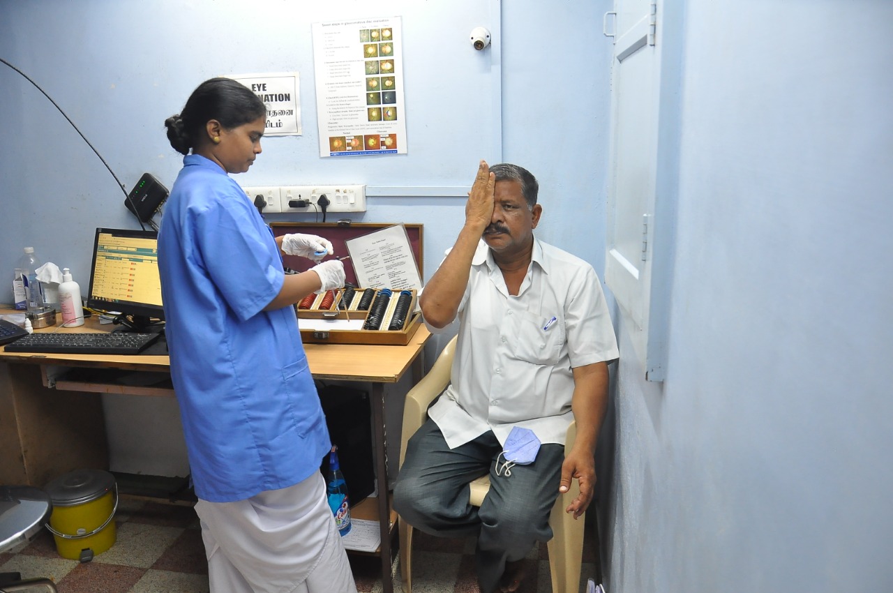
Digital image of the patient depicting visual acuity evaluation before performing a cycloplegic refraction in a young male. Contributed by Dr. Kirandeep Kaur, MBBS, DNB, FPOS, FICO, MRCS Ed, MNAMS
References
Gomez-Salazar F, Campos-Romero A, Gomez-Campaña H, Cruz-Zamudio C, Chaidez-Felix M, Leon-Sicairos N, Velazquez-Roman J, Flores-Villaseñor H, Muro-Amador S, Guadron-Llanos AM, Martinez-Garcia JJ, Murillo-Llanes J, Sanchez-Cuen J, Llausas-Vargas A, Alapizco-Castro G, Irineo-Cabrales A, Graue-Hernandez E, Ramirez-Luquin T, Canizalez-Roman A. Refractive errors among children, adolescents and adults attending eye clinics in Mexico. International journal of ophthalmology. 2017:10(5):796-802. doi: 10.18240/ijo.2017.05.23. Epub 2017 May 18 [PubMed PMID: 28546940]
Hashemi H, Fotouhi A, Yekta A, Pakzad R, Ostadimoghaddam H, Khabazkhoob M. Global and regional estimates of prevalence of refractive errors: Systematic review and meta-analysis. Journal of current ophthalmology. 2018 Mar:30(1):3-22. doi: 10.1016/j.joco.2017.08.009. Epub 2017 Sep 27 [PubMed PMID: 29564404]
Level 1 (high-level) evidenceVitale S, Ellwein L, Cotch MF, Ferris FL 3rd, Sperduto R. Prevalence of refractive error in the United States, 1999-2004. Archives of ophthalmology (Chicago, Ill. : 1960). 2008 Aug:126(8):1111-9. doi: 10.1001/archopht.126.8.1111. Epub [PubMed PMID: 18695106]
Sheeladevi S, Seelam B, Nukella PB, Borah RR, Ali R, Keay L. Prevalence of refractive errors, uncorrected refractive error, and presbyopia in adults in India: A systematic review. Indian journal of ophthalmology. 2019 May:67(5):583-592. doi: 10.4103/ijo.IJO_1235_18. Epub [PubMed PMID: 31007213]
Level 1 (high-level) evidenceSchiefer U, Kraus C, Baumbach P, Ungewiß J, Michels R. Refractive errors. Deutsches Arzteblatt international. 2016 Oct 14:113(41):693-702. doi: 10.3238/arztebl.2016.0693. Epub [PubMed PMID: 27839543]
Sun YY, Wei SF, Li SM, Hu JP, Yang XH, Cao K, Lin CX, Du JL, Guo JY, Li H, Liu LR, Morgan IG, Wang NL. Cycloplegic refraction by 1% cyclopentolate in young adults: is it the gold standard? The Anyang University Students Eye Study (AUSES). The British journal of ophthalmology. 2018 Jun 21:():. pii: bjophthalmol-2018-312199. doi: 10.1136/bjophthalmol-2018-312199. Epub 2018 Jun 21 [PubMed PMID: 29930099]
Krantz EM, Cruickshanks KJ, Klein BE, Klein R, Huang GH, Nieto FJ. Measuring refraction in adults in epidemiological studies. Archives of ophthalmology (Chicago, Ill. : 1960). 2010 Jan:128(1):88-92. doi: 10.1001/archophthalmol.2009.349. Epub [PubMed PMID: 20065223]
Level 2 (mid-level) evidenceSani RY, Hassan S, Habib SG, Ifeanyichukwu EP. Cycloplegic effect of atropine compared with cyclopentolate-tropicamide combination in children with hypermetropia. Nigerian medical journal : journal of the Nigeria Medical Association. 2016 May-Jun:57(3):173-7. doi: 10.4103/0300-1652.184065. Epub [PubMed PMID: 27397958]
Chen J, Xie A, Hou L, Su Y, Lu F, Thorn F. Cycloplegic and noncycloplegic refractions of Chinese neonatal infants. Investigative ophthalmology & visual science. 2011 Apr 14:52(5):2456-61. doi: 10.1167/iovs.10-5441. Epub 2011 Apr 14 [PubMed PMID: 21087952]
Erickson-Lamy KA, Polansky JR, Kaufman PL, Zlock DM. Cholinergic drugs alter ciliary muscle response and receptor content. Investigative ophthalmology & visual science. 1987 Feb:28(2):375-83 [PubMed PMID: 8591921]
Level 3 (low-level) evidenceSvoboda J, Popelikova A, Stuchlik A. Drugs Interfering with Muscarinic Acetylcholine Receptors and Their Effects on Place Navigation. Frontiers in psychiatry. 2017:8():215. doi: 10.3389/fpsyt.2017.00215. Epub 2017 Nov 9 [PubMed PMID: 29170645]
Crom DB, Crom WR. Memo on medications: atropine sulfate and homatropine hydrobromide: common mydriatic/cycloplegic agents. Journal of ophthalmic nursing & technology. 1982 Aug:1(2):38-40 [PubMed PMID: 6980991]
Hofmeister EM, Kaupp SE, Schallhorn SC. Comparison of tropicamide and cyclopentolate for cycloplegic refractions in myopic adult refractive surgery patients. Journal of cataract and refractive surgery. 2005 Apr:31(4):694-700 [PubMed PMID: 15899444]
Level 1 (high-level) evidenceAh-Kee EY, Egong E, Shafi A, Lim LT, Yim JL. A review of drug-induced acute angle closure glaucoma for non-ophthalmologists. Qatar medical journal. 2015:2015(1):6. doi: 10.5339/qmj.2015.6. Epub 2015 May 10 [PubMed PMID: 26535174]
Farkouh A, Frigo P, Czejka M. Systemic side effects of eye drops: a pharmacokinetic perspective. Clinical ophthalmology (Auckland, N.Z.). 2016:10():2433-2441 [PubMed PMID: 27994437]
Level 3 (low-level) evidenceRengstorff RH, Doughty CB. Mydriatic and cycloplegic drugs: a review of ocular and systemic complications. American journal of optometry and physiological optics. 1982 Feb:59(2):162-77 [PubMed PMID: 7039329]
. Homatropine. Drugs and Lactation Database (LactMed®). 2006:(): [PubMed PMID: 30000723]
Rajeev A, Gupta G, Adhikari KM, Yadav AK, Sathyamoorthy M. Neurotoxic Effects of Topical Cyclopentolate. Medical journal, Armed Forces India. 2010 Jul:66(3):288-9. doi: 10.1016/S0377-1237(10)80069-3. Epub 2011 Jul 21 [PubMed PMID: 27408323]
Portney GL, Purcell TW. The influence of tropicamide on intraocular pressure. Annals of ophthalmology. 1975 Jan:7(1):31-4 [PubMed PMID: 1111414]
Wallace DK, Morse CL, Melia M, Sprunger DT, Repka MX, Lee KA, Christiansen SP, American Academy of Ophthalmology Preferred Practice Pattern Pediatric Ophthalmology/Strabismus Panel. Pediatric Eye Evaluations Preferred Practice Pattern®: I. Vision Screening in the Primary Care and Community Setting; II. Comprehensive Ophthalmic Examination. Ophthalmology. 2018 Jan:125(1):P184-P227. doi: 10.1016/j.ophtha.2017.09.032. Epub 2017 Nov 4 [PubMed PMID: 29108745]
Major E, Dutson T, Moshirfar M. Cycloplegia in Children: An Optometrist's Perspective. Clinical optometry. 2020:12():129-133. doi: 10.2147/OPTO.S217645. Epub 2020 Aug 25 [PubMed PMID: 32904515]
Level 3 (low-level) evidenceCordero I. Understanding and looking after a retinoscope and trial lens set. Community eye health. 2017:30(98):40-41 [PubMed PMID: 29070929]
Level 3 (low-level) evidenceKlíma M, Juran J, Klimová A. [Cycloplegia and residual accommodation (author's transl)]. Klinische Monatsblatter fur Augenheilkunde. 1975 Jul:167(1):106-10 [PubMed PMID: 1242785]
Saxena R, Sharma P, Pediatric Ophthalmology Expert Group. National consensus statement regarding pediatric eye examination, refraction, and amblyopia management. Indian journal of ophthalmology. 2020 Feb:68(2):325-332. doi: 10.4103/ijo.IJO_471_19. Epub [PubMed PMID: 31957721]
Level 3 (low-level) evidenceYazdani N, Sadeghi R, Momeni-Moghaddam H, Zarifmahmoudi L, Ehsaei A. Comparison of cyclopentolate versus tropicamide cycloplegia: A systematic review and meta-analysis. Journal of optometry. 2018 Jul-Sep:11(3):135-143. doi: 10.1016/j.optom.2017.09.001. Epub 2017 Nov 11 [PubMed PMID: 29132914]
Level 1 (high-level) evidenceLin Z, Vasudevan B, Ciuffreda KJ, Zhou HJ, Mao GY, Wang NL, Liang YB. The difference between cycloplegic and non-cycloplegic autorefraction and its association with progression of refractive error in Beijing urban children. Ophthalmic & physiological optics : the journal of the British College of Ophthalmic Opticians (Optometrists). 2017 Jul:37(4):489-497. doi: 10.1111/opo.12381. Epub 2017 May 15 [PubMed PMID: 28503812]
Hadavand MB, Heidary F, Heidary R, Gharebaghi R. Role of ophthalmic nurses in prevention of ophthalmic diseases. Medical hypothesis, discovery & innovation ophthalmology journal. 2013 Winter:2(4):92-5 [PubMed PMID: 24822227]
Kyei S, Nketsiah AA, Asiedu K, Awuah A, Owusu-Ansah A. Onset and duration of cycloplegic action of 1% cyclopentolate - 1% tropicamide combination. African health sciences. 2017 Sep:17(3):923-932. doi: 10.4314/ahs.v17i3.36. Epub [PubMed PMID: 29085421]