Introduction
Manual refraction is the commonly performed optical investigation to understand the errors of refraction, and the procedure has been widely developed.[1] The retinoscopic procedure is time-consuming, subjective, and may not yield accurate results consistently. Not all optometrists or ophthalmologists can accomplish it accurately.[2]
The alternative to this refractometry. Refractometry or optometry is assessing refractive error with a refractometer or optometer instrument. Automated refractors or autorefractors are instruments designed to assess the refractive error and can vary based on the underlying principle.[3]
The last 200 years were focused on automating the process of refraction, but little success was achieved. The last few years have noticed the advent of successful autorefractors, and in the last 30 years, autorefractors have been manufactured which can objectively assess the patient's refractive status with a good level of reliability.[4]
As technology has improved, autorefractors have become reliable and sophisticated, and recently published literature has labeled them more reliable, repeatable, and accurate than subjective retinoscopy. Autorefractors were initially designed by NASA to assess the vision of the pilots. The growing popularity of these devices is due to increased speed, repeatability, accuracy, and repeatability.[5]
In high-volume tertiary eye care setups, autorefractors offer the speed and accuracy for patient assessment in a short period.[6] The autorefractors are based on Scheiner's principle and the optometer principle.[7]
The development of autorefractors can be grouped as early autorefractors and modern autorefractors. The early subjective autorefractors are the Badal optometer and Young's optometer. The early objective autorefractors are based on the optometer principle.[8] The limitations of earlier autorefractors were alignment issues, accommodation problems, and irregular astigmatism.[9]
The activity deals with the general comparison of objective and subjective autorefractors, types of autorefractors, general characteristics of autorefractors, commercially available autorefractors, their indications, and technique of performing autorefractometry, interfering factors, and clinical significance.
Anatomy and Physiology
Register For Free And Read The Full Article
 Search engine and full access to all medical articles
- 10 free questions in your specialty
- Free CME/CE Activities
- Free daily question in your email
- Save favorite articles to your dashboard
- Emails offering discounts
Learn more about a Subscription to StatPearls Point-of-Care
Anatomy and Physiology
Optical Principles
Schiener's Principle
Schiener, in 1619 was the first to describe that the eyes refractive error can be determined by employing a double pinhole aperture before the pupil. He observed that when parallel rays of light are passed through the eye from a distant object, they are limited to two small bundles when a double pinhole is placed in front of the eye. In hypermetropic eyes, the bundle of rays is intersected by the retina before they join, and hence two small light spots are observed.[10]
In myopic eyes, the two bundles of rays cross each other before falling on the retina, and thus two small spots of light are seen. The two points of light can be merged to form a single point by taking the double pinhole at the eye's far point. Hence, the ocular refractive error can be determined from the far point of the eye.[11]
Optometer Principle
In 1759, Porterfield was the first to describe an instrument called an optometer for assessing the limits of distant vision. The principle is known as the optometer principle. This instrument gives the leverage of power variation in the refracting apparatus. The autorefractors using this principle have a single converging lens placed at the focal length distance from the eye instead of interchangeable trail lenses.
Based on the target's position, light from the target situated on the far side of the lens enters the eyes with vergers of varying amounts. The light vergence at the focal plane of the optometer lens is directly related to the target's displacement. Hence a scale can be plotted with equal spacing showing the dioptres of correction needed.[12]
Optometers Development
For a long time, Schiener's and optometer's principle and their modifications have been used for automating the refraction. In today's era, autorefraction is a proven and tested well-established technique. Currently, computerized and electric autorefractors are widely used, and older ones are rarely used.[13] The development of optometers can be grouped as
- Early refractometers
- Modern refractometers
Early Subjective Optometers
These were first developed from 1895 to 1920. The subjective optometers required the patient to adjust to the instrument to get the best focus and alignment of the target. They become unpopular due to instrument accommodation. The examples were Badal and Young Optometers.[14]
Early Objective Optometers
They were designed as an alternative method for assessing the optical correction needed in the eye. However, these were less accurate as compared to retinoscopy. These objective optometers will depend on the examiner's decision when the image is transparent or need a coincident setting. They are more common in Europe and employ both optometers and Schiener's principle.[15]
Indications
- Myopia
- Hypermetropia
- Astigmatism
- Presbyopia
- Glass prescription
- Contact lens prescription
- The starting point for subjective refraction for ophthalmologists and optometrists
- Pediatric refraction
- People with a disability requiring glasses
Contraindications
- Mentally disabled patients
- Patients with postural problems
- Patients with gross vision loss
- Acute traumatic injury to the eye
- Conjunctivitis, keratitis, uveitis, episcleritis, corneal edema
- Anophthalmic socket
- Artificial prosthesis
- Phthisis bulbi
- Atrophic bulbi
- Very small children
- Patients with accommodation anomalies
Equipment
Common Features of Autorefractors
Fixation target: This feature is present in all autorefractor to control patient fixation and accommodation. This is also known as the phenomenon of proximal accommodation, which makes it difficult to assess the determination of appropriate refractive correction. Some autorefractors may have colored photos of outdoor scenes to make them understandable and comfortable for the patient.[16]
Source of Electromagnetic Radiation
Primary
In modern-day autorefractors, infrared radiation is used in the range of 780 nm and 950 nm as the primary radiation source. The near-infrared radiation (NIR) is used because of two reasons-
- NIR is reflected from the retina
- NIR is invisible to the patient[17]
Secondary
This is backscatter from the fundus. The method by which autorefractors determine the sphere power, cylinder power, and axis is determined by the secondary source used by the instruments' detection system.[18]
Nulling Principle
These autorefractors change their optical system until the refractor correction of the eye is neutralized. This is the point at which the null point is reached. These instruments are made to function with a high signal/noise ratio, as they can be optimized near the null point.[19][20]
Open Loop Principle
These autorefractors, also called non-nulling instruments, make measurements by analyzing the type of radiation. These instruments can determine the refractive state quickly as they are non-dependant on the optical system to move the null point.[21]
Ocular Refraction Allowance for Visible Light Versus Near Infrared Rays
Due to the eye being achromatic, an allowance has to be calculated for the difference between visible light and NIR. This approximates to around 800 to 900 nm, for which the eye is 0.75 to 1 diopter sphere hypermetropic shift relative to 500 nm.[22]
Allowance for Refraction Plane
The refraction plane may differ between visible light and infrared radiations, and in some cases, both may vary from the plane of the recipient layer of the retina. Thus an extra allowance of 0.50-0.75 D is made in addition to the effect of chromatic aberration.[23]
Vertex Distance
Autorefractors determine the refraction at the corneal plane of refraction. However, most autorefractors are inbuilt with an option to convert the spectacle plane of refraction by using the vertex distance.[23]
Modern Refractometers
A large number of novel autorefractors have prevailed in the market after 1960. The modern autorefractors have been grouped as objective and subjective.[24]
Comparison of Subjective and Objective Refractometer
|
S. No |
Characteristic |
Subjective Refractometer |
Objective Refractometer |
|
1 |
Light Source |
Visible light |
Infrared light |
|
2 |
Time required for refraction |
4-8 minutes |
2-4 minutes |
|
3 |
Details provided |
Provides more information and corrected distant visual acuity is obtained from refraction. |
It does not provide much information except Humphrey Automatic Refractor, which gives visual acuity. |
|
4 |
Patient cooperation |
More patient cooperation is needed, suitable for children above eight years |
Less patient cooperation is required, ideal for children above five years |
|
5 |
Ophthalmic pathologies |
Better results in patients with hazy ocular media when visual acuity is less than 20/60 |
Better result in macular diseases. |
|
6 |
Over refraction |
Comparatively easier in patients with spectacles, contact lenses, or IOL |
Difficult in patients with spectacles, contact lenses, or IOL |
|
7 |
Results |
Vision analyzer and SRN provide refined subjects results |
Provide preliminary refractive findings |
Objective Autorefractors
The objective autorefractors are recent devices also called autorefractors alone. These are modern devices with the facilities of electronic, electronic optical, charged coupled device cameras, and computer revolutions. Currently, autorefractors and automated keratometers are in high demand and use.[21]
Objective Autorefractors Available Commercially
The objective autorefractors work on the following principles
- Schiener principle[10]
- Optometer (retinoscopic principle)[25]
- Best focus principle[3]
- Image size principle[26]
- Ray-deflection principle[27]
- Knife-edge principle[5]
Subjective Autorefractors Available Commercially
Subjective Autorefractor-7
This has spherical optics. Since astigmatic correction refinement is impossible and visual acuity determination is possible only with spheres, this autorefractor is considered a screening instrument.[4]
Vision Analyzer
Humphrey first introduced this instrument in 1975. The instrument was combined with Humphrey Lens Analyzer to develop the over-refraction system. The vision analyzer employs an innovative optical system and methods for performing subjective refraction.[28]
SR-IV Programmed Subjective Refractor
This is based on the optometer principle. This instrument has a cylindrical lens that moves and achieves spherocylindrical power over a wide range. Many trials with SR-IV indicate that the Simulcross system provides nearly as accurate results as the conventional subjective technique.[29]
Personnel
The optometrists, ophthalmologists, mid-level ophthalmic personnel, and ophthalmic technicians are the personnel dealing with autorefractor in routine clinical ophthalmic practice. The ophthalmologist and paramedical staff should be aware of all the pearls and pitfalls of the autorefractors.[30]
Technique or Treatment
The patient should be educated regarding how to get seated on the instrument. An attendee can help the patient in the wheelchair. Before performing the investigation, the patient should be explained the procedure and the importance of autorefractometry.[16]
The patient should be described as an automated computerized machine that measures the baseline refraction and gives an indication of where to start the spectacle prescription. The patient should be told to remove glasses or contact lenses before performing the investigation.[31]
The patients with contact lenses will require two screenings, one with contact lenses and one without. The patient should be explained the alignment to get familiar with the instrument. There can be coarse alignment and fine alignment. The patient should be seated close to the instrument, and then only the adjustments should be performed. Each eye should be examined separately.[32]
The patient should be explained to keep the arms on the table, chin on the chinrest, and head should rest against the forehead rest. Then the chin adjustment should be made to align the visual axis. The height can be moved up and down with a height adjustment knob. Each step should be explained to the patient while operating the autorefractor.
The patient should be told that they will observe a hot air balloon in a starburst pattern, and that image will come in and out of sight. Blink and relax maneuvers should be explained to the patient. The fine horizontal alignment is performed by moving the joystick right and left and vertical up and down to get a proper focus.[31]
The joystick should be moved to bring the patient's eye in front of the monitor. The autorefractor depicts which eye is being examined. The joystick should be moved horizontally or vertically to set the patient's target, described as a bulls-eye appearance inside the pupil. The patient should be told to relax once they see the target.
The optometrist or the technician should explain all the steps to the patient and what is going on during the investigation. After completing the investigation, the patient should be complemented for good cooperation and told to take another seat and relax. The readings from the autorefractor can be automatically transferred to the computer or can be printed and stuck to the patient's case sheet. The investigation results should be explained to the patient by the doctor and not the technician or optometrist to avoid any discrepancy in counseling.[31][32]
Complications
- Proximal accommodation errors should not be used in young children with cycloplegic retinoscopic as proximal accommodation errors can occur, resulting in over minus refraction compared to subjective refraction.[33]
- Poor fixation- The patient blinks excessively and doesn't fix the primary gaze.[34]
- High refractive errors- The autorefractors may not be fairly accurate at high refractive errors.[4]
- Pupil dilatation- Small and constricted pupil may interfere with autorefractor results.[35]
- Media opacity – The results are unreliable, such as pterygium, adherent leucoma, corneal opacity, cataract, etc.[36]
- Involuntary eye movement- Nystagmus, opsoclonus, myoclonus, ocular bobbing, etc., all interfere with autorefractor readings.[37]
- Other factors- Pseudophakia, amblyopia, age-related macular degeneration, etc
- Lack of assessment of media opacity- Keratoconus, pterygium, cataract, etc
- Cost- They are relatively costly equipment's as compared to retinoscopy
- Non-portable- These instruments occupy more space compared to retinoscopy instruments
- Breakdown- The software or the electrical circuit can break down, sometimes interfering with the results.
Early Optometers Limitations
The limited acceptance of optometers in practice is due to alignment issues, irregular astigmatism, and accommodation.[38]
Clinical Significance
The autorefractors are widely used by ophthalmic clinicians and eye care practitioners globally to assess refractive error, accommodation, and prescription and dispense of spectacles. They are a highly repeatable, reliable, and accurate alternative to retinoscopy. Autorefractors are fairly precise in determining astigmatism. It is also helpful in children with cycloplegic retinoscopy and is fairly accurate compared to conventional retinoscopy.
Autorefractors can be operated by clinical ophthalmic assistants or mid-level ophthalmic personnel with good accuracy and don't always require an optometrist to perform them. Some of the autorefractors can be directly attached to a phoropter so that autorefractors results can be loaded into the phoropter if needed for comparison.[3]
Enhancing Healthcare Team Outcomes
Autorefraction is a basic investigation to assess the refractive error in a patient presenting to the routine outpatient setting. It serves as a start point for subjective refraction and helps optometrists and ophthalmologists to pinpoint the refractive error accurately.
Patient satisfaction and outcome result from the multidisciplinary approach of the ophthalmologist and the optometry team. Autorefractors also help to reduce patient waiting time at large volume centers.[39]
Nursing, Allied Health, and Interprofessional Team Interventions
The nursing staff also handles the autorefractor at most centers to assist the optometrists and ophthalmic technicians. They should be taught and be aware of this common ophthalmic investigation for rapid patient assessment and reduce chair time in a busy OPD.[40]
Nursing, Allied Health, and Interprofessional Team Monitoring
The senior nursing staff must assist, teach and monitor the technique of autorefraction being performed by the junior nursing staff. Junior nursing should be monitored and motivated daily to improvise their ophthalmic skill for better patient management and care.[41]
Media
(Click Image to Enlarge)
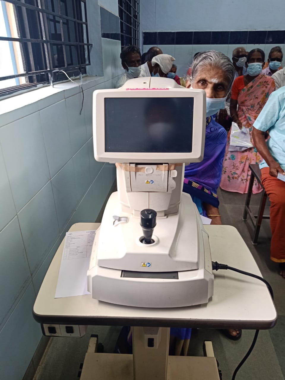
Digital image depicting a patient sitting on an auto-refractor for examination Contributed by Dr. Bharat Gurnani, MBBS, DNB, FCRS, FICO, MRCS Ed, MNAMS
References
Schiefer U, Kraus C, Baumbach P, Ungewiß J, Michels R. Refractive errors. Deutsches Arzteblatt international. 2016 Oct 14:113(41):693-702. doi: 10.3238/arztebl.2016.0693. Epub [PubMed PMID: 27839543]
Liu Z, Pazo EE, Ye H, Yu C, Xu L, He W. Comparing School-Aged Refraction Measurements Using the 2WIN-S Portable Refractor in Relation to Cycloplegic Retinoscopy: A Cross-Sectional Study. Journal of ophthalmology. 2021:2021():6612476. doi: 10.1155/2021/6612476. Epub 2021 May 21 [PubMed PMID: 34094595]
Level 2 (mid-level) evidenceXiong S, Lv M, Zou H, Zhu J, Lu L, Zhang B, Deng J, Yao C, He X, Xu X. Comparison of Refractive Measures of Three Autorefractors in Children and Adolescents. Optometry and vision science : official publication of the American Academy of Optometry. 2017 Sep:94(9):894-902. doi: 10.1097/OPX.0000000000001113. Epub [PubMed PMID: 28816868]
Kumar RS,Moe CA,Kumar D,Rackenchath MV,A V SD,Nagaraj S,Wittberg DM,Stamper RL,Keenan JD, Accuracy of autorefraction in an adult Indian population. PloS one. 2021; [PubMed PMID: 34010350]
Padhy D, Bharadwaj SR, Nayak S, Rath S, Das T. Does the Accuracy and Repeatability of Refractive Error Estimates Depend on the Measurement Principle of Autorefractors? Translational vision science & technology. 2021 Jan:10(1):2. doi: 10.1167/tvst.10.1.2. Epub 2021 Jan 5 [PubMed PMID: 33505769]
Pons J. Improving patient flow through an eye clinic. Community eye health. 2012:25(78):31-3 [PubMed PMID: 23139450]
Beverage JL, Schwiegerling J. A Shack-Hartmann-based autorefractor. Journal of refractive surgery (Thorofare, N.J. : 1995). 2006 Nov:22(9):932-7 [PubMed PMID: 17124892]
Nagra M, Akhtar A, Huntjens B, Campbell P. Open versus closed view autorefraction in young adults. Journal of optometry. 2021 Jan-Mar:14(1):86-91. doi: 10.1016/j.optom.2020.06.007. Epub 2020 Aug 11 [PubMed PMID: 32792330]
Hunt OA, Wolffsohn JS, Gilmartin B. Evaluation of the measurement of refractive error by the PowerRefractor: a remote, continuous and binocular measurement system of oculomotor function. The British journal of ophthalmology. 2003 Dec:87(12):1504-8 [PubMed PMID: 14660462]
Fitzke FW, Hayes BP, Hodos W, Holden AL. Electrophysiological optometry using Scheiner's principle in the pigeon eye. The Journal of physiology. 1985 Dec:369():17-31 [PubMed PMID: 4093879]
Level 3 (low-level) evidenceChen J, Lu F, Qu J, Li LP. [The development of a polarized vernier optometer for tonic accommodation measurement]. Zhongguo yi liao qi xie za zhi = Chinese journal of medical instrumentation. 2002 Jan:26(1):26-9 [PubMed PMID: 16104153]
Level 2 (mid-level) evidenceTeel DF, Copland RJ, Jacobs RJ, Wells T, Neal DR, Thibos LN. Design and validation of an infrared Badal optometer for laser speckle. Optometry and vision science : official publication of the American Academy of Optometry. 2008 Sep:85(9):834-42. doi: 10.1097/OPX.0b013e3181852742. Epub [PubMed PMID: 18772719]
Level 1 (high-level) evidenceOtero C, Aldaba M, Pujol J. Clinical evaluation of an automated subjective refraction method implemented in a computer-controlled motorized phoropter. Journal of optometry. 2019 Apr-Jun:12(2):74-83. doi: 10.1016/j.optom.2018.09.001. Epub 2018 Oct 30 [PubMed PMID: 30389250]
Hervella L, Villegas EA, Prieto PM, Artal P. Assessment of subjective refraction with a clinical adaptive optics visual simulator. Journal of cataract and refractive surgery. 2019 Jan:45(1):87-93. doi: 10.1016/j.jcrs.2018.08.022. Epub 2018 Oct 8 [PubMed PMID: 30309774]
Polse DA, Kerr KE. An automatic objective optometer. Description and clinical evaluation. Archives of ophthalmology (Chicago, Ill. : 1960). 1975 Mar:93(3):225-31 [PubMed PMID: 1094996]
Krishnacharya PS. Study on accommodation by autorefraction and dynamic refraction in children. Journal of optometry. 2014 Oct-Dec:7(4):193-202. doi: 10.1016/j.optom.2014.07.001. Epub 2014 Aug 15 [PubMed PMID: 25130066]
Level 2 (mid-level) evidenceTsai SR, Hamblin MR. Biological effects and medical applications of infrared radiation. Journal of photochemistry and photobiology. B, Biology. 2017 May:170():197-207. doi: 10.1016/j.jphotobiol.2017.04.014. Epub 2017 Apr 13 [PubMed PMID: 28441605]
Venkataraman AP, Sirak D, Brautaset R, Dominguez-Vicent A. Evaluation of the Performance of Algorithm-Based Methods for Subjective Refraction. Journal of clinical medicine. 2020 Sep 29:9(10):. doi: 10.3390/jcm9103144. Epub 2020 Sep 29 [PubMed PMID: 33003297]
Nguyen MT, Berntsen DA. Aberrometry Repeatability and Agreement with Autorefraction. Optometry and vision science : official publication of the American Academy of Optometry. 2017 Sep:94(9):886-893. doi: 10.1097/OPX.0000000000001107. Epub [PubMed PMID: 28727613]
Rauscher FG, Lange H, Yahiaoui-Doktor M, Tegetmeyer H, Sterker I, Hinz A, Wahl S, Wiedemann P, Ohlendorf A, Blendowske R. Agreement and Repeatability of Noncycloplegic and Cycloplegic Wavefront-based Autorefraction in Children. Optometry and vision science : official publication of the American Academy of Optometry. 2019 Nov:96(11):879-889. doi: 10.1097/OPX.0000000000001444. Epub [PubMed PMID: 31703049]
Galindo-Ferreiro A, De Miguel-Gutierrez J, González-Sagrado M, Galvez-Ruiz A, Khandekar R, Schellini S, Galindo-Alonso J. Validity of autorefractor based screening method for irregular astigmatism compared to the corneal topography- a cross sectional study. International journal of ophthalmology. 2017:10(9):1412-1418. doi: 10.18240/ijo.2017.09.14. Epub 2017 Sep 18 [PubMed PMID: 28944202]
Zhu Q, Xiao S, Hua Z, Yang D, Hu M, Zhu YT, Zhong H. Near Infrared (NIR) Light Therapy of Eye Diseases: A Review. International journal of medical sciences. 2021:18(1):109-119. doi: 10.7150/ijms.52980. Epub 2021 Jan 1 [PubMed PMID: 33390779]
Vinas M, Dorronsoro C, Cortes D, Pascual D, Marcos S. Longitudinal chromatic aberration of the human eye in the visible and near infrared from wavefront sensing, double-pass and psychophysics. Biomedical optics express. 2015 Mar 1:6(3):948-62. doi: 10.1364/BOE.6.000948. Epub 2015 Feb 24 [PubMed PMID: 25798317]
Carter J, Miller D. Automated objective refractometers. Annals of ophthalmology. 1984 Aug:16(8):712-5 [PubMed PMID: 6497216]
Asiedu K, Kyei S, Ampiah EE. Autorefraction, Retinoscopy, Javal's Rule, and Grosvenor's Modified Javal's Rule: The Best Predictor of Refractive Astigmatism. Journal of ophthalmology. 2016:2016():3584137 [PubMed PMID: 27803811]
Campbell CE, Suheimat M, Zacharovas S, Atchison DA. The use of autorefractors using the image-size principle in determining on-axis and off-axis refraction. Part 1: Analysis of optical principles of autorefractors. Ophthalmic & physiological optics : the journal of the British College of Ophthalmic Opticians (Optometrists). 2022 Mar:42(2):283-292. doi: 10.1111/opo.12933. Epub 2021 Dec 20 [PubMed PMID: 34927742]
Lebow KA, Campbell CE. A comparison of a traditional and wavefront autorefraction. Optometry and vision science : official publication of the American Academy of Optometry. 2014 Oct:91(10):1191-8. doi: 10.1097/OPX.0000000000000378. Epub [PubMed PMID: 25198541]
Kratz LD, Flom MC. The Humphrey Vision Analyzer tm: reliability and validity of refractive-error measures. American journal of optometry and physiological optics. 1977 Oct:54(10):653-9 [PubMed PMID: 605920]
Bannon RE, Waltuck MH. Clinical aspects of the SR-IV Programmed Subjective Refractor. American journal of optometry and physiological optics. 1982 Oct:59(10):815-20 [PubMed PMID: 7148974]
Prasad NM. Thoughts on establishing mid-level ophthalmic personnel for VISION 2020 in India. Community eye health. 2005 Oct:18(55):112 [PubMed PMID: 17491773]
Evans JR, Morjaria P, Powell C. Vision screening for correctable visual acuity deficits in school-age children and adolescents. The Cochrane database of systematic reviews. 2018 Feb 15:2(2):CD005023. doi: 10.1002/14651858.CD005023.pub3. Epub 2018 Feb 15 [PubMed PMID: 29446439]
Level 1 (high-level) evidenceConvergence Insufficiency Treatment Trial Study Group., Randomized clinical trial of treatments for symptomatic convergence insufficiency in children. Archives of ophthalmology (Chicago, Ill. : 1960). 2008 Oct; [PubMed PMID: 18852411]
Level 1 (high-level) evidenceHorwood AM, Riddell PM. Receding and disparity cues aid relaxation of accommodation. Optometry and vision science : official publication of the American Academy of Optometry. 2009 Nov:86(11):1276-86. doi: 10.1097/OPX.0b013e3181bb41de. Epub [PubMed PMID: 19770814]
Abusharha AA. Changes in blink rate and ocular symptoms during different reading tasks. Clinical optometry. 2017:9():133-138. doi: 10.2147/OPTO.S142718. Epub 2017 Nov 20 [PubMed PMID: 30214369]
Satou T, Takahashi Y, Niida T. Comparison of refractive value and pupil size under monocular and binocular conditions between the Spot Vision Screener and binocular open-field autorefractor. Strabismus. 2020 Dec:28(4):186-193. doi: 10.1080/09273972.2020.1832542. Epub 2020 Oct 16 [PubMed PMID: 33063575]
Rajavi Z, Sabbaghi H, Baghini AS, Yaseri M, Sheibani K, Norouzi G. Accuracy and Repeatability of Refractive Error Measurements by Photorefractometry. Journal of ophthalmic & vision research. 2015 Jul-Sep:10(3):221-8. doi: 10.4103/2008-322X.170360. Epub [PubMed PMID: 26730305]
Doustkouhi SM, Turnbull PRK, Dakin SC. The effect of refractive error on optokinetic nystagmus. Scientific reports. 2020 Nov 18:10(1):20062. doi: 10.1038/s41598-020-76865-x. Epub 2020 Nov 18 [PubMed PMID: 33208790]
Hussaindeen JR, Murali A. Accommodative Insufficiency: Prevalence, Impact and Treatment Options. Clinical optometry. 2020:12():135-149. doi: 10.2147/OPTO.S224216. Epub 2020 Sep 11 [PubMed PMID: 32982529]
Strang NC, Gray LS, Winn B, Pugh JR. Clinical evaluation of patient tolerance to autorefractor prescriptions. Clinical & experimental optometry. 1998 May-Jun:81(3):112-118 [PubMed PMID: 12482260]
Moradi M. Importance of Ophthalmic Nursing in Primary Healthcare Systems. Medical hypothesis, discovery & innovation ophthalmology journal. 2016 Spring:5(1):1-3 [PubMed PMID: 28289685]
Yip YC, Yip KH, Tsui WK. The Transformational Experience of Junior Nurses Resulting from Providing Care to COVID-19 Patients: From Facing Hurdles to Achieving Psychological Growth. International journal of environmental research and public health. 2021 Jul 10:18(14):. doi: 10.3390/ijerph18147383. Epub 2021 Jul 10 [PubMed PMID: 34299834]