Introduction
Wrist arthrography, first introduced by Kesseler and Silberman in 1961 to assess the carpal ligaments, is a valuable diagnostic modality used to evaluate the internal structures of the wrist.[1] This technique involves injecting contrast material into the wrist joint to enhance visualization during imaging examinations such as plain radiography, magnetic resonance imaging (MRI), and computed tomography (CT). Wrist arthrograms are particularly useful for assessing articular cartilage, ligaments, tendons, joint stability, and functional dynamics.[2] While advanced imaging techniques like MRI have gained prominence, wrist arthrography remains essential for diagnosing ligament tears, triangular fibrocartilage complex injuries, and other wrist pathologies (see Image. Wrist Arthrogram).
Anatomy and Physiology
Register For Free And Read The Full Article
 Search engine and full access to all medical articles
- 10 free questions in your specialty
- Free CME/CE Activities
- Free daily question in your email
- Save favorite articles to your dashboard
- Emails offering discounts
Learn more about a Subscription to StatPearls Point-of-Care
Anatomy and Physiology
Bony Structures of the Wrist
The wrist joint is formed by the articulation between the distal end of the radius and the articular disk with the proximal carpal row (excluding the pisiform). The carpal bones create a convex surface that matches the concave configuration of the distal radius and articular disk.[3] The distal ulna does not articulate directly with carpal bones due to the presence of the articular disk. The distal radioulnar joint articulates the distal ulna with the distal radius.[4]
Ligaments of the Wrist
The ligamentous attachments of the wrist are crucial for maintaining stability and facilitating coordinated movements.[5] There are 4 main ligaments situated at the wrist joint, each serving distinct functions:
- Palmar radiocarpal ligament: located on the volar aspect extending from the radius to both rows of carpal bones. This ligament enhances stability and ensures the synchronization of hand and forearm movements during supination.
- Dorsal radiocarpal ligament: runs from the radius to both carpal rows on the dorsal aspect of the wrist, contributing to wrist stability and coordinating hand and forearm motions during pronation.
- Ulnar collateral ligament: extending from the ulnar styloid process to the triquetrum and pisiform, effectively preventing excessive radial deviation of the hand.
- Radial collateral ligament: connecting the radial styloid process to the scaphoid and trapezium, inhibiting excessive ulnar deviation.[3]
Triangular Fibrocartilage Complex
The triangular fibrocartilage complex (TFCC) contributes significantly to the stability of the distal radioulnar joints. The TFCC is positioned on the ulnar aspect of the wrist between the ulna and the lunate and triquetrum of the proximal carpal row. The TFCC has an elongated triangular shape with its apex directed towards the radius.[6] The TFCC comprises various ligamentous and fibrocartilaginous components, including the triangular fibrocartilage disc proper and triangular ligament, volar and dorsal radioulnar ligaments, ulnocarpal ligaments (ulnotriquetral, ulnolunate, ulnocapitate), meniscal homologue, ulnar collateral ligament, and the extensor carpi ulnaris tendon sheath.[7] Functionally, the TFCC stabilizes the distal radius ulnar joint, cushions the ulnar carpus, accounts for 20% of axial wrist load, and is a crucial stabilizer for the ulnar carpus.[8][9]
Indications
The indications of performing a wrist arthrogram, whether conducted as a single investigation or in a combination of other imaging modalities, include but are not limited to:
- Diagnosis of wrist joint degenerative and inflammatory conditions such as osteoarthritis and rheumatoid arthritis
- Identification of traumatic ligamentous abnormalities in the scapholunate ligament, lunotriquetral ligament, and TFCC [10][11]
- Assessment of wrist joint articular cartilage for osteochondral defects [12]
- Investigation of persistent wrist pain [13]
- Suspected scaphoid fracture nonunion [14]
- Assessment for ulnar abutment [15]
Contraindications
A wrist arthrogram is generally a well-tolerated diagnostic procedure. However, there are certain contraindications that clinicians should consider before recommending or performing wrist arthrograms. These contraindications include:
Absolute Contraindications
- Previous allergic reactions to contrast material: A known allergy to the contrast material (usually iodine-based contrast agents) used in the arthrogram should preclude the procedure.
- Presence of an active infection: An active infection around the wrist joint can spread. For example, injecting contrast material through infected skin can lead to septic arthritis due to the deeper spreading of the infection.
Relative Contraindications
- Renal dysfunction: Patients with significant renal dysfunction may be at risk for contrast-induced nephropathy. The contrast dose needs to be adjusted accordingly.
- Coagulopathies: Individuals with bleeding disorders or those taking anticoagulant medications may be at an increased risk of bleeding complications during or after the arthrogram. In such cases, careful consideration and potential modification of medication regimens may be necessary.
- Uncontrolled diabetes: Uncontrolled diabetes may increase the risk of contrast-induced complications, and healthcare professionals may need to assess and manage the patient's diabetic status before proceeding with the arthrogram.
- Pregnancy: Pregnancy is generally considered a relative contraindication, and the risks and benefits of the procedure must be carefully weighed. Radiation exposure from imaging studies, such as x-rays, should be minimized during pregnancy.
Before recommending a wrist arthrogram, healthcare professionals must thoroughly assess the patient's medical history, current health status, and potential contraindications. In cases where contraindications are present, alternative diagnostic approaches or modifications to the procedure may be considered to ensure patient safety.
Equipment
The equipment required for performing a wrist arthrogram includes the components for injection of the local anesthesia and the contrast material into the wrist joint. This includes:
- Sterile drapes
- Local antiseptic solutions
- Blunt wide bore needle to withdraw the local anesthesia and contrast solutions
- Sharp fine needle to access the joint for injections
- 3-mL syringes for contrast solution injection
- 10-mL syringe for the local anesthetic solution preparation and injection
- Contrast agents, gadolinium or iodine-based
Due to its fast-acting anesthetic effect, 1% lidocaine is commonly used for local anesthesia. Lidocaine can be combined with a sodium bicarbonate solution as a buffering agent to minimize irritation caused by the acidic nature of the local anesthetic; this can be achieved by adding approximately 0.5 mL of sodium bicarbonate to 10 mL of 1% lidocaine. An emergency resuscitation cart should be accessible for use in case of an anaphylactic reaction to any of the injected materials.
Technique or Treatment
Informed consent must be obtained before performing wrist arthrography, and contraindications such as infection and contrast allergies should be ruled out before beginning the procedure. The patient is positioned supine on a fluoroscopy table with the wrist in a neutral position. The skin over the wrist is prepped with antiseptic, and an aseptic technique is employed to inject contrast. The injection is ideally performed from the side opposite to the wrist symptoms. For example, injection from the lateral side of ulnar wrist pain allows better differentiation between iatrogenic contrast leakage and leakage due to a capsular tear. Typically, contrast is injected into the joint under imaging guidance, often using an image intensifier.[15]
Wrist arthrography can be performed for 1 compartment (radiocarpal), 2 compartments (radiocarpal and midcarpal or radiocarpal and distal radioulnar), or more commonly for 3 compartments (radiocarpal, midcarpal, and distal radioulnar compartments).[16][17] To inject contrast into the midcarpal compartment, the space between the scaphoid and capitate or between the triquetrum and hamate is targeted. This allows contrast to flow into the scapholunate and lunotriquetral spaces. If the intrinsic ligaments between the scaphoid, lunate, and triquetrum are intact, the contrast will not flow into the radiocarpal joint. For contrast injection into the radiocarpal compartment from the ulnar side, the target is the space between the radial side of the pisiform and the proximal triquetrum. Injection from the radial side involves the space between the styloid process and the proximal pole of the scaphoid. The radial margin of the ulnar head is targeted to inject the distal radioulnar joint.[15]
Complications
Complications associated with wrist arthrogram may include:
- Allergic Reactions
- Some patients may develop allergic reactions to the contrast material used in the procedure. This is more common with iodine-based contrast agents.
- Adverse allergic reactions can range from mild skin irritation to severe anaphylaxis.
- Septic Arthritis
- There is a risk of introducing an intraarticular infection during the wrist arthrogram, particularly if aseptic techniques are not followed.
- Infections can present as local swelling, redness, pain, and systemic symptoms such as fever and sepsis.
- If the diagnosis of septic arthritis is confirmed, an urgent surgical wrist joint washout is mandatory to preserve the joint cartilage and prevent systemic sepsis.
- Bleeding: Bleeding at the injection site or within the joint may occur, especially in individuals with bleeding disorders or those taking anticoagulant medications. Clinicians may consider the benefits and risks of stopping anticoagulant treatment before wrist arthrogram.
- False-Positive Findings: Accidental gas injection during the performance of a wrist arthrogram can result in a false-positive diagnosis of loose intraarticular bodies, leading to misinterpretation of the diagnostic imaging results.
- Sterile Chemical Synovitis
- Postarthrography pain, often resulting from sterile chemical synovitis, is the most common complication of wrist arthrography.
- The introduction of contrast material causes an inflammatory reaction within the wrist joint. Symptoms typically begin about 4 hours postprocedure and peak at around 12 hours. Despite the absence of infection—hence the term "sterile"—the contrast agent's chemical irritant properties can inflame the synovial lining, causing pain, swelling, and discomfort.
- This complication can mimic septic arthritis, requiring close monitoring and management by healthcare professionals to ensure accurate diagnosis and treatment.[18]
- Nerve or Vascular Injury: In rare cases, the needle used for the injection may inadvertently damage nearby nerves or blood vessels, leading to neurological or vascular complications.
- Intraarticular Structures Damage: There is a small risk of causing damage to the joint structures during the injection, particularly if there are preexisting joint abnormalities or pathology.
- Pain or Discomfort: Some patients may experience temporary pain or discomfort at the injection site or within the joint, usually mild and short-lived.
- Radiation Exposure: If x-rays are used during the procedure, there is potential for radiation exposure. While the amount of radiation is generally low, it is a consideration, particularly for pregnant patients. Healthcare professionals should take the necessary precautions to minimize risks and follow safety protocols.
Clinical Significance
The wrist arthrogram is a valuable diagnostic tool for evaluating intraarticular pathology and structural abnormalities within the wrist joint, including:
TFCC Tears
The TFCC serves as a critical stabilizer for the lunate, triquetrum, and ulnar head while also functioning as a load-bearing structure on the ulnar side of the wrist. Injuries to the TFCC are commonly associated with activities that involve forced ulnar deviation or positive ulnar variance. Such injuries often occur when pressure is applied to the TFCC during wrist movements like wielding a racket, swinging a bat, or in cases of trauma such as falling on an outstretched hand or hyperextension injuries.[9][10][19]
Conventional MRI scans can struggle to differentiate between degenerative and traumatic TFCC tears due to their limited sensitivity and specificity.[20][21][22] However, combining MRI with an arthrogram (MR arthrography) significantly enhances diagnostic accuracy, with reported sensitivity and specificity rates of 97.1% and 96.4%, respectively, for identifying partial and complete TFCC tears.[23] In MR arthrography, TFCC perforations appear as areas of high signal intensity on fat-suppressed T1-weighted images, and contrast material may also be visualized in the distal radioulnar joint in cases of traumatic TFCC injury.[24]
Scapholunate Ligament Injury and Scapholunate Instability
Scapholunate ligament injury has the highest incidence of wrist ligament injuries. In advanced cases, injury of the scapholunate ligament can result in progressive scaphoid flexion, widening the articulation between the scaphoid and lunate bones in wrist anteroposterior radiographs. If a single compartment arthrogram is performed for the radiocarpal compartment and the contrast is identified in the midcarpal compartment, this is a sign of scapholunate ligament disruption. MR arthrography is more sensitive than MRI for detecting scapholunate ligament injuries' exact location and extension.[15]
Midcarpal Instability
These rare injuries involve disruption of the ligaments between the distal and proximal carpal row bones while the proximal row ligaments stay intact. MR arthrography has higher sensitivity in assessing each ligament and the exact extension of injury.[15]
Distal Radioulnar Joint Abnormalities
The wrist arthrogram allows further assessment of articular cartilage at the distal radioulnar joint and the joint capsule for any signs of tears or synovitis.[25]
Enhancing Healthcare Team Outcomes
Performing and interpreting a wrist arthrogram involves a collaborative effort among healthcare professionals, necessitating a spectrum of skills, strategic planning, ethical considerations, shared responsibilities, effective interprofessional communication, and meticulous care coordination to optimize patient-centered care, enhance outcomes, ensure patient safety, and improve team performance. Physicians must comprehensively understand wrist anatomy, pathology, and the diagnostic implications of wrist arthrogram. Advanced clinicians, including radiologists, need specialized skills in performing the procedure, ensuring accurate contrast injection, and interpreting imaging results. Nurses are crucial in preparing patients for the arthrogram, providing care before and after the procedure, and monitoring potential complications. Pharmacists contribute by ensuring appropriate contrast agents and medications and managing drug-related concerns.
Ethical considerations involve obtaining informed consent, ensuring patient privacy, and communicating clearly about the procedure and potential outcomes. Team members share responsibilities, with each professional contributing expertise to the overall patient care plan. Interprofessional communication is paramount to ensure that all team members are aligned in understanding the patient's condition, the purpose of the wrist arthrogram, and the subsequent care plan. This collaborative approach helps address concerns, share critical information, and foster a cohesive team dynamic. Care coordination involves scheduling appointments, communicating results among team members, and ensuring that the patient receives appropriate follow-up care, which is essential for a seamless patient experience. This coordinated effort enhances patient-centered care, contributes to positive outcomes, and reduces the risk of errors or oversights.
Media
(Click Image to Enlarge)
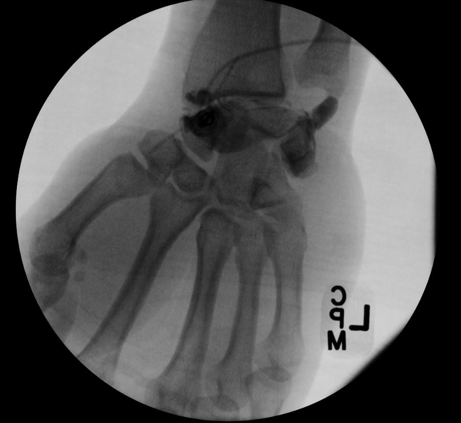
Wrist Arthrogram. The arthrogram shows an accumulation of contrast material on the ulnar side of the wrist joint, suggestive of a triangular fibrocartilage complex (TFCC) injury. Notably, there is no contrast leakage into the mid-carpal space or the distal radioulnar joint.
Contributed by D Tafti, MD
References
KESSLER I, SILBERMAN Z. An experimental study of the radiocarpal joint by arthrography. Surgery, gynecology & obstetrics. 1961 Jan:112():33-40 [PubMed PMID: 13752749]
Maloney E, Zbojniewicz AM, Nguyen J, Luo Y, Thapa MM. Anatomy and injuries of the pediatric wrist: beyond the basics. Pediatric radiology. 2018 Jun:48(6):764-782. doi: 10.1007/s00247-018-4111-2. Epub 2018 Mar 20 [PubMed PMID: 29557490]
Erwin J, Varacallo M. Anatomy, Shoulder and Upper Limb, Wrist Joint. StatPearls. 2024 Jan:(): [PubMed PMID: 30521200]
Lewis OJ, Hamshere RJ, Bucknill TM. The anatomy of the wrist joint. Journal of anatomy. 1970 May:106(Pt 3):539-52 [PubMed PMID: 4987391]
Taleisnik J. The ligaments of the wrist. The Journal of hand surgery. 1976 Sep:1(2):110-8 [PubMed PMID: 1018078]
Nakamura T, Takayama S, Horiuchi Y, Yabe Y. Origins and insertions of the triangular fibrocartilage complex: a histological study. Journal of hand surgery (Edinburgh, Scotland). 2001 Oct:26(5):446-54 [PubMed PMID: 11560427]
Martins T, Marappa-Ganeshan R. Anatomy, Shoulder and Upper Limb, Forearm Triangular Fibrocartilage Complex. StatPearls. 2024 Jan:(): [PubMed PMID: 32119451]
Palmer AK. Triangular fibrocartilage complex lesions: a classification. The Journal of hand surgery. 1989 Jul:14(4):594-606 [PubMed PMID: 2666492]
Casadei K, Kiel J. Triangular Fibrocartilage Complex. StatPearls. 2024 Jan:(): [PubMed PMID: 30725740]
Mirza A, Mirza JB, Zappia LC, Thomas TL. Ulnar-Sided Wrist Pain: A Diagnostic Evaluation Guide From 30-Plus Years of Experience. Cureus. 2024 Jan:16(1):e53332. doi: 10.7759/cureus.53332. Epub 2024 Jan 31 [PubMed PMID: 38435942]
Fransson SG. Wrist arthrography. Acta radiologica (Stockholm, Sweden : 1987). 1993 Mar:34(2):111-6 [PubMed PMID: 8452713]
Nischal N, Iyengar KP, Herlekar D, Botchu R. Imaging of Cartilage and Chondral Defects: An Overview. Life (Basel, Switzerland). 2023 Jan 28:13(2):. doi: 10.3390/life13020363. Epub 2023 Jan 28 [PubMed PMID: 36836719]
Level 3 (low-level) evidenceCooney WP. Evaluation of chronic wrist pain by arthrography, arthroscopy, and arthrotomy. The Journal of hand surgery. 1993 Sep:18(5):815-22 [PubMed PMID: 8228051]
Crema MD, Phan C, Miquel A, Quach C, Arrivé L, Menu Y. MDCT arthrography assessment of scaphoid nonunion advanced collapse: distribution of cartilage damage and relationship with scaphoid nonunion features. Skeletal radiology. 2018 Aug:47(8):1157-1165. doi: 10.1007/s00256-018-2907-7. Epub 2018 Mar 8 [PubMed PMID: 29520536]
Cerezal L, Abascal F, García-Valtuille R, Del Piñal F. Wrist MR arthrography: how, why, when. Radiologic clinics of North America. 2005 Jul:43(4):709-31, viii [PubMed PMID: 15893533]
Zinberg EM, Palmer AK, Coren AB, Levinsohn EM. The triple-injection wrist arthrogram. The Journal of hand surgery. 1988 Nov:13(6):803-9 [PubMed PMID: 3225404]
Kim S, Lee GY, Lee JS. Evaluation of the triangular fibrocartilage: comparison of two-compartment wrist CT arthrography using the distal radioulnar and radiocarpal joints and unicompartment wrist CT arthrography using the radiocarpal joint. The British journal of radiology. 2019 Oct:92(1102):20190298. doi: 10.1259/bjr.20190298. Epub 2019 Jul 19 [PubMed PMID: 31295006]
Oserowsky A, Layfield LJ, Crim J. Post-arthrogram synovitis: MRI and histopathologic findings. Skeletal radiology. 2022 Jan:51(1):219-223. doi: 10.1007/s00256-021-03877-7. Epub 2021 Jul 31 [PubMed PMID: 34331550]
Dunn JC, Polmear MM, Nesti LJ. Surgical Repair of Acute TFCC Injury. Hand (New York, N.Y.). 2020 Sep:15(5):674-678. doi: 10.1177/1558944719828007. Epub 2019 Feb 14 [PubMed PMID: 30762446]
Haims AH, Schweitzer ME, Morrison WB, Deely D, Lange R, Osterman AL, Bednar JM, Taras JS, Culp RW. Limitations of MR imaging in the diagnosis of peripheral tears of the triangular fibrocartilage of the wrist. AJR. American journal of roentgenology. 2002 Feb:178(2):419-22 [PubMed PMID: 11804907]
Ng WHA, Griffith JF, Ng ISH. How to Report: Wrist MRI. Seminars in musculoskeletal radiology. 2021 Oct:25(5):670-680. doi: 10.1055/s-0041-1736313. Epub 2021 Dec 3 [PubMed PMID: 34861712]
Daunt N, Couzens GB, Cutbush K, Green J, Ross M. Accuracy of magnetic resonance imaging of the wrist for clinically important lesions of the major interosseous ligaments and triangular fibrocartilage complex; correlation with radiocarpal arthroscopy. Skeletal radiology. 2021 Aug:50(8):1605-1616. doi: 10.1007/s00256-020-03701-8. Epub 2021 Jan 21 [PubMed PMID: 33474588]
Schmitt R, Christopoulos G, Meier R, Coblenz G, Fröhner S, Lanz U, Krimmer H. [Direct MR arthrography of the wrist in comparison with arthroscopy: a prospective study on 125 patients]. RoFo : Fortschritte auf dem Gebiete der Rontgenstrahlen und der Nuklearmedizin. 2003 Jul:175(7):911-9 [PubMed PMID: 12847645]
Jain DKA, Wahegaonkar AL. Ulnar-Side Wrist Pain Management Guidelines: All That Hurts is Not the TFCC! Indian journal of orthopaedics. 2021 Apr:55(2):310-317. doi: 10.1007/s43465-020-00319-9. Epub 2021 Jan 1 [PubMed PMID: 33927808]
Gilula LA, Hardy DC, Totty WG. Distal radioulnar joint arthrography. AJR. American journal of roentgenology. 1988 Apr:150(4):864-6 [PubMed PMID: 3258101]