Introduction
Tooth discoloration is a common complaint among patients seeking to improve the aesthetics of their anterior teeth. Tooth discoloration or stains are described as a change in color or translucency of 1 or more teeth. Depending on the etiology and location, tooth stains can be extrinsic or intrinsic. Understanding the specific etiology is crucial for determining the most appropriate treatment plan.
Extrinsic stains result from the accumulation of pigments on the enamel surface, caused by pigmented foods and beverages, such as coffee, tea, and wine, as well as lifestyle habits, such as tobacco smoking. Intrinsic stains occur within the enamel or underlying dentine, originating during tooth development, such as in cases of dental fluorosis and amelogenesis imperfecta, or appearing after tooth eruption due to factors such as pulpal trauma, necrosis, or the remnants of endodontic materials.[1]
Internal tooth whitening is a conservative procedure that improves the appearance of nonvital teeth with internal discoloration without requiring the loss of additional tooth structure. This procedure is also more economical compared to traditional treatments such as veneers or crowns. This procedure uses oxidizing agents to penetrate dental tissues and degrade organic pigments, thereby whitening the tooth structure.[2] The most common whitening agents are hydrogen peroxide and carbamide peroxide.[2]
The most commonly used methods for bleaching nonvital teeth include the walking bleach technique, inside-outside bleaching, and in-office bleaching.[3] The walking and inside-outside bleaching techniques are reported to be similarly effective for internal bleaching. The walking bleach approach is preferred as the access cavity is sealed, thereby avoiding the need for trays at home. In contrast, the success of the inside-outside technique depends on patient compliance with using bleaching trays and cleaning the access cavity with an interdental brush between office visits.[4] The results of internal bleaching are more predictable in discolored teeth caused by trauma or necrosis than material-related tooth discoloration.[2]
This activity reviews the factors involved in planning and performing internal tooth whitening and presents a case study of a 28-year-old man who underwent internal whitening of a nonvital upper central incisor using the walking bleach technique.
Indications
Register For Free And Read The Full Article
 Search engine and full access to all medical articles
- 10 free questions in your specialty
- Free CME/CE Activities
- Free daily question in your email
- Save favorite articles to your dashboard
- Emails offering discounts
Learn more about a Subscription to StatPearls Point-of-Care
Indications
Internal tooth whitening is indicated for managing nonvital teeth with internal discoloration following endodontic treatment. Common causes of such discoloration include pulp chamber hemorrhage, pulp necrosis, remnants of pulp tissue, and residual endodontic materials.[5] In these cases, byproducts with chromogenic capacity penetrate the dentinal tubules, leading to discoloration of the surrounding dentin.
Pulp bleeding may occur during pulp extirpation or after trauma. Blood products penetrate the dentinal tubules, causing a temporary pink discoloration of the crown.[6] Subsequently, hemolysis of red blood cells releases iron, which reacts with hydrogen sulfide produced by bacteria to form black ferric sulfide, resulting in grey discoloration.[6] In addition, the breakdown of proteins in a necrotic pulp may cause discoloration.[6] The longer the pulp remains necrotic, the greater the discoloration of the dentin.[5]
Incomplete preparation of the access cavity can lead to the retention of pulp remnants within the pulp chamber, especially in the pulp horns, causing discoloration at the coronal level.[7] Similarly, inadequate removal of root-filling materials from the pulp chamber over teeth can cause tooth discoloration. This inadequate removal typically occurs when the root-filling materials remain elevated above the level of the bone.[5] In such cases, the prognosis of internal tooth bleaching depends on the type and duration of contact with the material. For example, stains from metallic compounds are more challenging to remove.[5] The remaining materials must be entirely removed with burs before bleaching can proceed.[5]
Preparation
Patient Consent
Patients should be informed that complete recovery of natural tooth color cannot be guaranteed. Additionally, comprehensive information regarding potential complications should be provided.
Although rare, external cervical resorption is a noteworthy complication associated with internal tooth whitening. Studies on the etiology of external cervical resorption primarily attribute its development to orthodontic treatment (24.1%), surgical procedures (5.1%), and internal bleaching (3.9%).[8] External cervical resorption is usually asymptomatic and discovered incidentally on radiographic findings. Resorption appears as a radiolucent, dish-shaped near the cementoenamel junction (CEJ) on x-rays. Clinically, the affected tooth may respond well to sensitivity testing and bleed profusely upon probing. Additional clinical findings may include tenderness on percussion and swelling of the interdental papillae.[9]
The primary risk factors for cervical resorption include a history of trauma and bleaching. Although the exact cause of cervical resorption remains unclear, using a lower-concentration bleaching agent is recommended as a preventive measure.[3] In 2011, the Council of the European Union recommended using products that contain or release up to 6% hydrogen peroxide for teeth whitening. However, other countries still permit the use of higher concentrations of hydrogen peroxide.
Pretreatment Considerations
In the preliminary stages of treatment, it is crucial to evaluate the condition of the root canal treatment, as well as the presence of caries and the status of adjacent teeth.[5] The final shade of the tooth after bleaching cannot be predicted accurately. Defective restorations or caries in adjacent teeth should be addressed after completing the bleaching procedure rather than before. Pre- and post-treatment photographs are recommended for documentation and comparison purposes.[5]
Technique or Treatment
The active ingredient in tooth-whitening products is hydrogen peroxide, which can be applied directly or released from compounds such as carbamide peroxide or sodium perborate.[10] Regarding shade guide units, carbamide peroxide and hydrogen peroxide typically yield better outcomes in shade guide units compared to sodium perborate.[11] Hydrogen peroxide releases active oxygen molecules that break down chromophore molecules into smaller, more easily diffusible forms. These altered pigments reflect light differently, resulting in whitening effects. The action of hydrogen peroxide can be enhanced by heat and light.[10]
Walking Bleach Technique
A case study describes the application of the walking bleach technique. The tooth was isolated using a rubber dam, and existing composite materials were removed apically around the gingival margin. Sodium hypochlorite was used for disinfection, followed by placing a 2-mm layer of glass ionomer as a protective barrier. Intraorifice barriers are commonly used in the walking bleach technique to ensure proper coronal sealing, which reduces fracture resistance and prevents cervical resorption.[12][13][14] Common intraorifice barrier materials include glass ionomer cement, resin composite, and mineral trioxide aggregate (MTA). A damp cotton pellet soaked with 40% hydrogen peroxide was placed intracoronally. IRM was used to temporarily seal the access cavity, after which the rubber dam was removed. The patient was scheduled for follow-up every 2 weeks to assess and repeat the internal bleaching procedure until no further whitening was observed.
Clinical photographs were taken after each application using a Vita Classical Shade Guide. Postoperative radiographs were also taken to monitor for adverse effects, such as cervical resorption. Permanent adhesive restoration was completed during the final visit of the bleaching procedure.
Inside-Outside Bleaching Technique
The inside-outside bleaching method involves applying a bleaching agent to both the inner and outer surfaces of the tooth. Unlike the walking bleach technique, the access cavity remains open during treatment. The patient wears a vacuum-formed splint over the area being bleached. An impermeable base is used to seal the root canal filling. A small amount (pea-sized) of 10% carbamide peroxide is applied to the access cavity and the corresponding area of the splint using a syringe.[3] The patient is instructed to wear the splint overnight to protect the exposed tooth and follow up with the dentist every 3 days to monitor color changes. Once the target color or desired shade is achieved, the access cavity is cleaned and sealed with temporary materials such as glass ionomer cement. A permanent restoration is placed a week later.
The benefits of this technique include a low risk of cervical root resorption due to the low concentration of bleaching material and the absence of temporary restoration. However, it necessitates patients using bleaching trays, which can pose a risk of bacterial invasion if the patient neglects to return for access closure. Furthermore, an additional cost is associated with tray fabrication.
In-Office Bleaching Technique
In-office bleaching is a widely used method for whitening vital teeth and can also be applied to nonvital teeth. The initial steps of the procedure are identical to those of the walking bleach technique, including isolation, accessing the tooth, and sealing the root canal. After applying 35% hydrogen peroxide, the material is rinsed off 20 minutes later, and the process is repeated. This technique involves a more concentrated application compared to other methods, necessitating definitive access closure during a subsequent appointment.[3]
Clinical Significance
The walking bleach technique using hydrogen peroxide offers a promising and aesthetically pleasing solution for treating nonvital tooth discoloration. In addition, its conservative approach and economic advantages make it appealing to patients seeking satisfying results without removing additional tooth structure and at a fraction of the cost of a fixed restoration. This technique is effective for managing discoloration in teeth that have undergone endodontic treatment.
Case Report
A 28-year-old man with a medical history of controlled hypertension presented with the chief concern, "A previous dentist told me that I need a crown to fix my dark tooth." The patient underwent endodontic treatment on the upper right central incisor in July 2021 (see Image. Periapical x-ray), with discoloration reported 1 month after root canal therapy. The patient had previously undergone endodontic treatment on the upper right lateral incisor and received a porcelain fused metal (PFM) crown to improve discoloration after root canal therapy. Dissatisfied with the visible discoloration of his central incisor, the patient expressed motivation to pursue a crown, influenced by his positive experience with the treatment of the lateral incisor.
Crowns necessitate the loss of tooth structure, reducing the life cycle of teeth for future treatments, and are costly compared to noninvasive treatments.[10] Internal bleaching using the walking bleach technique was suggested as an alternative to a fixed prosthetic restoration, offering a more conservative and economical approach.
The discoloration on the upper right central incisor corresponded to a C4 VITA shade at the initial visit (see Image. Initial Discoloration). Hydrogen peroxide 40% was placed intracoronally in 2-week increments. Significant esthetic outcomes were observed after the second application. Tooth color was monitored regularly using a VITA shade guide, with the initial shade at C4 VITA progressing to a final shade of C1 VITA.
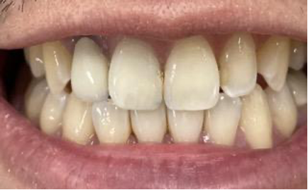
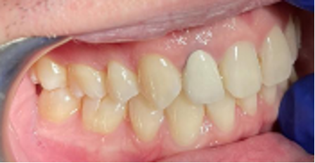
The initial discoloration corresponded to a C3 VITA shade after the first internal bleaching (see Image. Initial Bleaching), progressed to a C1 VITA shade after the second internal bleaching (see Image. Second Bleaching), and remained at a C1 VITA shade after the third internal bleaching (see Image. Third Bleaching). No discomfort or cervical resorption was reported during the initial visit, and the same C1 VITA shade was maintained at a 6-month follow-up visit (see Images. Follow-Up at 6 Months, VITA Shade Comparison, and Occlusial View at 6 Months). The patient expressed satisfaction with the treatment outcome.
Enhancing Healthcare Team Outcomes
Internal bleaching is a practical choice for managing intrinsic discoloration in nonvital root-filled teeth. This procedure offers a cost-effective, noninvasive alternative that avoids further loss of tooth structure, making it an economical and attractive option to restorative alternatives such as crowns and veneers.
Managing discolored endodontically treated teeth necessitates a multidisciplinary approach involving diverse healthcare professionals to ensure patient-centered care, optimal outcomes, safety, and efficient team performance. Interprofessional collaboration among dental professionals is essential for developing effective treatment plans, considering the integrity of the clinical crown and the type of discoloration. Ethical considerations in internal bleaching encompass informed consent, patient autonomy, and confidentiality. Dentists should offer comprehensive information about the procedure, potential risks, and alternative treatments, enabling patients to make informed decisions about their dental care.
Media
(Click Image to Enlarge)
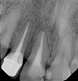
Periapical x-ray. The periapical radiographic image, taken at the initial visit, shows that the patient had previous endodontic treatment in the upper right central incisor.
Contributed by Y Jin, DDS
(Click Image to Enlarge)
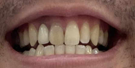
Initial Discoloration. A clinical photo of the patient from the initial visit shows discoloration on the upper right central incisor, corresponding to a C4 VITA shade.
Contributed by Y Jin, DDS
(Click Image to Enlarge)
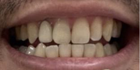
Initial Bleaching. After the first internal bleaching, the discoloration on the upper right central incisor corresponded to a C3 VITA shade.
Contributed by Y Jin, DDS
(Click Image to Enlarge)
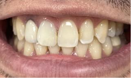
Second Bleaching. After the second internal bleaching, the discoloration on the upper right central incisor corresponded to a C1 VITA shade.
Contributed by Y Jin, DDS
(Click Image to Enlarge)
Third Bleaching. After the third internal bleaching, the discoloration on the upper right central incisor corresponded to a C1 VITA shade.
Contributed by Y Jin, DDS
(Click Image to Enlarge)
Follow-Up at 6 Months. At the 6-month follow-up visit, the discoloration on the upper right central incisor still corresponded to a C1 VITA shade.
Contributed by Y Jin, DDS
(Click Image to Enlarge)
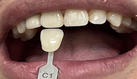
VITA Shade Comparison. The image is a comparison of the upper right central incisor at the 6-month follow-up visit, matching the C1 VITA shade.
Contributed by Y Jin, DDS
(Click Image to Enlarge)
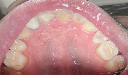
Occlusal View at 6 Months. An occlusal view showing the upper right central incisor at the 6-month follow-up visit, corresponding to the C1 VITA shade.
Contributed by Y Jin, DDS
References
Pedrollo Lise D, Siedschlag G, Bernardon JK, Baratieri LN. Randomized clinical trial of 2 nonvital tooth bleaching techniques: A 1-year follow-up. The Journal of prosthetic dentistry. 2018 Jan:119(1):53-59. doi: 10.1016/j.prosdent.2017.03.004. Epub 2017 May 5 [PubMed PMID: 28478984]
Level 1 (high-level) evidenceKnezevic N, Obradovic M, Dolic O, Veselinovic V, Kojic Z, Josipovic R, Arapovic-Savic M. Clinical Testing of Walking Bleach, In-Office, and Combined Bleaching of Endodontically Treated Teeth. Medicina (Kaunas, Lithuania). 2022 Dec 21:59(1):. doi: 10.3390/medicina59010018. Epub 2022 Dec 21 [PubMed PMID: 36676642]
Zimmerli B, Jeger F, Lussi A. Bleaching of nonvital teeth. A clinically relevant literature review. Schweizer Monatsschrift fur Zahnmedizin = Revue mensuelle suisse d'odonto-stomatologie = Rivista mensile svizzera di odontologia e stomatologia. 2010:120(4):306-20 [PubMed PMID: 20514558]
Leith R, Moore A, O'Connell AC. An effective bleaching technique for non-vital, discoloured teeth in children and adolescents. Journal of the Irish Dental Association. 2009 Aug-Sep:55(4):184-9 [PubMed PMID: 19753907]
Level 3 (low-level) evidencePlotino G, Buono L, Grande NM, Pameijer CH, Somma F. Nonvital tooth bleaching: a review of the literature and clinical procedures. Journal of endodontics. 2008 Apr:34(4):394-407. doi: 10.1016/j.joen.2007.12.020. Epub 2008 Feb 15 [PubMed PMID: 18358884]
Attin T, Paqué F, Ajam F, Lennon AM. Review of the current status of tooth whitening with the walking bleach technique. International endodontic journal. 2003 May:36(5):313-29 [PubMed PMID: 12752645]
BROWN G. FACTORS INFLUENCING SUCCESSFUL BLEACHING OF THE DISCOLORED ROOT-FILLED TOOTH. Oral surgery, oral medicine, and oral pathology. 1965 Aug:20():238-44 [PubMed PMID: 14319600]
Heithersay GS. Invasive cervical resorption: an analysis of potential predisposing factors. Quintessence international (Berlin, Germany : 1985). 1999 Feb:30(2):83-95 [PubMed PMID: 10356560]
Newton R, Hayes J. The association of external cervical resorption with modern internal bleaching protocols: what is the current evidence? British dental journal. 2020 Mar:228(5):333-337. doi: 10.1038/s41415-020-1317-0. Epub [PubMed PMID: 32170243]
Kahler B. Present status and future directions - Managing discoloured teeth. International endodontic journal. 2022 Oct:55 Suppl 4(Suppl 4):922-950. doi: 10.1111/iej.13711. Epub 2022 Mar 8 [PubMed PMID: 35188275]
Level 3 (low-level) evidenceFrank AC, Kanzow P, Rödig T, Wiegand A. Comparison of the Bleaching Efficacy of Different Agents Used for Internal Bleaching: A Systematic Review and Meta-Analysis. Journal of endodontics. 2022 Feb:48(2):171-178. doi: 10.1016/j.joen.2021.10.011. Epub 2021 Nov 9 [PubMed PMID: 34762968]
Level 1 (high-level) evidenceOskoee SS, Bahari M, Daneshpooy M, Ajami AA, Rahbar M. Effect of Different Intraorifice Barriers and Bleaching Agents on the Fracture Resistance of Endodontically Treated Anterior Teeth. Journal of endodontics. 2018 Nov:44(11):1731-1735. doi: 10.1016/j.joen.2018.07.025. Epub 2018 Sep 25 [PubMed PMID: 30266467]
Abduljalil M, Sakalli B, Basmaci F. Impact of different intraorifice barriers on fracture resistance of non-vital bleached teeth. Nigerian journal of clinical practice. 2023 Jan:26(1):95-101. doi: 10.4103/njcp.njcp_511_22. Epub [PubMed PMID: 36751830]
Sakalli B, Basmaci F, Dalmizrak O. Evaluation of the penetration of intracoronal bleaching agents into the cervical region using different intraorifice barriers. BMC oral health. 2022 Jun 30:22(1):266. doi: 10.1186/s12903-022-02300-4. Epub 2022 Jun 30 [PubMed PMID: 35773675]