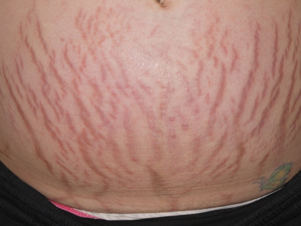
Stretch Marks

Earn CME/CE in your profession:


Continuing Education Activity

Striae, or stretch marks, are indented streaks that often affect the abdomen, buttocks, thighs, back, breasts, axillae and groin. They are divided into striae atrophicans (thinned skin), striae gravidarum (following pregnancy), striae distensae (stretched skin), striae rubrae (red), striae albae (white), striae nigra (black), and striae caerulea (dark blue). These common complaints are difficult to treat. This activity outlines the role of the interprofessional team in the evaluation and management of stretch marks.

Objectives:

  • Review the causes of stretch marks.

  • Describe the pathophysiology of stretch marks.

  • Summarize the treatment options for stretech marks

  • Explain the value of improving care coordination among the members of the interprofessional team to educate patients with stretch marks.

Introduction

Striae, or stretch marks, are a common complaint and can be distressing. They may affect the abdomen, buttocks, thighs, breasts, back, axillae and groin. They are classified according to appearance or epidemiology, as:

  • striae atrophicans (thinned skin)
  • striae gravidarum (following pregnancy)
  • striae distensae (stretched skin)
  • striae rubrae (red), striae albae (white)
  • striae nigra (black)
  • striae caerulea (dark blue). 

Striae are difficult to treat effectively. [1][2]

Etiology

Striae are a form of dermal scarring associated with stretching of the dermis. They often result from a rapid change in weight (gain and loss) or are associated with endogenous or exogenous corticosteroids. Proposed mechanisms relate to hormones, physical stretch, and structural alterations of dermal collagen and elastic tissue. Adrenocorticotropic hormones promote fibroblast activity and increase protein catabolism. Pregnancy-related hormones may also contribute. Serum relaxin has been described to be lower in women with striae distensae.[3] Deficiency of fibrillin has also been proposed. [4] Genetic factors are unexplored, except that decreased expression of collagen and fibronectin genes is reported to be associated with striae.

Epidemiology

Striae distensae occur in pregnancy (43% to 88%), puberty (6% to 86%) and obesity (43%). Striae atrophicans follow medical conditions, particularly Cushing syndrome/disease, and treatments, usually exogenous topical or systemic corticosteroids [5], or surgery. Other associated diseases are Marfan syndrome[6], anorexia nervosa[7], various febrile illnesses, and chronic liver disease. Medications associated with striae also include chemotherapy, prolonged antibiotic therapy, contraceptives[8], and neuroleptics.

Striae are more common in females than in males and may be more common in certain races. They can appear more prominent in dark-skinned individuals. A positive family history is a risk factor for striae. During pregnancy, striae are more common in younger women than in older women. Several studies have noted greater prevalence with large abdominal circumference and large weight gain (due to fetal size or polyhydramnios). [9] One study reported that striae were more prevalent in smokers than non-smokers.

Pathophysiology

Pathophysiology is thought to involve elastases released from mast cells and macrophage activity. [10] Elastolysis of the mid-dermis is followed by a reorganization of collagen and fibrillin.

Histopathology of striae rubrae reveals excessive fine elastic fibers in the papillary dermis with thicker tortuous fibers in the periphery, with perivascular lymphocytes, dilated dermal vessels and edema. There are reduction and reorganization of elastin and fibrillin fibers, and structural changes in collagen fibers, which are thicker and densely packed in parallel rows. Histopathology of striae albae shows epidermal atrophy, loss of rete ridges, less vascularity, and densely packed, thin and scar-like horizontal collagen bundles.[4] They appear similar to mature atrophic scars.

Electron microscopy studies have also reported mast cell degranulation, macrophage activation, and elastolysis of mid dermis.[11]

History and Physical

History may explain the appearance of striae by association with stretched skin, for example, in pregnancy, pubertal growth spurt, muscular exercise, or weight gain. Inquire about topical application of potent corticosteroid or prolonged systemic steroids during the recent or remote past. If striae are widespread and none of these explanations are relevant, take a full medical history and conduct a thorough clinical examination.[12]

The initial striae rubrae are slightly raised pink or violaceous linear marks (striae rubrae), which fade over months to years to hypopigmented, atrophic, wrinkled scars (striae albae). The marks are perpendicular to the direction of skin tension. They fade with time. In pregnancy, they occur on the abdomen, breasts, and thighs. In adolescents, they are common on thighs, buttocks, breasts (females) and back (males).

Striae rubrae are sometimes pruritic. Otherwise, striae are asymptomatic. Treatment is sought because of their unsightly appearance.

Evaluation

Authors have used various methods of assessing the type and severity of striae when discussing the efficacy of treatments. These are not standardized or validated. Dermoscopy shows increased melanization in striae rubrae and reduced melanization in striae albae. [13] A biopsy is not necessary or useful.

Treatment / Management

The aim of treatment is to reduce redness, swelling and irritation in striae rubrae, and to increase collagen and elastic fiber production, improve hydration, and reduce inflammation in striae albae.[14]

Topical management is commonly recommended to prevent and treat striae, with very little if any evidence of efficacy, according to several published comprehensive reviews. Clinical trials have been of low quality involving small numbers of subjects.

  • Many emollients and over-the-counter cosmeceuticals are marketed and used by pregnant women with the intention of preventing striae distensae or reducing their severity, resulting in considerable effort and expense, despite the considerable uncertainty whether any have benefit. [15]
  • Silicone gels are recommended for atrophic scars and may be used in striae distensae. Published results are difficult to interpret.[16]
  • Tretinoin cream has been reported to be possibly useful in striae rubrae when compared to placebo over a six-month period. It may result in irritation, redness, and peeling. It should not be applied during pregnancy due to its pregnancy category.[17][18]
  • Chemical peels using various acids have been used to treat striae. Efficacy is unknown. 

Physical treatments are also advocated but also have little evidence to support their use.

  • Massage is a component of topical therapy, and it is also used in scar management. [19]
  • Exposure to broadband ultraviolet radiation can cause repigmentation in striae alba but is reported to have temporary effect with loss of pigmentation within a few months.
  • Light and laser therapies have shown improvements in the appearance of striae, although it is uncertain which is best, and at what stage (rubrae, albae) they should be used. [19] Several laser types (e.g., pulse dye laser) target vascular chromophores in striae rubra and are reported to reduce redness and swelling.[20] Fractional lasers (e.g., Erbium-YAG) stimulate fibroblasts to produce collagen and elastin and can cause repigmentation in striae alba.[21]
  • Light and lasers cause short-term erythema and oedema. They should be avoided in darker skin types due to greater risk of adverse effects, particularly transient postinflammatory hyperpigmentation and persistent hypopigmentation. Non-ablative devices are safer than ablative lasers.
  • Radiofrequency energy devices emit high-frequency alternating electric current producing dermal heat. They are used to tighten the dermis, reduce wrinkles, and improve cellulite. Treatment promotes neocollagenesis, neoelastogenesis, and increased ground substances (proteoglycans) and should theoretically improve the appearance of striae. At least one published study has reported treatment was well tolerated, and the patients were satisfied with the results. A more recent advance has been the delivery of radiofrequency energy to a depth of 3.5 mm using a multiple needle delivery mode. Anecdotal reports are encouraging but proper studies are awaited. [22]
  • Other devices used have included microdermabrasion[23], galvanopuncture [24], needling [25], pulsed magnetic fields [22], and ultrasound devices.[26]

The effect of treatments is difficult to assess. For example, laser protocols use differing devices, fluence, pulse duration, spot size and treatment frequency and number.  The utility of combination strategies is unknown.[14]

Platelet-rich plasma injections are also under investigation.[27]

When using any of these modalities, it is important to have standardized pre-treatment and post-treatment photographs. Furthermore, it is important to take these photographs six and twelve months out after the course of treatments. Many published reports do not have standardized photographs, nor do they have long-term follow up. 

Differential Diagnosis

  • Anetoderma
  • Cutis Laxa
  • Linear Focal Elastosis
  • Mid-Dermal Elastosis
  • Pseudoxanthoma Elasticum
  • Striae from Topical Steroidal Abuse

Enhancing Healthcare Team Outcomes

Over time, many striae improve. The best time to perform a treatment (with proper photographs as discussed) is once the striae have stabilized. Many creams for striae are promoted on the internet with little evidence of the efficacy. The public should be made aware that the efficacy is very limited Newer lasers and radiofrequency treatments hold promise as they do try to modify the vascularity and the collagen and perhaps the elastin. Again, there is no clear leader among the many lasers that are promoted. The needle radiofrequency seems to show promise as, in theory, the deeper deliver of the energy may allow "shrinkage" of the striae in all directions, thereby improving the overall appearance. (Level V).


(Click Image to Enlarge)
<p>Striae Gravidarum Rubrae</p>

Striae Gravidarum Rubrae


DermNet New Zealand

Details

Updated:

8/7/2023 6:17:03 PM

Looking for an easier read?

Click here for a simplified version

References


[1]

Farahnik B, Park K, Kroumpouzos G, Murase J. Striae gravidarum: Risk factors, prevention, and management. International journal of women's dermatology. 2017 Jun:3(2):77-85. doi: 10.1016/j.ijwd.2016.11.001. Epub 2016 Dec 6     [PubMed PMID: 28560300]


[2]

Al-Himdani S, Ud-Din S, Gilmore S, Bayat A. Striae distensae: a comprehensive review and evidence-based evaluation of prophylaxis and treatment. The British journal of dermatology. 2014 Mar:170(3):527-47. doi: 10.1111/bjd.12681. Epub     [PubMed PMID: 24125059]


[3]

Lurie S, Matas Z, Fux A, Golan A, Sadan O. Association of serum relaxin with striae gravidarum in pregnant women. Archives of gynecology and obstetrics. 2011 Feb:283(2):219-22. doi: 10.1007/s00404-009-1332-5. Epub 2010 Jan 3     [PubMed PMID: 20047054]


[4]

Wang F, Calderone K, Smith NR, Do TT, Helfrich YR, Johnson TR, Kang S, Voorhees JJ, Fisher GJ. Marked disruption and aberrant regulation of elastic fibres in early striae gravidarum. The British journal of dermatology. 2015 Dec:173(6):1420-30. doi: 10.1111/bjd.14027. Epub 2015 Nov 8     [PubMed PMID: 26179468]


[5]

Neve S, Kirtschig G. Elastotic striae associated with striae distensae after application of very potent topical corticosteroids. Clinical and experimental dermatology. 2006 May:31(3):461-2     [PubMed PMID: 16681607]


[6]

Ledoux M, Beauchet A, Fermanian C, Boileau C, Jondeau G, Saiag P. A case-control study of cutaneous signs in adult patients with Marfan disease: diagnostic value of striae. Journal of the American Academy of Dermatology. 2011 Feb:64(2):290-5. doi: 10.1016/j.jaad.2010.01.032. Epub 2010 Nov 26     [PubMed PMID: 21112669]

Level 2 (mid-level) evidence

[7]

Strumia R. Skin signs in anorexia nervosa. Dermato-endocrinology. 2009 Sep:1(5):268-70     [PubMed PMID: 20808514]


[8]

Gupta M. Medroxyprogesterone acetate [Depo Provera] injections. Development of striae. The British journal of family planning. 2000 Apr:26(2):104-5     [PubMed PMID: 10773604]


[9]

Picard D, Sellier S, Houivet E, Marpeau L, Fournet P, Thobois B, Bénichou J, Joly P. Incidence and risk factors for striae gravidarum. Journal of the American Academy of Dermatology. 2015 Oct:73(4):699-700. doi: 10.1016/j.jaad.2015.06.037. Epub     [PubMed PMID: 26369842]


[10]

Sheu HM, Yu HS, Chang CH. Mast cell degranulation and elastolysis in the early stage of striae distensae. Journal of cutaneous pathology. 1991 Dec:18(6):410-6     [PubMed PMID: 1774350]


[11]

Zheng P, Lavker RM, Kligman AM. Anatomy of striae. The British journal of dermatology. 1985 Feb:112(2):185-93     [PubMed PMID: 3970840]


[12]

Kasielska-Trojan A, Sobczak M, Antoszewski B. Risk factors of striae gravidarum. International journal of cosmetic science. 2015 Apr:37(2):236-40. doi: 10.1111/ics.12188. Epub 2015 Jan 12     [PubMed PMID: 25440082]


[13]

Hermanns JF, Piérard GE. High-resolution epiluminescence colorimetry of striae distensae. Journal of the European Academy of Dermatology and Venereology : JEADV. 2006 Mar:20(3):282-7     [PubMed PMID: 16503888]


[14]

Forbat E, Al-Niaimi F. Treatment of striae distensae: An evidence-based approach. Journal of cosmetic and laser therapy : official publication of the European Society for Laser Dermatology. 2019:21(1):49-57. doi: 10.1080/14764172.2017.1418515. Epub 2018 Feb 16     [PubMed PMID: 29451986]


[15]

Rawlings AV, Bielfeldt S, Lombard KJ. A review of the effects of moisturizers on the appearance of scars and striae. International journal of cosmetic science. 2012 Dec:34(6):519-24. doi: 10.1111/j.1468-2494.2012.00751.x. Epub 2012 Sep 21     [PubMed PMID: 22994859]


[16]

Ud-Din S, McAnelly SL, Bowring A, Whiteside S, Morris J, Chaudhry I, Bayat A. A double-blind controlled clinical trial assessing the effect of topical gels on striae distensae (stretch marks): a non-invasive imaging, morphological and immunohistochemical study. Archives of dermatological research. 2013 Sep:305(7):603-17. doi: 10.1007/s00403-013-1336-7. Epub 2013 Apr 12     [PubMed PMID: 23579949]

Level 1 (high-level) evidence

[17]

Ud-Din S, McGeorge D, Bayat A. Topical management of striae distensae (stretch marks): prevention and therapy of striae rubrae and albae. Journal of the European Academy of Dermatology and Venereology : JEADV. 2016 Feb:30(2):211-22. doi: 10.1111/jdv.13223. Epub 2015 Oct 20     [PubMed PMID: 26486318]


[18]

Korgavkar K, Wang F. Stretch marks during pregnancy: a review of topical prevention. The British journal of dermatology. 2015 Mar:172(3):606-15. doi: 10.1111/bjd.13426. Epub 2015 Feb 8     [PubMed PMID: 25255817]


[19]

Timur Taşhan S, Kafkasli A. The effect of bitter almond oil and massaging on striae gravidarum in primiparaous women. Journal of clinical nursing. 2012 Jun:21(11-12):1570-6. doi: 10.1111/j.1365-2702.2012.04087.x. Epub     [PubMed PMID: 22594386]


[20]

Elsaie ML, Hussein MS, Tawfik AA, Emam HM, Badawi MA, Fawzy MM, Shokeir HA. Comparison of the effectiveness of two fluences using long-pulsed Nd:YAG laser in the treatment of striae distensae. Histological and morphometric evaluation. Lasers in medical science. 2016 Dec:31(9):1845-1853     [PubMed PMID: 27595152]


[21]

Zaleski-Larsen LA, Jones IT, Guiha I, Wu DC, Goldman MP. A Comparison Study of the Nonablative Fractional 1565-nm Er: glass and the Picosecond Fractional 1064/532-nm Nd: YAG Lasers in the Treatment of Striae Alba: A Split Body Double-Blinded Trial. Dermatologic surgery : official publication for American Society for Dermatologic Surgery [et al.]. 2018 Oct:44(10):1311-1316. doi: 10.1097/DSS.0000000000001555. Epub     [PubMed PMID: 29746426]

Level 1 (high-level) evidence

[22]

Dover JS, Rothaus K, Gold MH. Evaluation of safety and patient subjective efficacy of using radiofrequency and pulsed magnetic fields for the treatment of striae (stretch marks). The Journal of clinical and aesthetic dermatology. 2014 Sep:7(9):30-3     [PubMed PMID: 25276274]


[23]

Hersant B, Niddam J, Meningaud JP. Comparison between the efficacy and safety of platelet-rich plasma vs microdermabrasion in the treatment of striae distensae: clinical and histopathological study. Journal of cosmetic dermatology. 2016 Dec:15(4):565. doi: 10.1111/jocd.12246. Epub 2016 Jun 20     [PubMed PMID: 27320781]


[24]

Bitencourt S, Lunardelli A, Amaral RH, Dias HB, Boschi ES, de Oliveira JR. Safety and patient subjective efficacy of using galvanopuncture for the treatment of striae distensae. Journal of cosmetic dermatology. 2016 Dec:15(4):393-398. doi: 10.1111/jocd.12222. Epub 2016 Apr 19     [PubMed PMID: 27090205]


[25]

Aust M, Walezko N. [Acne scars and striae distensae: Effective treatment with medical skin needling]. Der Hautarzt; Zeitschrift fur Dermatologie, Venerologie, und verwandte Gebiete. 2015 Oct:66(10):748-52. doi: 10.1007/s00105-015-3662-5. Epub     [PubMed PMID: 26251169]


[26]

Kravvas G, Veitch D, Al-Niaimi F. The use of energy devices in the treatment of striae: a systematic literature review. The Journal of dermatological treatment. 2019 May:30(3):294-302. doi: 10.1080/09546634.2018.1506078. Epub 2018 Sep 7     [PubMed PMID: 30049244]

Level 1 (high-level) evidence

[27]

Gamil HD, Ibrahim SA, Ebrahim HM, Albalat W. Platelet-Rich Plasma Versus Tretinoin in Treatment of Striae Distensae: A Comparative Study. Dermatologic surgery : official publication for American Society for Dermatologic Surgery [et al.]. 2018 May:44(5):697-704. doi: 10.1097/DSS.0000000000001408. Epub     [PubMed PMID: 29701622]

Level 2 (mid-level) evidence