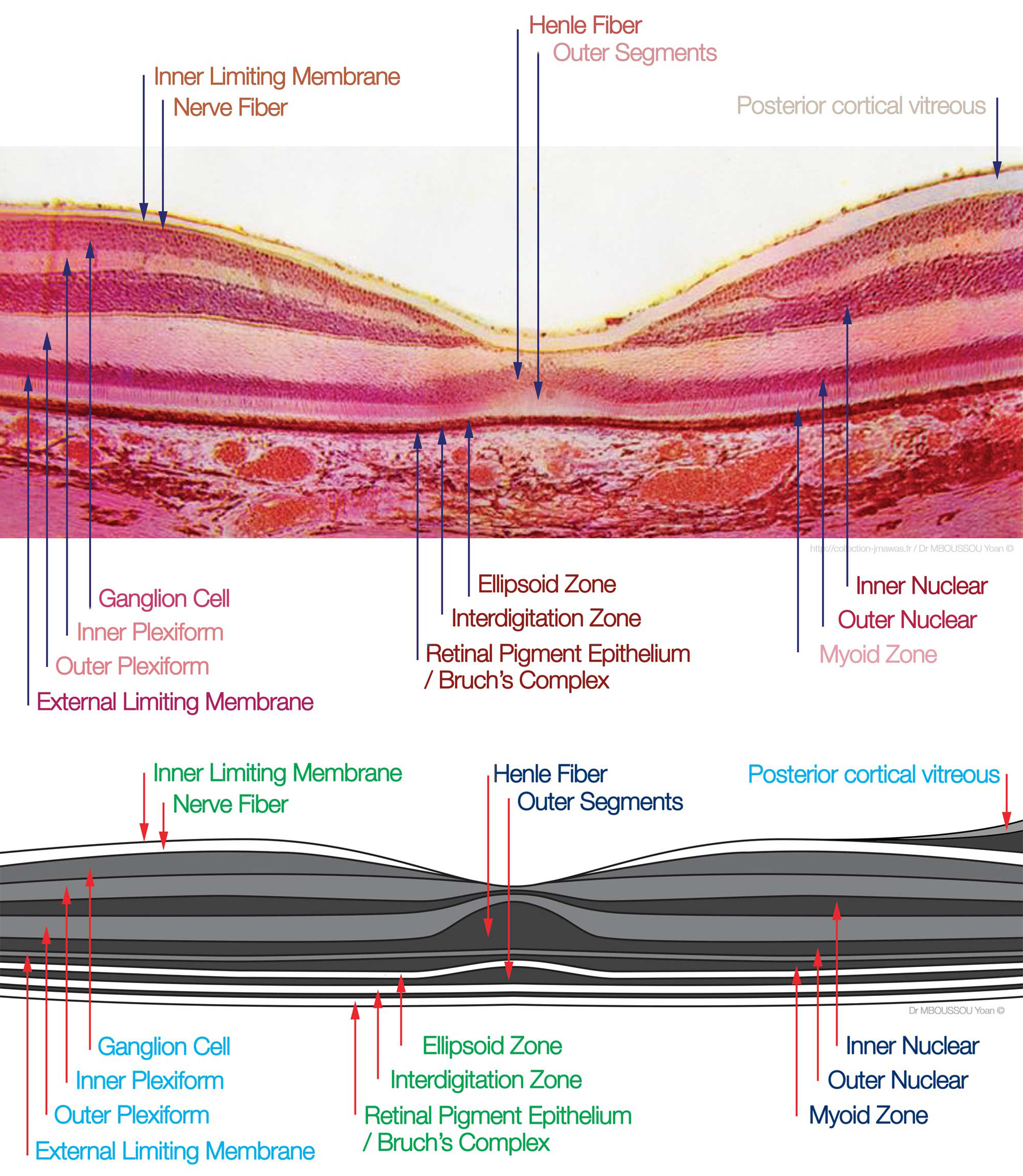
[1]
Hoon M, Okawa H, Della Santina L, Wong RO. Functional architecture of the retina: development and disease. Progress in retinal and eye research. 2014 Sep:42():44-84. doi: 10.1016/j.preteyeres.2014.06.003. Epub 2014 Jun 28
[PubMed PMID: 24984227]
[2]
Chapot CA, Euler T, Schubert T. How do horizontal cells 'talk' to cone photoreceptors? Different levels of complexity at the cone-horizontal cell synapse. The Journal of physiology. 2017 Aug 15:595(16):5495-5506. doi: 10.1113/JP274177. Epub 2017 May 18
[PubMed PMID: 28378516]
[3]
Lukowski SW, Lo CY, Sharov AA, Nguyen Q, Fang L, Hung SS, Zhu L, Zhang T, Grünert U, Nguyen T, Senabouth A, Jabbari JS, Welby E, Sowden JC, Waugh HS, Mackey A, Pollock G, Lamb TD, Wang PY, Hewitt AW, Gillies MC, Powell JE, Wong RC. A single-cell transcriptome atlas of the adult human retina. The EMBO journal. 2019 Sep 16:38(18):e100811. doi: 10.15252/embj.2018100811. Epub 2019 Aug 22
[PubMed PMID: 31436334]
[4]
Matsui K, Hosoi N, Tachibana M. Active role of glutamate uptake in the synaptic transmission from retinal nonspiking neurons. The Journal of neuroscience : the official journal of the Society for Neuroscience. 1999 Aug 15:19(16):6755-66
[PubMed PMID: 10436033]
[5]
Coughlin BA, Feenstra DJ, Mohr S. Müller cells and diabetic retinopathy. Vision research. 2017 Oct:139():93-100. doi: 10.1016/j.visres.2017.03.013. Epub 2017 Sep 5
[PubMed PMID: 28866025]
[6]
Puller C, Manookin MB, Neitz M, Neitz J. Specialized synaptic pathway for chromatic signals beneath S-cone photoreceptors is common to human, Old and New World primates. Journal of the Optical Society of America. A, Optics, image science, and vision. 2014 Apr 1:31(4):A189-94. doi: 10.1364/JOSAA.31.00A189. Epub
[PubMed PMID: 24695169]
[7]
Wensel TG,Zhang Z,Anastassov IA,Gilliam JC,He F,Schmid MF,Robichaux MA, Structural and molecular bases of rod photoreceptor morphogenesis and disease. Progress in retinal and eye research. 2016 Nov;
[PubMed PMID: 27352937]
[8]
Mustafi D, Engel AH, Palczewski K. Structure of cone photoreceptors. Progress in retinal and eye research. 2009 Jul:28(4):289-302. doi: 10.1016/j.preteyeres.2009.05.003. Epub 2009 Jun 6
[PubMed PMID: 19501669]
[9]
Dowling JE, Boycott BB. Organization of the primate retina: electron microscopy. Proceedings of the Royal Society of London. Series B, Biological sciences. 1966 Nov 15:166(1002):80-111
[PubMed PMID: 4382694]
[10]
Boycott BB, Wässle H. The morphological types of ganglion cells of the domestic cat's retina. The Journal of physiology. 1974 Jul:240(2):397-419
[PubMed PMID: 4422168]
[11]
Dowling JE. Synaptic organization of the frog retina: an electron microscopic analysis comparing the retinas of frogs and primates. Proceedings of the Royal Society of London. Series B, Biological sciences. 1968 Jun 11:170(1019):205-28
[PubMed PMID: 4385244]
[12]
Uga S, Smelser. Comparative study of the fine structure of retinal Müller cells in various vertebrates. Investigative ophthalmology. 1973 Jun:12(6):434-48
[PubMed PMID: 4541022]
Level 2 (mid-level) evidence
[13]
Johnston RL, Tufail A, Lightman S, Luthert PJ, Pavesio CE, Cooling RJ, Charteris D. Retinal and choroidal biopsies are helpful in unclear uveitis of suspected infectious or malignant origin. Ophthalmology. 2004 Mar:111(3):522-8
[PubMed PMID: 15019330]
