Continuing Education Activity
Disseminated superficial actinic porokeratosis (DSAP) is a disorder of keratinization and 1 of 6 porokeratosis variants, characterized by widespread lesions predominantly in sun-exposed areas. Risk factors include genetic predisposition, immunosuppression, and ultraviolet light exposure. Some individuals have a familial predisposition to DSAP, which follows an autosomal-dominant pattern and involves mutations in genes associated with cholesterol synthesis, including those affecting mevalonate kinase.
Clinically, DSAP presents as pink or brown papules and macules with raised borders, often asymptomatic or mildly pruritic. Lesions can resemble actinic keratosis and superficial basal cell carcinoma, necessitating further evaluation in some cases. Early recognition remains crucial due to the potential for malignant transformation to squamous cell carcinoma. Diagnosis relies on clinical examination, skin biopsy, and additional testing to distinguish DSAP from other etiologies. Multiple treatments, including topical and procedural interventions, have demonstrated efficacy.
This activity for healthcare professionals is designed to enhance learners' proficiency in evaluating and managing DSAP. Participants will deepen their understanding of the condition's etiology, risk factors, pathophysiology, and clinical features. Evidence-based diagnostic and therapeutic recommendations will also be discussed in depth. Improved skills will equip clinicians to collaborate within an interprofessional team caring for affected individuals, improving outcomes.
Objectives:
Differentiate disseminated superficial actinic porokeratosis from other cutaneous disorders, including actinic keratosis and superficial basal cell carcinoma, based on clinical and diagnostic features.
Select the appropriate diagnostic tools to evaluate potential cases of disseminated superficial actinic porokeratosis.
Implement personalized, evidence-based treatment strategies for disseminated superficial actinic porokeratosis cases.
Collaborate with all members of the interprofessional team, including specialists such as dermatologists and pathologists, to provide efficient, comprehensive, and coordinated care for individuals with disseminated superficial actinic porokeratosis.
Introduction
Disseminated superficial actinic porokeratosis (DSAP) is a disease of disordered keratinization. This condition represents 1 of 6 porokeratosis variants and involves more extensive distribution than most others. Linear porokeratosis, porokeratosis of Mibelli, punctate porokeratosis, porokeratosis palmaris et plantaris disseminata, and disseminated superficial porokeratosis comprise the remaining 5 variants. Other rare forms include porokeratosis ptychotropica, facial porokeratosis, giant porokeratosis, hypertrophic verrucous porokeratosis, reticulate porokeratosis, and eruptive pruritic papular porokeratosis. The eruptive form has been linked to malignancy, immunosuppression, and a pro-inflammatory state. Lesions develop across the body. Risk factors for porokeratosis include genetic predisposition, immunosuppression, and ultraviolet light exposure.
A defining feature of all porokeratosis variants is the cornoid lamella, which appears histologically as a column of parakeratotic cells. This structure is characterized by a raised ridge circumscribing porokeratotic lesions. DSAP lesions begin as pink to brown papules and macules with a raised border in sun-exposed areas. These lesions may be asymptomatic or mildly pruritic. DSAP is considered precancerous, with a 7.5% to 10% risk of malignant transformation to squamous cell carcinoma (SCC) or basal cell carcinoma (BCC). Multiple treatment options exist, including topical diclofenac, photodynamic therapy (PDT), 5-fluorouracil (5-FU), imiquimod, vitamin D analogs (eg, calcipotriene), retinoids, and lasers.[1][2]
Etiology
Genetics, ultraviolet radiation, trauma, infection, and immunosuppression (including posttransplant immune dysfunction) contribute to porokeratosis. Cases have been reported in recipients of kidney transplants, children with acute leukemia, and individuals receiving multiple medications, such as hydroxyurea or other immunosuppressive agents.[3][4][5][6][7] A familial form of DSAP follows an autosomal dominant inheritance pattern with incomplete penetrance. Mutations in the mevalonate kinase (MVK) gene on chromosome 12q24 have been identified in individuals with DSAP. MVK encodes mevalonate kinase, an enzyme involved in cholesterol synthesis that plays a role in protecting cells from ultraviolet light-induced damage.[8]
DSAP has the highest incidence among porokeratosis variants. The potential for malignant transformation arises from p53 overexpression, particularly in chronic or large lesions and those affecting older adults or immunocompromised individuals.[9][10]
Epidemiology
DSAP is the most common porokeratosis variant, accounting for 56% of cases. Although DSAP shows a slight female predominance, porokeratosis as a whole is more common in male individuals. Onset typically occurs in the 30s and 40s.
Histopathology
A skin biopsy should include the lesion’s border, where a column of parakeratotic cells corresponding to the raised edge is observed. This column, known as the cornoid lamella, overlies a granular layer that may be thinned or absent. Dyskeratosis occurs in the underlying epidermis, and spongiosis may be present (see Image. Cornoid Lamellae on Histopathology).
History and Physical
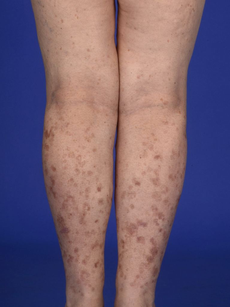
Lesions present as asymptomatic or pruritic annular erythematous or brown macules, papules, or plaques with a raised hyperkeratotic border. Bilateral involvement can occur. DSAP most commonly appears in the 3rd or 4th decade and affects sun-exposed areas, with the legs, forearms, shoulders, and back being the most frequently involved sites (see Image. Disseminated Superficial Actinic Porokeratosis). Facial involvement is rare, and the palms and soles remain unaffected. Sun exposure often exacerbates DSAP, with pruritus potentially intensifying.[11] Dermoscopy may reveal a double margination of the peripheral white border.[12] Unlike actinic keratosis, DSAP lesions exhibit scaling along the periphery rather than centrally. Individuals with DSAP or lesions suspicious for DSAP should undergo a full-body skin examination to assess for additional lesions or skin cancer.
Evaluation
The characteristic appearance of DSAP allows for a clinical diagnosis. Dermoscopy and physical examination may be used for monitoring, reducing the need for a skin biopsy. However, when uncertainty exists, a biopsy may be performed. Nicola et al identified key dermoscopic features of porokeratotic lesions, including a white border encircling the lesion, a homogeneous central white scar-like area, brownish globules or dots, and vascular structures, such as pinpoint or irregular linear vessels crossing the lesion.[13]
Treatment / Management
Treatment options for DSAP aim to reduce lesion burden, alleviate symptoms, and prevent malignant transformation. While no single therapy is universally effective, various topical, procedural, and systemic interventions have shown efficacy in managing the condition.
Topical Diclofenac
Diclofenac, a nonsteroidal anti-inflammatory drug (NSAID) that inhibits cyclooxygenase 2 (COX-2), is approved for actinic keratosis and has been used in DSAP with variable success. This medication has a favorable safety profile and has been effective in stabilizing lesions, including those in genital areas, while also providing symptomatic relief.[14]
Ingenol Mebutate
Ingenol mebutate, approved for actinic keratosis, has been used in DSAP to reduce hyperkeratosis but has limited effects on atrophy and hyperpigmentation. This agent's action mechanism involves inducing cell death, which is presumed to contribute to its therapeutic effect. Ingenol mebutate is available as a 0.05% gel.[15]
Topical Vitamin D Analog
Vitamin D3 analogs have demonstrated favorable responses after 6 to 8 weeks of use. These drugs induce the transcription of genes that regulate keratinocyte differentiation and proliferation.
5-Fluorouracil
5-FU inhibits thymidylate synthase, disrupting DNA synthesis and targeting rapidly proliferating cells. The use of this agent often triggers a pronounced inflammatory reaction, which may include erythema, ulceration, and dermatitis. Clinical improvements are typically temporary.
Imiquimod
Imiquimod stimulates the immune response by activating toll-like receptors 7 and 8, leading to cytokine induction and immune cell recruitment. This drug has primarily been used for porokeratosis of Mibelli and porokeratosis palmaris et plantaris. Treatment can trigger an inflammatory response similar to that seen with 5-FU.
Photodynamic Therapy
PDT has been employed in the management of actinic keratosis, BCC, and SCC in situ. This treatment modality involves the application of a photosensitizer, which is selectively taken up by atypical keratinocytes. The most commonly used PDT photosensitizers are 5-aminolevulinic acid and methyl aminolevulinate. Upon exposure to light, these agents generate reactive oxygen species that induce cellular destruction. Some studies suggest that methyl aminolevulinate may be more effective than 5-aminolevulinic acid due to its greater lipophilicity.
Retinoids
Retinoids, derived from vitamin A, are used in conditions characterized by abnormal keratinocyte proliferation. Topical formulations are preferred over systemic retinoids due to the latter’s increased risk of side effects and teratogenicity. Relapse is common after discontinuation.[16]
Cryotherapy and Other Surgical Dermatology Procedures
Cryotherapy, excision, curettage, and dermabrasion have demonstrated some effectiveness but are generally not used for extensive disease. Cryotherapy, in particular, often results in scarring, and recurrence is common.
Lasers
Several laser modalities, including carbon dioxide (CO2), Q-switched ruby (QSR), neodymium:yttrium-aluminum-garnet (Nd:YAG), fractional photothermolysis, and Grenz ray, have been used in the treatment of DSAP. The carbon dioxide laser utilizes pulsed or scattered infrared light at wavelengths between 9.4 and 10.6 μm, targeting intracellular water to induce tissue vaporization. While effective, this approach may result in posttreatment hyperpigmentation. The Q-switched ruby laser selectively targets melanin, reducing hyperpigmentation without destroying the cornoid lamella, and offers deeper penetration than the Nd:YAG laser.
The Nd:YAG laser, operating at 532 nm, removes the superficial papillary dermis and has been shown to decrease hyperpigmentation while obliterating the cornoid lamella. Fractional photothermolysis creates small zones of thermal necrosis, minimizing damage, redness, and pain while promoting faster healing. Grenz ray therapy, which utilizes low-energy electromagnetic radiation similar to x-rays, inhibits cell proliferation by suppressing DNA synthesis.[17]
Immunosuppressive Agents
Medications that suppress the immune response, such as topical corticosteroids, are generally ineffective for DSAP, which is not an inflammatory disease. However, these agents may provide symptomatic relief for associated pruritus.[18][19][20][21][22]
Topical Statin-Cholesterol
The combination of 2% simvastatin or lovastatin with 2% cholesterol cream has been shown to reduce lesion count, scaling, and erythema.[23][24] Since DSAP is often linked to mutations in the mevalonate pathway, which regulates cholesterol synthesis, supplementation with cholesterol and modulation of this pathway may help improve the condition or slow its progression.
Differential Diagnosis
The differential diagnosis of DSAP is broad and includes various papulosquamous conditions. Actinic keratosis is a primary consideration, distinguished by its centrally located scale, whereas the scale in DSAP is more peripheral. Actinic keratosis also occurs exclusively in sun-exposed areas, while DSAP may not be limited to these regions. Superficial BCC is another important differential, but it typically does not present with numerous lesions. Other conditions to consider include guttate psoriasis, other forms of psoriasis, granuloma annulare, lichen planus, seborrheic keratosis, tinea corporis, and flat warts.
Prognosis
Malignant transformation can occur in porokeratosis, leading to the development of SCC, SCC in situ, or BCC. Between 6.4% and 16.4% of porokeratosis lesions undergo malignant transformation, though the true rate may be closer to 6.9% to 11.6%. DSAP has been specifically reported to carry a 3.4% risk, though this rate may be underestimated.[25]
Complications
Complications of porokeratosis include symptomatic irritation and an increased risk of malignant transformation, with DSAP being one of the most frequently studied subtypes in this context.[26] Without treatment, progression may lead to malignancy.
Deterrence and Patient Education
Patients should be educated on monitoring lesions for recurrence and new development. Proper skin care for DSAP should be emphasized, including regular sunscreen use, adherence to sun safety measures, and compliance with treatment. However, individuals with genetic forms of the condition may not have a known history of sun exposure. These patients should conduct self-examinations and undergo regular skin evaluations to ensure timely assessment of lesions.
Enhancing Healthcare Team Outcomes
An interprofessional team is best suited to manage DSAP. Primary care providers should refer patients to dermatologists for confirmation of the diagnosis or specialized treatment. Oncologists and transplant physicians should maintain a high level of suspicion for DSAP, given its potential for malignant transformation. Clinicians should recognize DSAP as a premalignant condition and emphasize sun avoidance, including the risks of tanning spas. Any changes in lesion characteristics warrant a biopsy. Pharmacists play a key role in educating patients about medications, checking for drug-drug interactions, and ensuring adherence to treatment.