Urea Breath Test


Introduction

Helicobacter pylori (H. pylori) is a ubiquitous, microaerophilic, gram-negative, bacillus.[1][2] It affects more than 50% of the population worldwide and is one of the most prevalent chronic bacterial infections.[3][1][2] Drs. Barry Marshall and Robin Warren first isolated H. pylori in 1983.[4] The prevalence of H. pylori varies between 19% and 88% and depends on geographical location, the patient's age, sanitation, and socioeconomic status.[3][1][5] Patients often acquire H. pylori infection during childhood, and if untreated, persist throughout life.[1][5][6]

Procedures

Tests for H. pylori include both invasive and noninvasive methods.[7] Each method has its advantages and limitations.[2] The selection of a test or combination of tests depends on clinical context, cost, availability, and their sensitivities and specificities.[7][2][8] 

Endoscopy

The gold standard investigation for confirmation of H. pylori is esophagogastroduodenoscopy (EGD) with histopathological examination of the biopsy.[9][6] Further testing methods such as immunohistochemical staining, rapid urease test, bacterial culture, and PCR increase the diagnostic yield of EGD.[10][9] EGD also offers the advantage of evaluating the long-term complications associated with H. pylori.[2] EGD is recommended in the evaluation of dyspepsia in patients older than 60 and in the presence of alarm symptoms such as significant weight loss, gastrointestinal bleeding, abdominal mass, iron deficiency anemia, and difficulty in swallowing.[11][12] However, EGD is invasive and expensive, limiting its utility as the first investigation of choice in younger (less than 60 years of age) patients with dyspepsia and without alarm symptoms.[11][13][2][14] An EGD evaluation may be falsely negative in patients with a patchy gastric distribution of H. pylori.[2][6][10]. EGD could also be considered for younger patients younger than 60 who have a higher risk of malignancy, such as a family history of gastric cancer or living in places with a high prevalence of gastric cancer.[12]

Noninvasive Testing

The popular noninvasive methods include urea breath test (UBT), H. pylori stool antigen test (SAT), and serological tests. Noninvasive tests, such as UBT or SAT, are preferred for patients who do not need an endoscopic evaluation but have diseases strongly associated with H. pylori. This includes patients with peptic ulcer disease (PUD) or a past medical history of PUD but no documented eradication.[5][15] For patients younger than 60 with dyspepsia, a noninvasive H. pylori test could be offered in the absence of alarming symptoms, which include GI bleeding, anemia, significant weight loss, loss of appetite, dysphagia, significant emesis, family history of GI malignancy, and history of GI cancer. H. pylori treatment should be offered if the test result is positive.[16][12][5][15][17]

Serology

H. pylori IgG serology is not recommended in places with prevalence rates of less than 30%.[15][11] With the lower prevalence of H. pylori, the pretest probability of diagnosing a true infection is lower.[16][15] Also, the antibody against H. pylori persists indefinitely and does not differentiate past from present infections.[7][16][8] For the same reason, serological methods are not useful for determining eradication after treatment.[11][1] However, serological testing for H. pylori is useful for epidemiological studies and screening larger populations in places with a higher prevalence rate.[8][2] Antigen-specific serological tests in whole blood and saliva are not recommended due to their lower predictive value.[15][11]

Stool Antigen Test 

The most reliable noninvasive methods include UBT and SAT, with UBT being the most accurate.[8][18] However, the SAT is less expensive and has comparable accuracy to the UBT, with sensitivity and specificity exceeding 93%.[15][7][19] The most reliable SAT uses laboratory-based monoclonal antibody enzyme immunoassay to detect H. pylori antigens directly in the stool. Polyclonal immunoassay and rapid in-office monoclonal immunochromatographic tests are less commonly used due to lower sensitivities. SAT involves stool collection, which some patients may not prefer.[11] 

Urea Breath Test

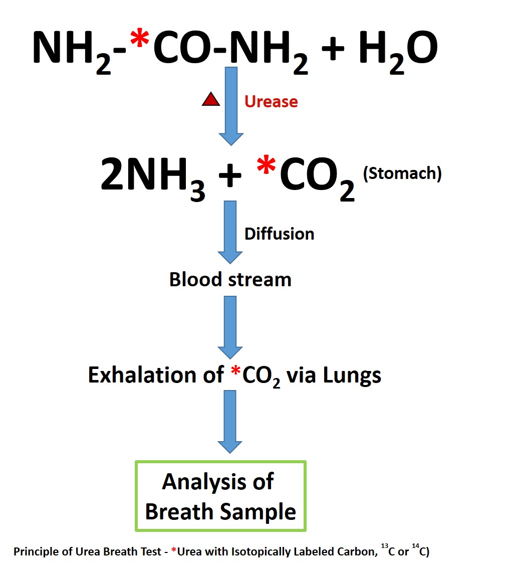
The UBT is useful for both the initial diagnosis of H. pylori (test-and-treat strategy) and the evaluation of post-treatment eradication of infection.[7][20][18] Urease is not present in mammalian cells, and its presence indicates urease-containing microbes. H. pylori utilizes urease to break down urea into ammonia and carbon dioxide (CO2).[10] H. pylori utilizes ammonia to neutralize gastric acids and help in its colonization.[8] Bacteria other than H. pylori are usually absent in the stomach, with a few exceptions, including patients with achlorhydria.[21][22] UBT uses urease, abundantly present in H. pylori, to hydrolyze urea into ammonia and carbon dioxide (CO2).[20] This CO2 enters the bloodstream and is later exhaled via the lungs (see Image. Urea Breath Test Pathway). Using urea as a substrate with a labeled carbon (C) isotope, the exhaled CO2 with the labeled C can be measured and quantified to diagnose H. pylori. Testing commonly uses 2 isotopes of carbon for UBT: 14C (a radioactive isotope) and 13C (a naturally occurring, stable non-radioactive isotope). The performance of both UBTs is similar, with sensitivities above 95% and specificities above 93%.[16][23][24]

Before the UBT is performed, patients should discontinue antibiotics and bismuth compounds for at least 4 weeks and proton pump inhibitors (PPIs) and sucralfate for at least 2 weeks.[25][11][20][2] These medications can reduce the urease activity of H. pylori and generate a false negative result.[26] In these scenarios, treatment should begin if the testing result is positive. If the result is negative, these medications should be discontinued as recommended above and repeat the UBT.[25] Histamine-2 receptor antagonists should be ideally discontinued 24 to 48 hours before the testing to avoid a false negative result.[11][16][27] The use of antacids is permitted and does not seem to interfere with the UBT results.[16] Following H. pylori treatment, UBT should also be performed to evaluate eradication at least 4 weeks after the completion of the treatment.[11]

14C-UBT: The commercial brand available for 14C-UBT in the U.S. is PYtest. Current testing dose exposes patients to a small dose of 1 microcurie, which is equivalent to natural environmental background radiation received in about 24 hours.[20] The labeled urea with 14C is an oral capsule, which the patient takes after fasting for 6 hours. Lukewarm water is used to swallow. The capsule should not be opened or crushed in the mouth to avoid contamination with oral flora, which may have urease activity. Ten minutes after the capsule ingestion, the patient is instructed to blow through a straw, and the breath sample is collected in a balloon. Measurement of the labeled 14CO2 in the collected sample is by scintillation technique. In the presence of H. pylori, an increase in 14CO2 is noted due to hydrolysis of 14C-urea by the urease. Without H. pylori, there will be no hydrolysis of 14C-urea. The unhydrolyzed 14C-urea is absorbed in the stomach, enters the bloodstream, and is excreted by the kidneys. Appropriate safety precautions are necessary for storing, handling, and disposing of the radioactive test ingredients.[20] The overall cost of 14C-UBT is cheaper when compared to 13C-UBT.[11][20] Hence, 14C-UBT is more popular in resource-limited settings.[2] 

13C-UBT: Although 14C-UBT is safe, the current preference is for the 13C isotope as it is non-radioactive, especially in children, women of childbearing age, and during pregnancy.[28][6][2][20][16] Graham et al first developed the 13C-UBT.[29] Historically, a gas-isotype ratio mass spectrometer was utilized to measure the labeled 13-CO2.[29][8][20] This method is costly and also needs technical expertise.[8][20] The newer methods utilize easier and less costly techniques such as infrared spectrophotometry or laser-assisted ratio analysis for analyzing the labeled 13CO2.[7][8][20] 

The test is easily performed in clinical settings and is also reproducible. 13C-UBTs can be administered in children as young as 3. Since 13C is natural and present everywhere, a baseline breath sample of 13CO2 is noted before the administration of labeled 13C-urea, and the baseline value is expressed as a ratio of 13CO2/12CO2. Unlike in 14C-UBT, a test meal is required to delay gastric emptying, and one containing citric acid is the most commonly used test meal given with 13C-urea.[7][20] 

Citric acid reduces the duodenal pH, decreasing the antral motility and relaxing the fundus of the stomach.[20] 13C-urea with the citric acid mixture is ingested using a straw to minimize contamination with oral bacteria with urease activity. Fifteen minutes later, breath is collected again for analysis. An increase in the 13CO2 value from the baseline indicates urease activity, a surrogate for the presence of H. pylori. A post-urea ratio of 13CO2/12CO2 is noted. The difference in the ratios between the post-urea and the baseline is called delta over baseline (DOB). There is no consensus on the cut-off value for DOB to differentiate between positive and negative results.[2] The threshold values for positivity vary widely, with the DOB ranging from 2.7 to 7, depending on sociodemographic disparities, patient characteristics (age, gender), bacterial factors, and laboratory specifications.[30][31] In the absence of urease, the baseline and post-urea sample 13CO2/12CO2 ratios will be the same. Unlike the 14C isotope, another advantage of using a 13C isotope is that the breath samples can be sent safely from the clinics by mail to the laboratory for breath analysis.[20] 

Indications

Careful consideration is given to the indication for testing for H. pylori, as treatment is indicated for a positive test result. H. pylori has strong associations with many non-neoplastic (peptic ulcer disease, chronic atrophic gastritis) and neoplastic (adenocarcinoma of the stomach, gastric lymphoma arising from mucosa-associated lymphoid tissue or MALT) conditions of the gastrointestinal (GI) tract.[25][11][7][2][5] Due to this strong association with malignant conditions, the World Health Organization (WHO) categorized H. pylori as a class I (definite) carcinogen in 1994.[32] Early diagnosis of H. pylori and successful eradication cures chronic gastritis and can reduce the progression to long-term complications.[11] H. pylori gastritis is also implicated in non-ulcer dyspepsia.[14][11] 

Hematologic disorders such as unexplained iron deficiency anemia (after a thorough negative evaluation) and immune thrombocytopenic purpura in adults may also have links with H. pylori, but the exact pathogenic mechanisms are unclear.[33] Consideration should be given to H. pylori testing before the initiation of long-term non-steroidal anti-inflammatory drugs (NSAID) or aspirin therapy due to increased chances of development of peptic ulcer disease (PUD) and its complications.[5]

Interfering Factors

False Positive Test Results

  1. Helicobacter heilmannii also has urease activity and can produce a positive result in UBT.[34]
  2. Patients with achlorhydria can also have false-positive UBT results.[21] The acidic environment usually inhibits the growth of bacteria other than H. pylori. In patients with achlorhydria, other urease-producing bacteria such as Proteus mirabilis, Citrobacter freunii, and Staphylococcus aureus have been reported to cause false positive UBT.[21][22][10]
  3. Some oral flora have urease activity, and contamination with oral flora could also result in false-positive results.[35] This contamination can be prevented or minimized by swallowing the 14C-urea in a gelatin capsule. Similarly, in the 13C-UBT method, the test meal and 13C-urea are recommended to be ingested using a straw.

 False Negative Test Results

  1. Recent use of medications that interfere with H. pylori, such as antibiotics, bismuth compounds, and PPIs, may give a false-negative result.[26] As mentioned earlier, patients should discontinue antibiotics and bismuth compounds for at least 4 weeks and proton pump inhibitors (PPIs) and sucralfate for at least 2 weeks before the testing.[25][11]
  2. Following the H. pylori eradication, the UBT should be performed at least 4 weeks after the completion of treatment. If completed earlier, the result could be falsely negative.[11]
  3. In patients with H. pylori infection predominantly in the body (corpus) of the stomach, data shows a higher proportion of false-negative results with 13C-UBT.[36]

Clinical Significance

Advantages of UBT

  1. Noninvasive, safer, and cost-effective when compared to endoscopic evaluation.[16] UBT does not carry the risk of sampling errors and is more sensitive than EGD in patients with a patchy distribution of H. pylori infection.[20]
  2. UBT is convenient and preferred by many patients compared to stool collection for SAT.[16]
  3. UBT is overall more accurate than the SAT and serological tests.[1]
  4. UBT is helpful for initial diagnosis (test-and-treat strategy) and to confirm infection eradication.
  5. 13C-UBT can be easily repeated and administered safely in children (3 years or older) and pregnant women.[20]
  6. 13C-UBTs are helpful in the epidemiological evaluation of H. pylori in children and adults.[7]  

Limitations of UBT

  1. Helicobacter heilmannii, another urease-containing bacteria, may cause a false-positive test.[2]
  2. False-positive test results in achlorhydria and oral contamination of bacteria with urease activity.[7]  
  3. A recent history of upper GI bleeding could affect the accuracy of UBT.[1][2]
  4. 13C-UBT is less accurate in the pediatric population, with sensitivity and specificity ranging from 75% to 100%.[24][7]  
  5. Low sensitivity in patients with a history of gastric surgery.[10][2] EGD with rapid urease test and histology are preferable in this context.[2]


(Click Image to Enlarge)
<p>Urea Breath Test Pathway</p>

Urea Breath Test Pathway

Contributed by Senthilkumar Sankararaman, MD

Details

Editor:

Leila Moosavi

Updated:

2/23/2024 7:57:31 AM

References


[1]

Kalali B, Formichella L, Gerhard M. Diagnosis of Helicobacter pylori: Changes towards the Future. Diseases (Basel, Switzerland). 2015 Jun 29:3(3):122-135. doi: 10.3390/diseases3030122. Epub 2015 Jun 29     [PubMed PMID: 28943614]


[2]

Wang YK, Kuo FC, Liu CJ, Wu MC, Shih HY, Wang SS, Wu JY, Kuo CH, Huang YK, Wu DC. Diagnosis of Helicobacter pylori infection: Current options and developments. World journal of gastroenterology. 2015 Oct 28:21(40):11221-35. doi: 10.3748/wjg.v21.i40.11221. Epub     [PubMed PMID: 26523098]


[3]

Hooi JKY, Lai WY, Ng WK, Suen MMY, Underwood FE, Tanyingoh D, Malfertheiner P, Graham DY, Wong VWS, Wu JCY, Chan FKL, Sung JJY, Kaplan GG, Ng SC. Global Prevalence of Helicobacter pylori Infection: Systematic Review and Meta-Analysis. Gastroenterology. 2017 Aug:153(2):420-429. doi: 10.1053/j.gastro.2017.04.022. Epub 2017 Apr 27     [PubMed PMID: 28456631]

Level 1 (high-level) evidence

[4]

Warren JR, Marshall B. Unidentified curved bacilli on gastric epithelium in active chronic gastritis. Lancet (London, England). 1983 Jun 4:1(8336):1273-5     [PubMed PMID: 6134060]


[5]

Chey WD, Leontiadis GI, Howden CW, Moss SF. ACG Clinical Guideline: Treatment of Helicobacter pylori Infection. The American journal of gastroenterology. 2017 Feb:112(2):212-239. doi: 10.1038/ajg.2016.563. Epub 2017 Jan 10     [PubMed PMID: 28071659]


[6]

Delvin EE, Brazier JL, Deslandres C, Alvarez F, Russo P, Seidman E. Accuracy of the [13C]-urea breath test in diagnosing Helicobacter pylori gastritis in pediatric patients. Journal of pediatric gastroenterology and nutrition. 1999 Jan:28(1):59-62     [PubMed PMID: 9890470]


[7]

Lopes AI, Vale FF, Oleastro M. Helicobacter pylori infection - recent developments in diagnosis. World journal of gastroenterology. 2014 Jul 28:20(28):9299-313. doi: 10.3748/wjg.v20.i28.9299. Epub     [PubMed PMID: 25071324]


[8]

Sabbagh P, Mohammadnia-Afrouzi M, Javanian M, Babazadeh A, Koppolu V, Vasigala VR, Nouri HR, Ebrahimpour S. Diagnostic methods for Helicobacter pylori infection: ideals, options, and limitations. European journal of clinical microbiology & infectious diseases : official publication of the European Society of Clinical Microbiology. 2019 Jan:38(1):55-66. doi: 10.1007/s10096-018-3414-4. Epub 2018 Nov 9     [PubMed PMID: 30414090]


[9]

Best LM, Takwoingi Y, Siddique S, Selladurai A, Gandhi A, Low B, Yaghoobi M, Gurusamy KS. Non-invasive diagnostic tests for Helicobacter pylori infection. The Cochrane database of systematic reviews. 2018 Mar 15:3(3):CD012080. doi: 10.1002/14651858.CD012080.pub2. Epub 2018 Mar 15     [PubMed PMID: 29543326]

Level 1 (high-level) evidence

[10]

Patel SK, Pratap CB, Jain AK, Gulati AK, Nath G. Diagnosis of Helicobacter pylori: what should be the gold standard? World journal of gastroenterology. 2014 Sep 28:20(36):12847-59. doi: 10.3748/wjg.v20.i36.12847. Epub     [PubMed PMID: 25278682]


[11]

Malfertheiner P, Megraud F, O'Morain CA, Gisbert JP, Kuipers EJ, Axon AT, Bazzoli F, Gasbarrini A, Atherton J, Graham DY, Hunt R, Moayyedi P, Rokkas T, Rugge M, Selgrad M, Suerbaum S, Sugano K, El-Omar EM, European Helicobacter and Microbiota Study Group and Consensus panel. Management of Helicobacter pylori infection-the Maastricht V/Florence Consensus Report. Gut. 2017 Jan:66(1):6-30. doi: 10.1136/gutjnl-2016-312288. Epub 2016 Oct 5     [PubMed PMID: 27707777]

Level 3 (low-level) evidence

[12]

Moayyedi P, Lacy BE, Andrews CN, Enns RA, Howden CW, Vakil N. ACG and CAG Clinical Guideline: Management of Dyspepsia. The American journal of gastroenterology. 2017 Jul:112(7):988-1013. doi: 10.1038/ajg.2017.154. Epub 2017 Jun 20     [PubMed PMID: 28631728]


[13]

Ling D. Carbon-13 urea breath test for Helicobacter pylori infection in patients with uninvestigated ulcer-like dyspepsia: an evidence-based analysis. Ontario health technology assessment series. 2013:13(19):1-30     [PubMed PMID: 24228082]


[14]

Ford AC, Qume M, Moayyedi P, Arents NL, Lassen AT, Logan RF, McColl KE, Myres P, Delaney BC. Helicobacter pylori "test and treat" or endoscopy for managing dyspepsia: an individual patient data meta-analysis. Gastroenterology. 2005 Jun:128(7):1838-44     [PubMed PMID: 15940619]


[15]

Crowe SE. Helicobacter pylori Infection. The New England journal of medicine. 2019 Mar 21:380(12):1158-1165. doi: 10.1056/NEJMcp1710945. Epub     [PubMed PMID: 30893536]


[16]

Chey WD, Wong BC, Practice Parameters Committee of the American College of Gastroenterology. American College of Gastroenterology guideline on the management of Helicobacter pylori infection. The American journal of gastroenterology. 2007 Aug:102(8):1808-25     [PubMed PMID: 17608775]


[17]

Talley NJ, Vakil NB, Moayyedi P. American gastroenterological association technical review on the evaluation of dyspepsia. Gastroenterology. 2005 Nov:129(5):1756-80     [PubMed PMID: 16285971]


[18]

Skrebinska S, Mégraud F, Bessède E. Diagnosis of Helicobacter pylori infection. Helicobacter. 2018 Sep:23 Suppl 1():e12515. doi: 10.1111/hel.12515. Epub     [PubMed PMID: 30203584]


[19]

Choi J, Kim CH, Kim D, Chung SJ, Song JH, Kang JM, Yang JI, Park MJ, Kim YS, Yim JY, Lim SH, Kim JS, Jung HC, Song IS. Prospective evaluation of a new stool antigen test for the detection of Helicobacter pylori, in comparison with histology, rapid urease test, (13)C-urea breath test, and serology. Journal of gastroenterology and hepatology. 2011 Jun:26(6):1053-9. doi: 10.1111/j.1440-1746.2011.06705.x. Epub     [PubMed PMID: 21362044]


[20]

Savarino V, Vigneri S, Celle G. The 13C urea breath test in the diagnosis of Helicobacter pylori infection. Gut. 1999 Jul:45 Suppl 1(Suppl 1):I18-22     [PubMed PMID: 10457031]


[21]

Osaki T, Mabe K, Hanawa T, Kamiya S. Urease-positive bacteria in the stomach induce a false-positive reaction in a urea breath test for diagnosis of Helicobacter pylori infection. Journal of medical microbiology. 2008 Jul:57(Pt 7):814-819. doi: 10.1099/jmm.0.47768-0. Epub     [PubMed PMID: 18566138]


[22]

Borriello SP, Reed PJ, Dolby JM, Barclay FE, Webster AD. Microbial and metabolic profile of achlorhydric stomach: comparison of pernicious anaemia and hypogammaglobulinaemia. Journal of clinical pathology. 1985 Aug:38(8):946-53     [PubMed PMID: 4031106]


[23]

Ferwana M, Abdulmajeed I, Alhajiahmed A, Madani W, Firwana B, Hasan R, Altayar O, Limburg PJ, Murad MH, Knawy B. Accuracy of urea breath test in Helicobacter pylori infection: meta-analysis. World journal of gastroenterology. 2015 Jan 28:21(4):1305-14. doi: 10.3748/wjg.v21.i4.1305. Epub     [PubMed PMID: 25632206]

Level 1 (high-level) evidence

[24]

Leal YA, Flores LL, Fuentes-Pananá EM, Cedillo-Rivera R, Torres J. 13C-urea breath test for the diagnosis of Helicobacter pylori infection in children: a systematic review and meta-analysis. Helicobacter. 2011 Aug:16(4):327-37. doi: 10.1111/j.1523-5378.2011.00863.x. Epub     [PubMed PMID: 21762274]

Level 1 (high-level) evidence

[25]

Laine L, Estrada R, Trujillo M, Knigge K, Fennerty MB. Effect of proton-pump inhibitor therapy on diagnostic testing for Helicobacter pylori. Annals of internal medicine. 1998 Oct 1:129(7):547-50     [PubMed PMID: 9758575]


[26]

Bravo LE, Realpe JL, Campo C, Mera R, Correa P. Effects of acid suppression and bismuth medications on the performance of diagnostic tests for Helicobacter pylori infection. The American journal of gastroenterology. 1999 Sep:94(9):2380-3     [PubMed PMID: 10483995]


[27]

Klein PD, Malaty HM, Martin RF, Graham KS, Genta RM, Graham DY. Noninvasive detection of Helicobacter pylori infection in clinical practice: the 13C urea breath test. The American journal of gastroenterology. 1996 Apr:91(4):690-4     [PubMed PMID: 8677930]


[28]

Logan RP. Urea breath tests in the management of Helicobacter pylori infection. Gut. 1998 Jul:43 Suppl 1(Suppl 1):S47-50     [PubMed PMID: 9764040]


[29]

Graham DY, Klein PD, Evans DJ Jr, Evans DG, Alpert LC, Opekun AR, Boutton TW. Campylobacter pylori detected noninvasively by the 13C-urea breath test. Lancet (London, England). 1987 May 23:1(8543):1174-7     [PubMed PMID: 2883491]


[30]

Perets TT, Gingold-Belfer R, Leibovitzh H, Itskoviz D, Schmilovitz-Weiss H, Snir Y, Dickman R, Dotan I, Levi Z, Boltin D. Optimization of (13) C-urea breath test threshold levels for the detection of Helicobacter pylori infection in a national referral laboratory. Journal of clinical laboratory analysis. 2019 Feb:33(2):e22674. doi: 10.1002/jcla.22674. Epub 2018 Sep 17     [PubMed PMID: 30221401]


[31]

Mauro M, Radovic V, Zhou P, Wolfe M, Kamath M, Bercik P, Croitoru K, Armstrong D. 13C urea breath test for (Helicobacter pylori): determination of the optimal cut-off point in a Canadian community population. Canadian journal of gastroenterology = Journal canadien de gastroenterologie. 2006 Dec:20(12):770-4     [PubMed PMID: 17171195]


[32]

Houghton J, Wang TC. Helicobacter pylori and gastric cancer: a new paradigm for inflammation-associated epithelial cancers. Gastroenterology. 2005 May:128(6):1567-78     [PubMed PMID: 15887152]


[33]

Santambrogio E, Orsucci L. Helicobacter pylori and hematological disorders. Minerva gastroenterologica e dietologica. 2019 Sep:65(3):204-213. doi: 10.23736/S1121-421X.19.02580-7. Epub 2019 Apr 16     [PubMed PMID: 30994322]


[34]

Garza-González E, Perez-Perez GI, Maldonado-Garza HJ, Bosques-Padilla FJ. A review of Helicobacter pylori diagnosis, treatment, and methods to detect eradication. World journal of gastroenterology. 2014 Feb 14:20(6):1438-49. doi: 10.3748/wjg.v20.i6.1438. Epub     [PubMed PMID: 24587620]


[35]

Hamlet AK, Erlandsson KI, Olbe L, Svennerholm AM, Backman VE, Pettersson AB. A simple, rapid, and highly reliable capsule-based 14C urea breath test for diagnosis of Helicobacter pylori infection. Scandinavian journal of gastroenterology. 1995 Nov:30(11):1058-63     [PubMed PMID: 8578164]


[36]

Capurso G, Carnuccio A, Lahner E, Panzuto F, Baccini F, Delle Fave G, Annibale B. Corpus-predominant gastritis as a risk factor for false-negative 13C-urea breath test results. Alimentary pharmacology & therapeutics. 2006 Nov 15:24(10):1453-60     [PubMed PMID: 17032284]