Anatomy, Head and Neck, Tensor Veli Palatini Muscle


Introduction

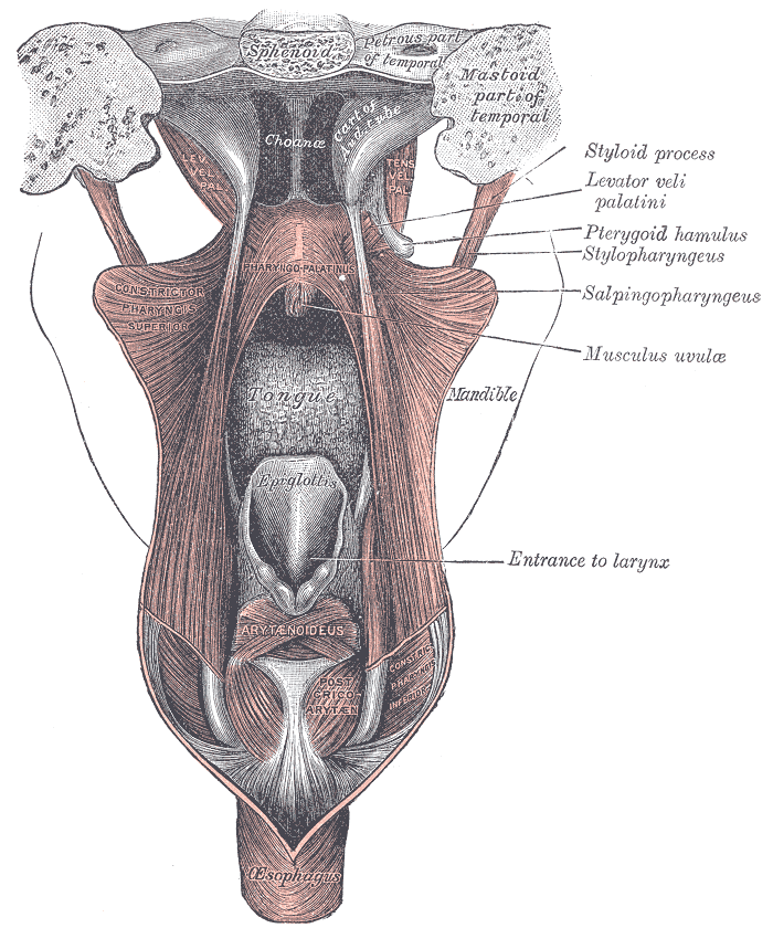
Otitis media is a very common condition in the pediatric population. This diagnosis can include effusion, depending on the patient. Otitis media is a primary problem of the Eustachian tube. Control of the Eustachian tube’s dilation is by the tensor veli palatini (TVP) and the levator veli palatini (LVP).[1]  Both of these muscles reside in the head, specifically near the palatal region, and they both technically originate from the Eustachian tube itself. In particular, the TVP has been targeted more as being the muscle with the most influence on the Eustachian tube’s ventilation and function. Many studies do not agree on the importance of the LVP, and some even argue that it is not relevant to the Eustachian tube’s function other than the LVP's anatomical derivation. Some reports note the TVP’s significance in connecting the soft and hard palates via the palatal aponeurosis, but this functionality is not why the TVP is classically notable. Because of this structuring of the TVP, when the muscle constricts, it acts to aid in swallowing.[2] See Image. Fauces Anatomy. 

Structure and Function

The tensor veli palatini originates from the inferior surface of the cartilage of the Eustachian tube, the scaphoid fossa, and the sphenoid bone’s spine. The muscle then wraps around the pterygoid hamulus, becoming tendinous, becoming a fan-like aponeurosis attaching to the palatine aponeurosis and horizontal aspect of the palatine bone.[3][4][5] Hypomochlia that control the force vectors dictate the muscle's functions. The hypomochlia include the following: pterygoid hamulus, lateral Ostmann fat pad, and the medial pterygoid muscle. The muscle has 2 layers. The lateral layer increases drainage and protection when contracted, while the medial layer increases ventilation.[6][4] The TVP’s ventilation ability of the Eustachian tube involves equilibrating air pressure to atmospheric pressure.[7] The portion of the TVP responsible for the ventilation is called dilator tubae because of its ability to dilate and open the Eustachian tube.[4]

The tensor veli palatini is also a component of the soft palate at its midline.[3] Some studies argue that an additional function is velar movement; if the TVP contracts, this increases the swallowing mechanism and decreases airway resistance.[4] Others think that the TVP should be considered a muscle of mastication because, like the masticators, they all have the same embryological origin.[5] These other functions are integral to the head and neck region, but only limited studies discuss the TVP’s additional functionality.

Embryology

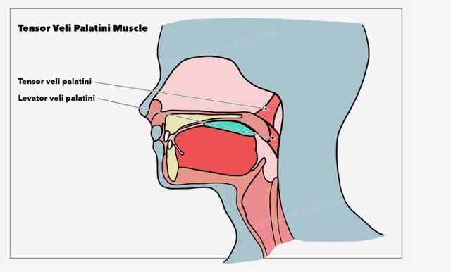
The tensor veli palatini begins to develop as early as 6 weeks into gestation. It originates from the first pharyngeal arch’s mesenchyme, specifically from the medial blastema.[5][8] Because the TVP is near the pharyngotympanic tube, it is considered a paratubal or peritubal muscle. The tensor tympani is a muscle that is also considered a paratubal/peritubal muscle because of its proximity to the pharyngotympanic tube. Like the TVP, the tensor tympani is also developed at the same time in gestation.[8] See Image. Muscles Around the Pharyngotympanic Tube. 

Blood Supply and Lymphatics

The tensor veli palatini receives its blood supply from the maxillary artery, a branch of the external carotid artery just below the mastoid bone. The lesser palatine branch of the maxillary artery enters through the maxillary tubercle and emerges through a foramen in the lesser palatine. In some patients, the lesser palatine branch anastomoses with the greater palatine branch.[9]

Nerves

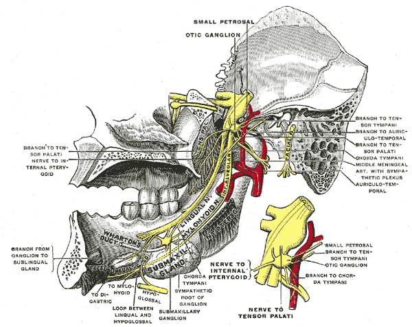
This muscle's innervation is by the motor part of the mandibular nerve. The mandibular nerve is the third division of the trigeminal nerve, CNV3 (see Image. Trigeminal Nerve).[6][5] This nerve also supplies the medial pterygoid.[10] The medial pterygoid and the TVP can work together to open the Eustachian tube and play a role in mastication. The TVP's innervation allows for an understanding of the vast functionality of the muscle.

Muscles

The muscle is at the base of the skull, in the shape of an inverse triangle. The motion of the muscle is activated when a patient swallows or yawns.[6] This motion opens the Eustachian tube because this tube normally remains closed.[1] The TVP has a close association with the tensor tympani. Recent studies have shown that in some patients, the TVP and the tensor tympani are connected, forming a complex or a digastric muscle.[8] In these patients, the tensor tympani has similar functions as the TVP. 

Physiologic Variants

There is controversy over the tensor veli palatini’s association with the hamulus. Some studies have shown bursa involvement, others have shown attachments, and some even argue that there may be ethnic properties involved. When considering cleft palate patients, the fanned aponeurosis only reaches the hard palate and fails to hit the soft palate.[4] Research study results have shown some lateral bundles of the TVP to be continuous with the buccinator muscle. Some medial bundles of the TVP were responsible for creating the palatine aponeurosis.[11] These variants don’t typically alter the function of the TVP.

Surgical Considerations

Some surgeons, when performing palatoplasties, actually cut the tendinous portion of the tensor veli palatini. Palatoplasties are done to surgically correct cleft palates. Interestingly enough, in this patient population, the TVP is anatomically altered.[4]

Clinical Significance

Middle ear disease, like otitis media, has various probable mechanisms; one mechanism, in particular, involves the tensor veli palatini. Because the TVP is effective in opening the nasopharyngeal orifice, it can cause Eustachian tube dysfunction via obstruction if the muscle loses its function.[12] Other forms of obstruction are inflammation, invasion, and paralysis.[10] Because the TVP is called dilator tubae in specific anatomical regions, this emphasizes its significant role in the Eustachian tube's function.

Because of TVP’s minor role in the palatal function, the TVP is theorized to be dysfunctional in a patient with cleft palates. Results from a recent study found that in these patients with cleft palates, the TVP is positioned more medially, as well as has a decrease in the volume of the muscle and the length.[2] This positioning may be important in the future to aid in diagnosing more mild cleft palate deformities.

Other Issues

Of other clinical importance is the ability to use electromyography on the tensor veli palatini muscle. There would be a decrease in activity in the TVP’s electromyography results if the ear were subject to a current bout of otitis media.[7] This technique can theoretically be used in the future to dictate if a child has an otitis media diagnosis and may help quantify the number of occurrences to discuss myringotomies with parents. 


(Click Image to Enlarge)
<p>Trigeminal Nerve

Trigeminal Nerve. The mandibular division of the trifacial nerve is seen from the middle line; the small figure is an enlarged view of the otic ganglion.


Henry Vandyke Carter, Public Domain, via Wikimedia Commons


(Click Image to Enlarge)
<p>Fauces Anatomy

Fauces Anatomy. The illustration depicts a dissection of the muscles of the palate from behind, revealing the larynx, tongue, pharyngopalatinus, constrictor pharyngis superior, and mandible.


Henry Vandyke Carter, Public Domain, via Wikimedia Commons


(Click Image to Enlarge)
<p>Muscles Around the Pharyngotympanic Tube

Muscles Around the Pharyngotympanic Tube. This illustration shows the tensor veli palatini and levator veli palatini, which assist in opening the pharyngotympanic tube, aka eustachian or auditory tube.


Illustration by E Gregory

Details

Editor:

Judith Borger

Updated:

1/2/2023 8:09:54 PM

References


[1]

Gyanwali B, Li H, Xie L, Zhu M, Wu Z, He G, Tang A. The role of tensor veli palatini muscle (TVP) and levetor veli palatini [corrected] muscle (LVP) in the opening and closing of pharyngeal orifice of Eustachian tube. Acta oto-laryngologica. 2016:136(3):249-55. doi: 10.3109/00016489.2015.1107192. Epub 2015 Dec 1     [PubMed PMID: 26624574]


[2]

George TN, Kotlarek KJ, Kuehn DP, Sutton BP, Perry JL. Differences in the Tensor Veli Palatini Between Adults With and Without Cleft Palate Using High-Resolution 3-Dimensional Magnetic Resonance Imaging. The Cleft palate-craniofacial journal : official publication of the American Cleft Palate-Craniofacial Association. 2018 May:55(5):697-705. doi: 10.1177/1055665617752802. Epub 2018 Jan 23     [PubMed PMID: 29360409]


[3]

Cho JH, Kim JK, Lee HY, Yoon JH. Surgical anatomy of human soft palate. The Laryngoscope. 2013 Nov:123(11):2900-4. doi: 10.1002/lary.24067. Epub 2013 May 17     [PubMed PMID: 23686451]


[4]

Schönmeyr B, Sadhu P. A review of the tensor veli palatine function and its relevance to palatoplasty. Journal of plastic surgery and hand surgery. 2014 Feb:48(1):5-9. doi: 10.3109/2000656X.2013.793603. Epub 2013 May 28     [PubMed PMID: 23710786]


[5]

De la Cuadra Blanco C, Peces Peña MD, Rodríguez-Vázquez JF, Mérida-Velasco JA, Mérida-Velasco JR. Development of the human tensor veli palatini: specimens measuring 13.6-137 mm greatest length; weeks 6-16 of development. Cells, tissues, organs. 2012:195(5):392-9. doi: 10.1159/000329253. Epub 2011 Sep 9     [PubMed PMID: 21912075]


[6]

Leuwer R. Anatomy of the Eustachian Tube. Otolaryngologic clinics of North America. 2016 Oct:49(5):1097-106. doi: 10.1016/j.otc.2016.05.002. Epub 2016 Jul 25     [PubMed PMID: 27468634]


[7]

Picciotti PM, Della Marca G, D'Alatri L, Lucidi D, Rigante M, Scarano E. Tensor veli palatini electromyography for monitoring Eustachian tube rehabilitation in otitis media. The Journal of laryngology and otology. 2017 May:131(5):411-416. doi: 10.1017/S0022215117000482. Epub 2017 Mar 15     [PubMed PMID: 28294083]


[8]

Rodríguez-Vázquez JF, Sakiyama K, Abe H, Amano O, Murakami G. Fetal Tendinous Connection Between the Tensor Tympani and Tensor Veli Palatini Muscles: A Single Digastric Muscle Acting for Morphogenesis of the Cranial Base. Anatomical record (Hoboken, N.J. : 2007). 2016 Apr:299(4):474-83. doi: 10.1002/ar.23310. Epub 2016 Jan 27     [PubMed PMID: 26744237]


[9]

Huang MH, Lee ST, Rajendran K. Clinical implications of the velopharyngeal blood supply: a fresh cadaveric study. Plastic and reconstructive surgery. 1998 Sep:102(3):655-67     [PubMed PMID: 9727428]


[10]

Su CY, Hsu SP, Chee CY. Electromyographic study of tensor and levator veli palatini muscles in patients with nasopharyngeal carcinoma. Implications for eustachian tube dysfunction. Cancer. 1993 Feb 15:71(4):1193-200     [PubMed PMID: 8435792]


[11]

Okada R, Muro S, Eguchi K, Yagi K, Nasu H, Yamaguchi K, Miwa K, Akita K. The extended bundle of the tensor veli palatini: Anatomic consideration of the dilating mechanism of the Eustachian tube. Auris, nasus, larynx. 2018 Apr:45(2):265-272. doi: 10.1016/j.anl.2017.05.014. Epub 2017 Jun 16     [PubMed PMID: 28625531]


[12]

Sapci T, Mercangoz E, Evcimik MF, Karavus A, Gozke E. The evaluation of the tensor veli palatini muscle function with electromyography in chronic middle ear diseases. European archives of oto-rhino-laryngology : official journal of the European Federation of Oto-Rhino-Laryngological Societies (EUFOS) : affiliated with the German Society for Oto-Rhino-Laryngology - Head and Neck Surgery. 2008 Mar:265(3):271-8     [PubMed PMID: 17851675]