Pectus Excavatum

Earn CME/CE in your profession:


Continuing Education Activity

Approximately 95 percent of congenital chest wall anomalies are attributed to pectus deformities, with pectus excavatum being the most common. Pectus excavatum is characterized by a depression of the anterior chest wall resulting in a "funnel chest". While the defect involves the third to seventh costocartilages or ribs, the most severe aspect of the deformity occurs in the area of the xiphisternum. Although the deformity may be symmetrical, it is more commonly asymmetrical and may involve other aspects of the thorax. A pectus defomity may be appreciated in an infant at birth or develop it later during childhood. This activity examines the differential of pectus excavatum and how to properly evaluate a patient with pectus excavatum, and highlights the role of the interprofessional team in caring for patients with this condition.

Objectives:

  • List the hypothesized etiologies for pectus excavatum.
  • Describe a complete evaluation of pectus excavatum, including any appropriate diagnostic imaging.
  • Discuss when surgery is necessary in pectus excavatum management, and review the various surgical options available.
  • Describe how an optimally functioning interprofessional team would coordinate care to enhance outcomes for patients with pectus excavatum.

Introduction

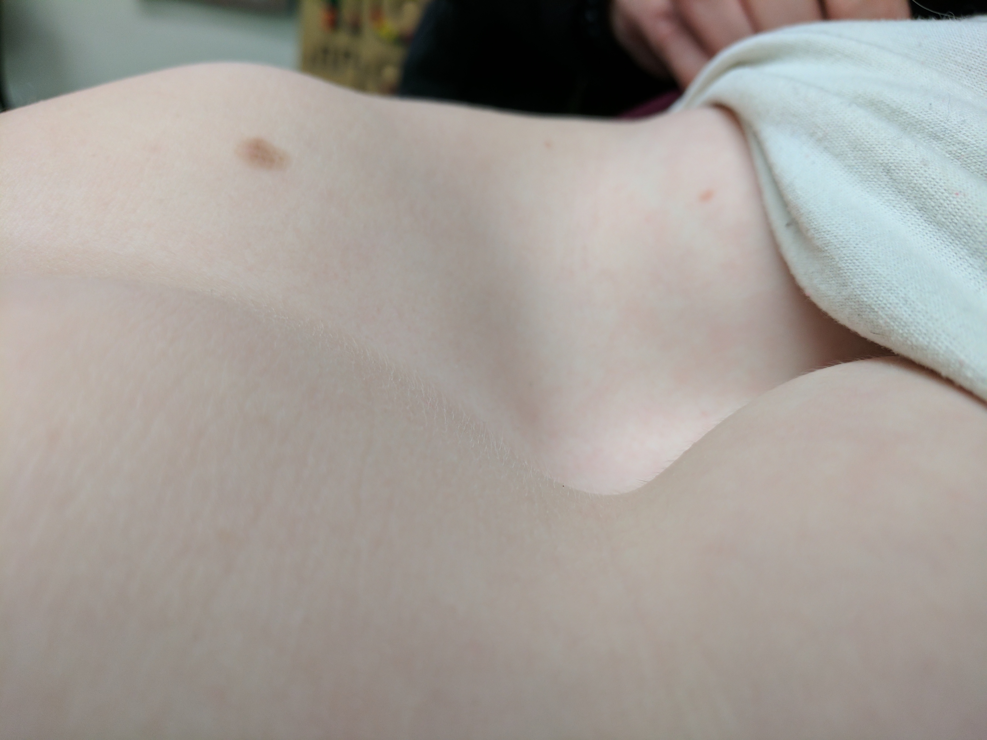
Approximately 95% of congential chest wall anomalies are attributed to pectus deformities, with pectus excavatum being the most common.  A depression of the anterior chest wall results in a "funnel chest".  While the defect involves the third to seventh costocartilages or ribs, the most severe aspect of the deformity occurs in the area of the xiphisternum. (Figure 1).  Although the deformity may be symmetrical, it is more commonly asymmetrical and may involve other aspects of the thorax. A pectus defomity may be appreciated in an infant at birth or develop it later during childhood. [1][2][3]

Etiology

Several hypotheses exists regarding the underlying etiology that results in pectus excavatum.  The funnel - formed chest has been attributed to a weakness and abnormal flexibility of the sternum, overgrowth of the ribs, and developmental failure of the bony thorax.[4]  Regardless of the mechanism, the result is depression and dorsal deviation of the sternum and adjacent ribs or costal cartilages of varying degrees. Although a specific genetic defect has not been identified, a genetic predisposition is supported by the presence of a positive family history in more than 40% of cases.  

Epidemiology

Reported prevalence has been 1/300 to 1/1000 live births; with a 5:1male to female ratio.[5] Pectus excavatum constitutes 90% of all chest wall deformities.  Most defects are appreciated within the first year of life, with severe deformities present at birth.  The funnel - formed chest tends to become more pronounced during the pubertal growth spurt.[6]  Pectus excavatum may present as an isolated anomaly or as a part of a multitude of congenital disorders. Amongst these clinical syndromes, connective tissue disorders are rarely (less than 1%) associated with pectus excavatum.[7]

History and Physical

The presenting patient is commonly a thin, tall male that appears to be slouching, and thoracic scoliosis may also be appreciated.  Sternal depression and rotation may depress the heart, causing a slew of cardiopulmonary signs and symptoms, including exercise intolerance. An audible murmur can be attributed to a defective mitral valve (i.e., prolapse, regurgitation).  Exercise intolerance, though its exact cause is unclear, is one of the most common presenting complaints. Additionally, the psychological impact on patients can be significant, especially during the adolescent period. 

Evaluation

The lateral view of the chest radiograph clearly demonstrates the sternal defect.  Additional imaging studies may reveal displaced vertebral bodies and varying degrees of scoliosis.  Patients with mild pectus excavatum may have minimal or no symptoms; however, cardiopulmonary evaluation to establish a baseline as well as assess for progression is warranted every one to two years.[3][8][9][4][5]  Complete evaluation includes chest radiograph, pulmonary function testing (spirometry, plethysmography, respiratory muscle strength assessment), electrocardiogram, and echocardiogram to assess for the secondary or associated anomalies of a clinical syndrome. Pulmonary function testing may reveal obstructive or restrictive lung disease in older patients, as well as air trapping with increase in residual volume (RV). The air trapping may be caused by a mechanical disadvantage impairing respiratory muscle function. The presence of axis deviation on electrocardiogram (EKG) suggests leftward cardiac deviation.  Arrhythmias in the form of first-degree heart block, right bundle branch block, and Wolff–Parkinson–White occur in 16% in this patient cohort.  Cardiopulmonary exercise testing may reveal cardiopulmonary limitation not appreciated in a resting state.  Echocardiography should be performed to assess for cardiac compression, valvular defects, and myocardial function.  Patient with pectus excavatum may have a cardiac deviation to the left and conduction defects.[4]

The Haller index (HI) is the standard upon which to quantify the severity of a pectus excavatum deformity.  It is defined as the ratio of the transverse diameter and anteroposterior diameter.  The measurements are obtained from computed tomography (CT) scan; normal value is 2.5 or less.  Measurement above 3.2 are considered severe.[5]  Patients with a Haller Index of greater than 7 are four times more likely to have a restrictive pattern on pulmonary function testing. [10] MRI including breath-hold MRI has been used for the morphologic assessment of thoracic deformities for the preoperative assessment of pectus excavatum. A better insight in the functioning of lungs of the patients with pectus excavatum deformity may be obtained using recently introduced techniques such as oculo-electronic plethysmography (OEP),which can show that the depressed portion of the sternum and adjacent chest wall do not move with respiration and there is a reduction in lung volume.[11]

Treatment / Management

Early surgical correction of pectus excavatum was based on the aggressive resection and chest wall reconstruction performed for asphyxiating thoracic dystrophy (Jeune's syndrome). The current trend is to delay the repair until pubertal growth, and the technique is modified to perform limited cartilage resection. Impaired cardiopulmonary function, not cosmesis, is the most common indication for surgical correction.  It is pertinent that surgical repair occur after the child's pubertal growth spurt.[12][13][14]

The first documented surgical repair of pectus excavatum was performed by Ravitch in 1949.  Based on the aggressive chest wall resection used to manage asphyxiating thoracic dystrophy (Jeune's syndrome), Ravitch performed a subperichondrial resection of all deformed costal cartilages and the xiphoid process, along with a transverse sternotomy.[15]  Rehbein and Wernicke modified the procedure six years later, implanting a variety of metallic bars  longitudinally and/or transversely to stabilize the sternum.[16]  This repair can be approached via a median longitudinal incision long the sternum or a submammary skin incision, which is preferable in female patients.  Patients return one year later for removal of the implanted bars.  Modernization of the Ravitch procedure utilizes orthopedic metal plates and screws. These devices can be modelled specifically to the patient's defect, and don't require a second operative procedure for hardware removal.  

The minimally invasive technique described by Nuss and Kelly involves inserting a metal bar under the sternum at the most depressed zone and most everted costal line on both sides of the chest.[17]  The retrosternal bar in placed thoracoscopically, via approximately 3cm incisions along the mid-axillary lines.  Skin flaps are raised and tennelled up to the previously identified most-everted intercostal spaces.  A metal introducer is guided under thoracoscopic guidance to dissect a retrosternal plane between the anterior pericardium and the posterior sternal table.  The guide is exteriorized through the left sided incision, in order to be tied to titanium rod and subsequently withdrawn.  The retrosternal curve var is rotated such that the concave side is directed backwards, thus pushing the sternum ventrally.  Metallic stabilizers and/or subperiostal cable wires are used to prevent migration of the bar.  The initial stabilization period was 2 years; however, it is now more common to leave the bar in situ for 3 years.

Additional modifications to both the Ravitch and Nuss procedures have been described.  The Leonard modification to Ravitch's procedure entails excision of the lower costal cartilage, leaving the perichondirum in place, after mobilization of the pectoralis muscles.[18]  Robicsek describes posterior stabilization of the sternum after the transverse osteotomy with fixation of a piece of Marlex mesh to the remnants of costal cartilage.[19] Minimal cartilage resection defines Erlangen's adaptation of the Nuss procedure,[20] with transternal implantation of an elastic metal bar through stitch incisions.  Intraoperative tensiometry is utilized to minimize the cartilage excision.  The magnetic mini-procedure employs magnetic forces between a sternal magnet and another on a prescribed brace worn by the patient to pull the sternum anteriorly.[21] This modification described by Lacquet is the only technique that avoids the use of prosthetic material, correcting the defect with a sternochondroplasty.  This procedure appears to offer results superior to the Nuss procedure for asymmetric defects.  

The application of negative pressure to the thorax offers a non-surgical alternative to pectus excavatum.[22]  A vacuum bell is applied to the chest wall defect and negative pressure applied by the patient via a handpump.  While long term results are lacking, this may be a viable option for the management of less severe defects in the future.  Additionally, this therapy would be applicable in younger symptomatic patients, in which prepubertal surgical correction is not desired.

Differential Diagnosis

  • Ehlers-Danlos syndrome
  • Marfan syndrome
  • Noonan syndrome
  • Scoliosis

Complications

Minor pectus excavatum defects are usually asymptomatic, thus without complications.  More severe defects can, however, negatively impact cardiopulmonary function, presenting with variety of symptoms:

  • chest pain
  • fatigue
  • exertional dyspnea
  • recurrent respiratory tract infections
  • asthma
  • palpitations
  • heart murmur (mitral regurgitation)
  • arrythmia
  • syncope

Complications are more commonly appreciated in patients who undergo surgical correction of the defect: 

  • migration of the sternal bar
  • infection
  • pneumothorax
  • bleeding
  • cardiac laceration
  • chronic pain
  • recurrence

Overall morbidity and length of stay are comparable for Ravitch and Nuss procedures, according to a recent meta-analysis.[23] However, the incidence of postoperative hemothorax, pneumothorax, and return to OR rates are higher with the Nuss procedure.  The comparison done by Antonoff et al included the Ravitch, Nuss, and Leonard procedures.  The analysis clearly demontrated increased LOS, costs, narcotic use, and complications with the Nuss cohort.[24]  Subjectively, the Nuss and Ravitch procedures appear to be comparable, based on similar health-related quality-of-life (HRQL) outcomes.[25]

Enhancing Healthcare Team Outcomes

Overall, congential chest wall defects are rare.  As the most common defect, pectus excavatum can be appreciated on physical examination by health care providers at every level.  Pediatricians are well equipped to follow these patients, and determine the need for comprehensive cardiopulmonary assessment.  There is a misconception that pectus chest wall deformities are merely cosmetic defects, even in the absence of abnormal pulmonary function and cardiac testing. Asymptomatic patients or those with minor defects may not need any treatment, be offered physical therapy consultation, or can be considered for negative pressure therapy. Surgical (pediatric or cardiothoracic) consultation should be sought for all patients with a Haller Index of 2.5 or greater, as well as for patients with concomitant cardiac defects that need surgical repair or decreased pulmonary reserve.  Regardless of the Haller index measure, impaired cardiopulmonary function is an indication for surgical correction. Surgical correction of the funnel chest attributed to a pectus excavatum defect significant improves pulmonary function at rest and VO2 max in cases in which the Hallex index measures greater than 3.2.  Subjective improvements tracked my quality measurements have also been attributed to operative correction.[4][6][26] (Level I, II, IV, V)


(Click Image to Enlarge)
Pectus excavatum in a 12 yr old female
Pectus excavatum in a 12 yr old female Contributed by Girish Sharma, MD
Details

Author

Girish Sharma

Updated:

7/17/2023 9:15:28 PM

Looking for an easier read?

Click here for a simplified version

References


[1]

Rha EY, Kim JH, Yoo G, Ahn S, Lee J, Jeong JY. Changes in thoracic cavity dimensions of pectus excavatum patients following Nuss procedure. Journal of thoracic disease. 2018 Jul:10(7):4255-4261. doi: 10.21037/jtd.2018.06.159. Epub     [PubMed PMID: 30174871]


[2]

Fortmann C, Petersen C. Surgery for Deformities of the Thoracic Wall: No More than Strengthening the Patient's Self-Esteem? European journal of pediatric surgery : official journal of Austrian Association of Pediatric Surgery ... [et al] = Zeitschrift fur Kinderchirurgie. 2018 Aug:28(4):355-360. doi: 10.1055/s-0038-1668131. Epub 2018 Aug 21     [PubMed PMID: 30130828]


[3]

Schwabegger AH. Deformities of the Thoracic Wall: Don't Forget the Plastic Surgeon. European journal of pediatric surgery : official journal of Austrian Association of Pediatric Surgery ... [et al] = Zeitschrift fur Kinderchirurgie. 2018 Aug:28(4):361-368. doi: 10.1055/s-0038-1668129. Epub 2018 Aug 15     [PubMed PMID: 30112747]


[4]

Brochhausen C, Turial S, Müller FK, Schmitt VH, Coerdt W, Wihlm JM, Schier F, Kirkpatrick CJ. Pectus excavatum: history, hypotheses and treatment options. Interactive cardiovascular and thoracic surgery. 2012 Jun:14(6):801-6. doi: 10.1093/icvts/ivs045. Epub 2012 Mar 5     [PubMed PMID: 22394989]


[5]

Abdullah F, Harris J. Pectus Excavatum: More Than a Matter of Aesthetics. Pediatric annals. 2016 Nov 1:45(11):e403-e406. doi: 10.3928/19382359-20161007-01. Epub     [PubMed PMID: 27841924]


[6]

Kelly RE Jr, Daniel A. Outcomes, quality of life, and long-term results after pectus repair from around the globe. Seminars in pediatric surgery. 2018 Jun:27(3):170-174. doi: 10.1053/j.sempedsurg.2018.05.003. Epub     [PubMed PMID: 30078488]

Level 2 (mid-level) evidence

[7]

Colombani PM. Preoperative assessment of chest wall deformities. Seminars in thoracic and cardiovascular surgery. 2009 Spring:21(1):58-63. doi: 10.1053/j.semtcvs.2009.04.003. Epub     [PubMed PMID: 19632564]


[8]

Sujka JA, St Peter SD. Quantification of pectus excavatum: Anatomic indices. Seminars in pediatric surgery. 2018 Jun:27(3):122-126. doi: 10.1053/j.sempedsurg.2018.05.006. Epub 2018 May 16     [PubMed PMID: 30078482]


[9]

De Feria AE, Bajaj NS, Polk DM, Desai AS, Blankstein R, Vaduganathan M. Pectus Excavatum and Right Ventricular Compression in a Young Athlete with Syncope. The American journal of medicine. 2018 Nov:131(11):e451-e453. doi: 10.1016/j.amjmed.2018.07.033. Epub 2018 Aug 1     [PubMed PMID: 30076820]


[10]

Lawson ML, Mellins RB, Paulson JF, Shamberger RC, Oldham K, Azizkhan RG, Hebra AV, Nuss D, Goretsky MJ, Sharp RJ, Holcomb GW 3rd, Shim WK, Megison SM, Moss RL, Fecteau AH, Colombani PM, Moskowitz AB, Hill J, Kelly RE Jr. Increasing severity of pectus excavatum is associated with reduced pulmonary function. The Journal of pediatrics. 2011 Aug:159(2):256-61.e2. doi: 10.1016/j.jpeds.2011.01.065. Epub 2011 Mar 22     [PubMed PMID: 21429515]


[11]

Kelly RE Jr, Obermeyer RJ, Nuss D. Diminished pulmonary function in pectus excavatum: from denying the problem to finding the mechanism. Annals of cardiothoracic surgery. 2016 Sep:5(5):466-475     [PubMed PMID: 27747180]


[12]

Emil S. Current Options for the Treatment of Pectus Carinatum: When to Brace and When to Operate? European journal of pediatric surgery : official journal of Austrian Association of Pediatric Surgery ... [et al] = Zeitschrift fur Kinderchirurgie. 2018 Aug:28(4):347-354. doi: 10.1055/s-0038-1667297. Epub 2018 Aug 15     [PubMed PMID: 30112746]


[13]

Velazco CS, Arsanjani R, Jaroszewski DE. Nuss procedure in the adult population for correction of pectus excavatum. Seminars in pediatric surgery. 2018 Jun:27(3):161-169. doi: 10.1053/j.sempedsurg.2018.05.002. Epub     [PubMed PMID: 30078487]


[14]

Goretsky MJ, McGuire MM. Complications associated with the minimally invasive repair of pectus excavatum. Seminars in pediatric surgery. 2018 Jun:27(3):151-155. doi: 10.1053/j.sempedsurg.2018.05.001. Epub     [PubMed PMID: 30078485]


[15]

Ravitch MM. The Operative Treatment of Pectus Excavatum. Annals of surgery. 1949 Apr:129(4):429-44     [PubMed PMID: 17859324]


[16]

REHBEIN F, WERNICKE HH. The operative treatment of the funnel chest. Archives of disease in childhood. 1957 Feb:32(161):5-8     [PubMed PMID: 13403703]


[17]

Nuss D, Kelly RE Jr. Minimally invasive surgical correction of chest wall deformities in children (Nuss procedure). Advances in pediatrics. 2008:55():395-410     [PubMed PMID: 19048741]

Level 3 (low-level) evidence

[18]

Wolf WM, Fischer MD, Saltzman DA, Leonard AS. Surgical correction of pectus excavatum and carinatum. Minnesota medicine. 1987 Aug:70(8):447-53     [PubMed PMID: 3657762]


[19]

Robicsek F. Repair of pectus excavatum. The Annals of thoracic surgery. 2010 Jul:90(1):363. doi: 10.1016/j.athoracsur.2010.01.024. Epub     [PubMed PMID: 20609833]


[20]

Weber PG, Hümmer HP. [The "new" Erlangen technique of funnel chest correction - minimalization of a well working procedure]. Zentralblatt fur Chirurgie. 2006 Dec:131(6):493-8     [PubMed PMID: 17206569]


[21]

Lacquet LK. [Surgical treatment of anterior chest wall deformities by sternochondroplasty and internal fixation without prosthesis]. Acta chirurgica Belgica. 1982 Nov-Dec:82(6):541-9     [PubMed PMID: 7158183]


[22]

Haecker FM, Sesia S. Vacuum bell therapy. Annals of cardiothoracic surgery. 2016 Sep:5(5):440-449     [PubMed PMID: 27747177]


[23]

Nasr A, Fecteau A, Wales PW. Comparison of the Nuss and the Ravitch procedure for pectus excavatum repair: a meta-analysis. Journal of pediatric surgery. 2010 May:45(5):880-6. doi: 10.1016/j.jpedsurg.2010.02.012. Epub     [PubMed PMID: 20438918]

Level 1 (high-level) evidence

[24]

Antonoff MB, Erickson AE, Hess DJ, Acton RD, Saltzman DA. When patients choose: comparison of Nuss, Ravitch, and Leonard procedures for primary repair of pectus excavatum. Journal of pediatric surgery. 2009 Jun:44(6):1113-8; discussion 118-9. doi: 10.1016/j.jpedsurg.2009.02.017. Epub     [PubMed PMID: 19524726]


[25]

Lam MW, Klassen AF, Montgomery CJ, LeBlanc JG, Skarsgard ED. Quality-of-life outcomes after surgical correction of pectus excavatum: a comparison of the Ravitch and Nuss procedures. Journal of pediatric surgery. 2008 May:43(5):819-25. doi: 10.1016/j.jpedsurg.2007.12.020. Epub     [PubMed PMID: 18485946]

Level 2 (mid-level) evidence

[26]

Kelly RE Jr, Mellins RB, Shamberger RC, Mitchell KK, Lawson ML, Oldham KT, Azizkhan RG, Hebra AV, Nuss D, Goretsky MJ, Sharp RJ, Holcomb GW 3rd, Shim WK, Megison SM, Moss RL, Fecteau AH, Colombani PM, Cooper D, Bagley T, Quinn A, Moskowitz AB, Paulson JF. Multicenter study of pectus excavatum, final report: complications, static/exercise pulmonary function, and anatomic outcomes. Journal of the American College of Surgeons. 2013 Dec:217(6):1080-9. doi: 10.1016/j.jamcollsurg.2013.06.019. Epub     [PubMed PMID: 24246622]

Level 2 (mid-level) evidence