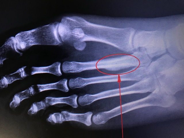
March Fracture (Metatarsal Stress Fractures)

Earn CME/CE in your profession:


Continuing Education Activity

March fractures, also known as metatarsal stress fractures, were first documented in 1855 when Prussian soldiers experienced debilitating foot pain and swelling during long marches, hence the name "March fractures." The primary cause of these fractures is repetitive stress. Diagnosis involves a combination of historical features, comprehensive physical evaluation, and imaging studies. Remarkably, these fractures can manifest prodromal symptoms before appearing on plain radiographs, often leading to delayed diagnosis.

This course discusses the conservative management of March fractures, which is the typical approach. The activity highlights the critical instances where nonunion complicates the healing process, necessitating surgical intervention. The interprofessional healthcare team is instrumental in caring for patients affected by March fractures, ensuring a comprehensive approach to their treatment and recovery.

Objectives:

  • Identify clinical signs and imaging findings specific to march fractures, distinguishing them from similar injuries for precise diagnosis.

  • Select and proficiently use imaging modalities to aid diagnosis and accurately evaluate fracture progression.

  • Implement evidence-based treatment strategies, incorporating rest, activity modification, and potential orthopedic interventions tailored to fracture severity.

  • Collaborate with orthopedic specialists, physical therapists, and trainers for comprehensive march fracture management and follow-up.

Introduction

March fractures, ie, metatarsal stress fractures, were first described in 1855, after the foot pain and swelling experienced by Prussian soldiers on long marches.[1] March fractures are metatarsal fractures caused by repetitive stress.[2] Intrinsic patient and extrinsic environmental risk factors contribute to the development of these fractures. A combination of historical features and physical evaluation with imaging can help make the diagnosis. However, prodromal symptoms are common before evidence of a stress fracture is seen on plain radiographs. Radiographs may be negative for 2 to 4 weeks after the onset of symptoms.[3] These stress fractures are typically managed conservatively but can be complicated by nonunion. In such instances, surgical fixation may be indicated.

Etiology

March fractures, ie, metatarsal stress fractures, are most commonly second and third metatarsal fractures caused by overuse injuries.[1] They result from a sudden increase in the intensity, duration, and frequency of activity without sufficient rest.[4] The repetitive impact on the metatarsals with weight-bearing exercises causes microfractures, which consolidate into stress fractures and are not the result of a single traumatic event.[5] The most common location of metatarsal stress fractures is the second metatarsal neck. The second metatarsal is prone to injury due to its limited motion from strong ligamentous attachment to the first and second cuneiforms and length compared to the other foot metatarsals.[6]

Epidemiology

Metatarsal stress fractures are common in athletes and the military, accounting for 25% of all stress fractures and 20% of all sports medicine clinic visits.[5][7] Forty percent of all athletes will suffer a stress fracture at some point in their careers.[8] Overall data suggest that females have a higher incidence of march fractures than men. In addition, those with prior stress fractures are more likely to develop another. Sixty percent of patients with a stress fracture will have had a previous one.[9]

Pathophysiology

Stress fractures are the result of either bone fatigue or bone insufficiency.[7] Bone fatigue occurs when a normal bone cannot resist excessive mechanical demands. Two different theories may explain the development of a stress fracture. One theory suggests that when osteoblastic activity lags behind osteoclastic activity, it leaves bone susceptible to injury during a sudden increased demand. A second theory suggests that stress fractures develop due to the bone failing, specifically at muscle insertion points where repetitive load and stress are highest.[5]

Classification Systems 

Stress fractures can fall into two broad categories: high-risk and low-risk injuries. Stress fractures of the metatarsals are considered low-risk fractures because they are common and tend to heal well with activity modification while weight-bearing.[7]

Kaeding and Miller’s 5-Tier Grading System [10]

Grade of Stress Fracture/Radiographic Finding

  1. Asymptomatic radiographic findings
  2. Pain with no fracture on imaging
  3. Nondisplaced fracture on imaging
  4. Displaced fracture on imaging
  5. Sclerotic nonunion on imaging

Nattiv Grading System [11]

Evaluation of bone marrow edema and periosteal reaction in bone stress injuries on MRI. 

  • Grade 1: mild marrow edema or periosteal edema on fat-suppressed T2WI (but not on T1WI) 
  • Grade 2: moderate marrow edema or periosteal edema on fat-suppressed T2WI (but not on T1WI) 
  • Grade 3: severe marrow edema or periosteal edema on both fat-suppressed T2WI and T1WI, without a fracture line on T1WI or T2WI
  • Grade 4: severe marrow edema or periosteal edema on both fat-suppressed T2WI and T1WI, with a fracture line on T1WI or T2WI.

Biomechanics

There are two types of cortical stress fracture described in the literature. The two types depend on whether the stress is localized to the tensile or compression side of the bone under stress. Compression stress fractures correspond to a fracture line occurring on the concave side of the bone. These fractures run parallel to the bone axis. In comparison, tensile stress fractures appear on the convex side of the stressed bone, and these fracture lines develop perpendicular to the bone axis. Compression stress fractures tend to be less common than their traction counterparts.[12] Metatarsal length and sagittal plane position can disrupt the even distribution of weight across the forefoot. Metatarsals that are longer or more plantarflexed than the surrounding metatarsals will bear more biomechanical load. Foot type may also play a role. A cavovarus foot puts an increased load on the lateral column and is associated with fourth and fifth metatarsal stress fractures.[13][14] Extrinsic risk factors, such as the training type, intensity, or environment, as well as athletic shoe type or age, can also contribute to an increased risk of metatarsal stress fractures.

Histopathology

Objective information on stress fracture healing is negligible as biopsies are rarely taken as part of routine treatment.[15] Research is limited to animal studies often focused on forearm stress fractures. Animal studies have found that at the cellular level, stress fractures are characterized by irregular bone changes. At 2 weeks, this irregular woven bone develops islands of cartilage and active resorption cavities along the periosteal margin. At 4 weeks, the woven bone is remodeled, starting from the periosteal surface and progressing along the fracture plane and into the medullary cavity.[16] A hallmark sign of healing is the formation of periosteal hard callous.[15] Slow healing rates and recurrent stress fractures may be explained by the failure or slow healing of the most central portions of a stress fracture.[16]

History and Physical

During an interview, patients may describe an insidious, new onset of pain that improves transiently with rest but increases again with activity. Questions should focus on new exercise routines or routines that may have increased in intensity, duration, or frequency over the last 6 to 8 weeks. Changes in terrain or shoe gear should also be noted, as these may elucidate changes in force distributions through the foot.[4] Pain may be nonspecific in location but is often described as dull, aching, and worse with any weight-bearing activities.[17]

Obtaining a thorough medical history with particular attention to intrinsic and extrinsic risk factors is important. Intrinsic risk factors may include age, endocrine disorders, body composition, bone mineral density, and history of a stress fracture.[8] Advanced age and a low body mass index, specifically in women, are associated with increased rates of stress fractures.[18] Prior stress fracture increases the chances of re-fracture due to an increased likelihood of associated intrinsic factors or architectural changes in the bone from previous injury. Questions about extrinsic risk factors should elucidate more than changes in activity level and include diet content, eating behavior, medication regimens, and foot biomechanics.[8] The female triad, which is characterized by abnormal menstrual cycles (amenorrhea), low bone mass (osteoporosis), and disordered eating, is one of the most well-documented risk factors for stress fractures.[8] Certain medications, such as bisphosphonates and glucocorticoids, have shown an increased risk of metatarsal fracture when taken at any dose.[19]

Physical examination should include a complete biomechanical evaluation with palpation and localization of the site of pain. If the fracture is in the proximity of a joint, the joint motion will aggravate the pain. Patients may have a limping gait with weight bearing. A biomechanical exam should also evaluate foot type with attention to a more supinated or less pronated foot, which has an increased risk for second metatarsal stress fracture secondary to increased load across the forefoot.[20]

Evaluation

March fractures are diagnosed based on historical clues and physical examination and are confirmed with diagnostic imaging. Plain radiographs are the initial imaging modality of choice. However, plain radiographs have a high rate of false negatives for metatarsal stress fractures early on and may not demonstrate fractures until 2 to 4 weeks after the onset of pain.[21][3] While an apparent fracture through the metatarsal may be visible on a radiograph, subtle periosteal reactions and blurring of the cortex may be the only clues of a stress fracture. The “gray cortex” sign indicates an ill-defined cortical lucency suggestive of an early stress fracture.[22] More mature fractures may demonstrate callus formation or cortical lucency of this incomplete, nondisplaced fracture.[23]

Occult, suspected fractures not visible on plain radiographs can be imaged using three-phase bone scans with technetium-99, magnetic resonance imaging (MRI), or computed tomography (CT). Both bone scans and MRIs have been proven to be sensitive to these fractures up to 24 hours after the onset of pain. While bone scans are considered sensitive but not specific, MRI is sensitive and specific for metatarsal stress fractures and is considered the gold standard of diagnosis with advanced imaging.[6][24] Furthermore, CT may illustrate fracture characteristics but is less sensitive than MRI.[21]

Lastly, thorough testing of the patient’s intrinsic risk factors may be needed. For example, measuring serum vitamin D concentration can be considered in those suspected of nutritional deficiencies.[6]

Treatment / Management

In most cases, analgesia and rest are the only steps needed for metatarsal stress fracture healing. Acetaminophen and ice are implemented for pain and swelling. The effect of nonsteroidal anti-inflammatory drugs (NSAIDs) on fracture healing remains contested.[25]

Immobilization is often unnecessary, and a stiff-soled orthopedic boot or walking boot may be used for 4 to 8 weeks.[6] Weight-bearing may be allowed as long as the patient is pain-free. Low-impact exercises such as swimming, cycling, or deep-water running may supplement exercise routines during recovery. Fractures at the base of the fifth metatarsal and the neck of the second metatarsal are at high risk for nonunion. If advanced imaging is negative in the pain setting, treatment should include a period of non-weight bearing for these injury locations.[9][5][6] Modifiable risk factors should be thoroughly addressed to optimize healing potential and reduce re-injury risk. Nutritional deficiencies such as low vitamin D levels, calcium, or calories should be supplemented.[9] Training intensity, duration, and frequency should be reduced, and patients may consider supportive or shock-absorbing shoes and insoles to modify and evenly distribute forces during weight-bearing exercises.

Differential Diagnosis

Differentials for march fracture include the following conditions:[3]

  • Acute metatarsal fracture
  • Acute sesamoid fracture
  • Common peroneal nerve injury
  • Hallux rigidus
  • Jones fracture
  • Posterior tibial nerve injury
  • Proximal fifth metatarsal avulsion fracture
  • Saphenous nerve injury
  • Sesamoid stress fracture
  • Sural nerve injury

On advanced imaging, stress fractures may present similarly to several other differential diagnoses, including malignancies with an osteoid matrix or periosteal reaction (osteosarcoma, Ewing sarcoma, metastasis) or relatively more benign presentations of bone marrow edema or hyperintense signal changes (chronic osteomyelitis, osteoid osteoma). MRI has a reported 93% to 98% accuracy in differentiating between a stress fracture and a pathologic fracture.[7]

Prognosis

Although most stress fractures heal with conservative care and rest, optimization of intrinsic and extrinsic factors gives patients the best chance at avoiding delayed healing, nonunion, or repeat injury.

Complications

If pain and advanced imaging show no improvement of the stress fracture at 6 to 8 weeks, patients should be referred to a foot and ankle surgeon. Nonunion is a complication of march fractures with symptoms of chronic pain, swelling, or instability in 20% to 67% of patients.[6] Surgical options such as medullary curettage, autologous bone grafting, bridge plating, or intramedullary nailing may be warranted in such cases.[6][26][27] Postoperatively, healing can be arduous, sometimes taking months to years. Other treatment options include bone stimulation, shockwave, or ultrasound. Injectable bone cement options (calcium sulfate hydroxyapatite) have not had favorable outcomes as a stand-alone treatment.[28] Although pulsed ultrasound may be noninvasive, it has not significantly decreased overall healing time.[29] Shockwave has shown some promising outcomes secondary to increased bone turnover, osteoblast stimulation, and neovascularization and offers a noninvasive adjunct to treatment.[30]

Postoperative and Rehabilitation Care

Patients should not return to exercise activity until they are pain-free for 5 consecutive days with activities of daily living.[31] A graded return to activity should be employed as a March fracture heals. When the patient is fully weight-bearing and pain-free with low-intensity exercise, they may gradually increase their exercise intensity by 10% weekly to avoid further stress fractures.[6] Physical therapy may also aid swelling, strength, balance, and a controlled progression back to full activity. March fractures can be avoided with a regimented exercise routine that allows optimal recovery and a progressive increase in intensity. Activity-specific shoe wear and insoles may also help prevent biomechanical overload to high-risk areas of the foot.

Consultations

Consultations typically involve:

  • Podiatry
  • Orthopedic surgery
  • Physical therapy
  • Athletic training
  • Sports medicine

Deterrence and Patient Education

March fractures are small, nondisplaced bone breaks that occur in the metatarsals of the feet. Various causes include overuse, overtraining, incorrect shoes, pedal deformities such as pes planus and overpronation, osteopenia/osteoporosis, and incorrect training habits or surfaces. When left untreated, a complete fracture can occur.

Symptoms can include pain, redness, swelling, and bruising. Symptoms are typically worse with weight-bearing activities. X-rays and MRI of the foot can help the diagnosis. Typical treatment can include rest, immobilization, decreased athletics, nutrition counseling, and occasionally surgery to stabilize and repair the fracture.

March fractures can be avoided with a regimented exercise routine that allows optimal recovery and a progressive increase in intensity. Activity-specific shoe wear and insoles may also help prevent biomechanical overload to high-risk areas of the foot.

Pearls and Other Issues

Key facts regarding march fractures are listed below.

  • The etiology of march fractures (metatarsal stress fractures) is multifactorial, and extrinsic and intrinsic factors should be evaluated. 
  • Radiographic evidence of a stress fracture may be negative for 2 to 4 weeks after symptom onset.
  • In most cases, conservative care, including analgesia and rest, are the only indicated options.

Enhancing Healthcare Team Outcomes

March fractures are best managed by an interprofessional team that includes nurses and physical therapists. The key is prevention. Patients need to be educated on the importance of stretching before exercise. In addition, correcting predisposing biomechanical conditions, such as gait training and arch supports, can be beneficial. The outcomes for most patients are good, but recurrence is common. Failure to diagnose a march fracture can lead to non-union and moderate disability.


(Click Image to Enlarge)
<p>March Fracture of Second Metatarsal</p>

March Fracture of Second Metatarsal


Contributed by Mark A. Dreyer, DPM, FACFAS

Details

Author

Alex Y. Koo

Editor:

Dedra R. Tolson

Updated:

11/22/2023 10:59:10 PM

Looking for an easier read?

Click here for a simplified version

References


[1]

Jacobs JM, Cameron KL, Bojescul JA. Lower extremity stress fractures in the military. Clinics in sports medicine. 2014 Oct:33(4):591-613. doi: 10.1016/j.csm.2014.06.002. Epub     [PubMed PMID: 25280611]


[2]

Matheson GO, Clement DB, McKenzie DC, Taunton JE, Lloyd-Smith DR, MacIntyre JG. Stress fractures in athletes. A study of 320 cases. The American journal of sports medicine. 1987 Jan-Feb:15(1):46-58     [PubMed PMID: 3812860]

Level 3 (low-level) evidence

[3]

Patel DR. Stress fractures: diagnosis and management in the primary care setting. Pediatric clinics of North America. 2010 Jun:57(3):819-27. doi: 10.1016/j.pcl.2010.03.004. Epub     [PubMed PMID: 20538158]


[4]

Shi E,Oloff LM,Todd NW, Stress Injuries in the Athlete. Clinics in podiatric medicine and surgery. 2023 Jan;     [PubMed PMID: 36368842]


[5]

Sanderlin BW, Raspa RF. Common stress fractures. American family physician. 2003 Oct 15:68(8):1527-32     [PubMed PMID: 14596439]


[6]

Pegrum J, Dixit V, Padhiar N, Nugent I. The pathophysiology, diagnosis, and management of foot stress fractures. The Physician and sportsmedicine. 2014 Nov:42(4):87-99. doi: 10.3810/psm.2014.11.2095. Epub     [PubMed PMID: 25419892]


[7]

Mandell JC, Khurana B, Smith SE. Stress fractures of the foot and ankle, part 2: site-specific etiology, imaging, and treatment, and differential diagnosis. Skeletal radiology. 2017 Sep:46(9):1165-1186. doi: 10.1007/s00256-017-2632-7. Epub 2017 Mar 25     [PubMed PMID: 28343329]


[8]

Abbott A, Bird ML, Wild E, Brown SM, Stewart G, Mulcahey MK. Part I: epidemiology and risk factors for stress fractures in female athletes. The Physician and sportsmedicine. 2020 Feb:48(1):17-24. doi: 10.1080/00913847.2019.1632158. Epub 2019 Jul 11     [PubMed PMID: 31213104]


[9]

Patel DS, Roth M, Kapil N. Stress fractures: diagnosis, treatment, and prevention. American family physician. 2011 Jan 1:83(1):39-46     [PubMed PMID: 21888126]


[10]

Kaeding CC, Miller T. The comprehensive description of stress fractures: a new classification system. The Journal of bone and joint surgery. American volume. 2013 Jul 3:95(13):1214-20. doi: 10.2106/JBJS.L.00890. Epub     [PubMed PMID: 23824390]


[11]

Nattiv A, Kennedy G, Barrack MT, Abdelkerim A, Goolsby MA, Arends JC, Seeger LL. Correlation of MRI grading of bone stress injuries with clinical risk factors and return to play: a 5-year prospective study in collegiate track and field athletes. The American journal of sports medicine. 2013 Aug:41(8):1930-41. doi: 10.1177/0363546513490645. Epub 2013 Jul 3     [PubMed PMID: 23825184]


[12]

Saunier J, Chapurlat R. Stress fracture in athletes. Joint bone spine. 2018 May:85(3):307-310. doi: 10.1016/j.jbspin.2017.04.013. Epub 2017 May 13     [PubMed PMID: 28512006]


[13]

Rongstad KM, Tueting J, Rongstad M, Garrels K, Meis R. Fourth metatarsal base stress fractures in athletes: a case series. Foot & ankle international. 2013 Jul:34(7):962-8. doi: 10.1177/1071100713475613. Epub 2013 Feb 5     [PubMed PMID: 23386752]

Level 2 (mid-level) evidence

[14]

Saxena A, Krisdakumtorn T, Erickson S. Proximal fourth metatarsal injuries in athletes: similarity to proximal fifth metatarsal injury. Foot & ankle international. 2001 Jul:22(7):603-8     [PubMed PMID: 11503989]


[15]

Uthgenannt BA, Kramer MH, Hwu JA, Wopenka B, Silva MJ. Skeletal self-repair: stress fracture healing by rapid formation and densification of woven bone. Journal of bone and mineral research : the official journal of the American Society for Bone and Mineral Research. 2007 Oct:22(10):1548-56     [PubMed PMID: 17576168]


[16]

Kidd LJ, Stephens AS, Kuliwaba JS, Fazzalari NL, Wu AC, Forwood MR. Temporal pattern of gene expression and histology of stress fracture healing. Bone. 2010 Feb:46(2):369-78. doi: 10.1016/j.bone.2009.10.009. Epub 2009 Oct 15     [PubMed PMID: 19836476]


[17]

Welck MJ, Hayes T, Pastides P, Khan W, Rudge B. Stress fractures of the foot and ankle. Injury. 2017 Aug:48(8):1722-1726. doi: 10.1016/j.injury.2015.06.015. Epub 2015 Sep 15     [PubMed PMID: 26412591]


[18]

Kale NN, Wang CX, Wu VJ, Miskimin C, Mulcahey MK. Age and Female Sex Are Important Risk Factors for Stress Fractures: A Nationwide Database Analysis. Sports health. 2022 Nov-Dec:14(6):805-811. doi: 10.1177/19417381221080440. Epub 2022 Mar 4     [PubMed PMID: 35243941]


[19]

West TA, Pollard JD, Chandra M, Hui RL, Weintraub MR, King CM, Grimsrud CD, Lo JC. The Epidemiology of Metatarsal Fractures Among Older Females With Bisphosphonate Exposure. The Journal of foot and ankle surgery : official publication of the American College of Foot and Ankle Surgeons. 2020 Mar-Apr:59(2):269-273. doi: 10.1053/j.jfas.2019.02.008. Epub     [PubMed PMID: 32130989]


[20]

Dixon S, Nunns M, House C, Rice H, Mostazir M, Stiles V, Davey T, Fallowfield J, Allsopp A. Prospective study of biomechanical risk factors for second and third metatarsal stress fractures in military recruits. Journal of science and medicine in sport. 2019 Feb:22(2):135-139. doi: 10.1016/j.jsams.2018.06.015. Epub 2018 Jul 26     [PubMed PMID: 30057365]


[21]

Greaser MC. Foot and Ankle Stress Fractures in Athletes. The Orthopedic clinics of North America. 2016 Oct:47(4):809-22. doi: 10.1016/j.ocl.2016.05.016. Epub 2016 Aug 9     [PubMed PMID: 27637667]


[22]

Mulligan ME. The "gray cortex ": an early sign of stress fracture. Skeletal radiology. 1995 Apr:24(3):201-3     [PubMed PMID: 7610412]


[23]

Burr DB, Forwood MR, Fyhrie DP, Martin RB, Schaffler MB, Turner CH. Bone microdamage and skeletal fragility in osteoporotic and stress fractures. Journal of bone and mineral research : the official journal of the American Society for Bone and Mineral Research. 1997 Jan:12(1):6-15     [PubMed PMID: 9240720]


[24]

Marshall RA, Mandell JC, Weaver MJ, Ferrone M, Sodickson A, Khurana B. Imaging Features and Management of Stress, Atypical, and Pathologic Fractures. Radiographics : a review publication of the Radiological Society of North America, Inc. 2018 Nov-Dec:38(7):2173-2192. doi: 10.1148/rg.2018180073. Epub     [PubMed PMID: 30422769]


[25]

Bosch DJ, Nieuwenhuijs-Moeke GJ, van Meurs M, Abdulahad WH, Struys MMRF. Immune Modulatory Effects of Nonsteroidal Anti-inflammatory Drugs in the Perioperative Period and Their Consequence on Postoperative Outcome. Anesthesiology. 2022 May 1:136(5):843-860. doi: 10.1097/ALN.0000000000004141. Epub     [PubMed PMID: 35180291]


[26]

Murakami R, Sanada T, Fukai A, Yoshitomi H, Honda E, Goto H, Iwaso H. Less Invasive Surgery With Autologous Bone Grafting for Proximal Fifth Metatarsal Diaphyseal Stress Fractures. The Journal of foot and ankle surgery : official publication of the American College of Foot and Ankle Surgeons. 2022 Jul-Aug:61(4):807-811. doi: 10.1053/j.jfas.2021.11.022. Epub 2021 Dec 7     [PubMed PMID: 34973864]


[27]

Morio F, Morimoto S, Onishi S, Tachibana T, Iseki T. Nonunion of a Stress Fracture at the Base of the Second Metatarsal in a Soccer Player Treated by Osteosynthesis with the Bridging Plate Fixation Technique. Case reports in orthopedics. 2020:2020():6649443. doi: 10.1155/2020/6649443. Epub 2020 Dec 22     [PubMed PMID: 33489396]

Level 3 (low-level) evidence

[28]

McKissack HM, He JK, Montgomery TP, Wilson JT, Jha AJ, Moraes LV, Shah A. Is Use of Bone Cement for Treatment of Second Metatarsal Stress Fractures Safe? A Case Report. Cureus. 2018 Oct 9:10(10):e3436. doi: 10.7759/cureus.3436. Epub 2018 Oct 9     [PubMed PMID: 30546983]

Level 3 (low-level) evidence

[29]

Rue JP, Armstrong DW 3rd, Frassica FJ, Deafenbaugh M, Wilckens JH. The effect of pulsed ultrasound in the treatment of tibial stress fractures. Orthopedics. 2004 Nov:27(11):1192-5     [PubMed PMID: 15566133]


[30]

Leal C, D'Agostino C, Gomez Garcia S, Fernandez A. Current concepts of shockwave therapy in stress fractures. International journal of surgery (London, England). 2015 Dec:24(Pt B):195-200. doi: 10.1016/j.ijsu.2015.07.723. Epub 2015 Aug 25     [PubMed PMID: 26318502]


[31]

Warden SJ, Edwards WB, Willy RW. Optimal Load for Managing Low-Risk Tibial and Metatarsal Bone Stress Injuries in Runners: The Science Behind the Clinical Reasoning. The Journal of orthopaedic and sports physical therapy. 2021 Jul:51(7):322-330. doi: 10.2519/jospt.2021.9982. Epub 2021 May 7     [PubMed PMID: 33962529]