Left Atrial Enlargement

Earn CME/CE in your profession:


Continuing Education Activity

Left atrial enlargement (LAE) refers to an abnormal increase in the left atrium size, typically resulting from prolonged elevation of left atrial pressure. Although not a disease, LAE is a clinically significant finding, often detected through echocardiography or chest imaging. Its presence is frequently associated with a variety of cardiovascular diseases, such as hypertension, mitral valve disorders, and heart failure. Beyond its association with these conditions, LAE is an independent prognostic marker, with studies indicating its ability to predict adverse cardiovascular outcomes, including atrial fibrillation, stroke, and heart failure. Early recognition and evaluation of underlying etiologies are essential to prevent potentially life-threatening complications.

This activity provides an in-depth review of LAE, emphasizing its evaluation, underlying causes, and management strategies. It highlights the critical role of the interprofessional healthcare team—including physicians, nurses, pharmacists, and allied health professionals—in diagnosing, treating, and educating patients with LAE. By fostering collaborative efforts, this activity aims to enhance understanding, improve patient outcomes, and reduce the risk of severe complications associated with LAE.

Objectives:

  • Identify the key clinical signs and imaging findings indicative of left atrial enlargement during patient evaluations.

  • Assess the prognostic implications of left atrial enlargement, including its association with atrial fibrillation, heart failure, and stroke risk.

  • Select individualized treatment plans for patients with left atrial enlargement.

  • Communicate the significance of LAE and associated risks to patients clearly, empathetically, and informally.

Introduction

The left atrium (LA) is a crucial component of cardiac physiology. It collects blood into the heart and modulates left ventricular filling during systole and diastole.[1] Left atrial enlargement (LAE) marks the structural remodeling process in response to chronic pressure and volume overload. It is most commonly associated with diastolic dysfunction, left ventricular hypertrophy, mitral valvular disease, and systemic hypertension.[2][3]

The dimensions of the left atrium hold significant prognostic value. Recent studies have shown that LA enlargement may independently predict several serious cardiovascular conditions, including heart failure.[4][5] 

In the absence of mitral valvular disease, atrial fibrillation (AF), and high cardiac output states, LAE is a strong indicator of left ventricular diastolic dysfunction. As such, evaluating LAE is crucial and can be performed using various imaging modalities.

Etiology

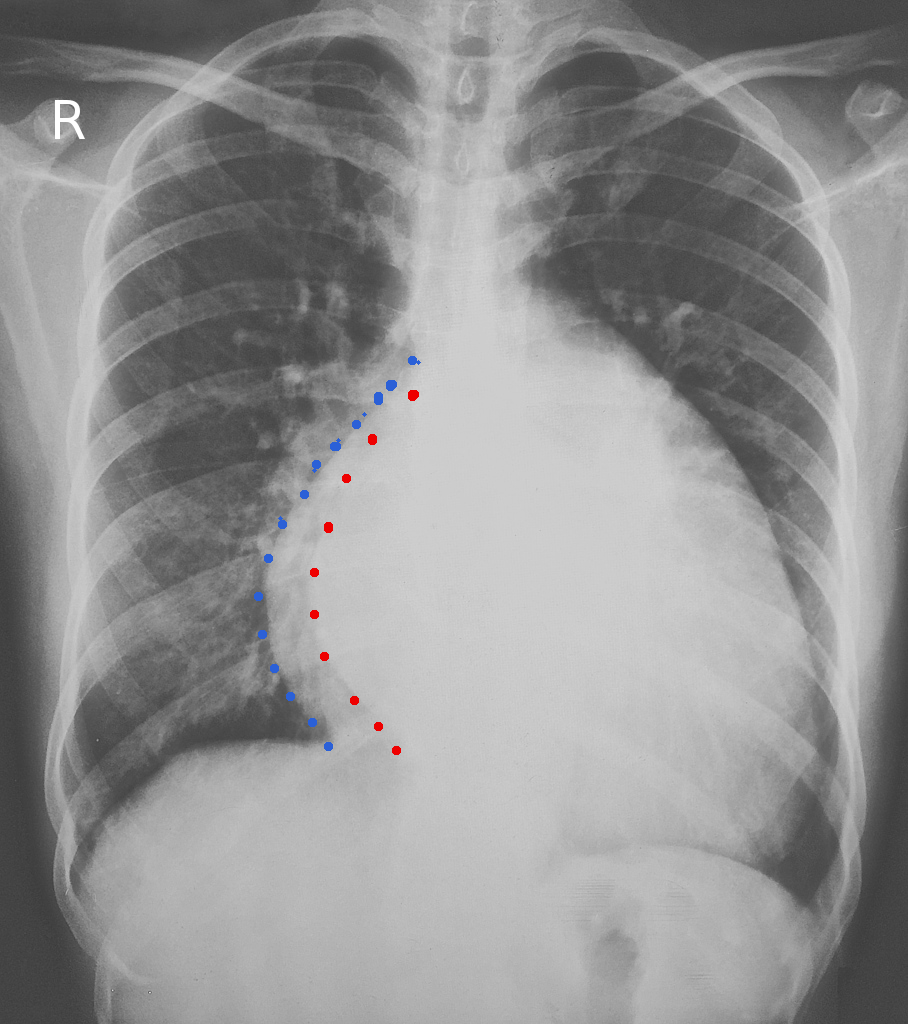
LAE occurs due to chronic pressure and volume overload, driving structural changes in the heart (see Image. Left Atrial Enlargement). Both congenital and acquired conditions can contribute to its development. Some of the key causes include:[4][6]

  • Left ventricular diastolic dysfunction: Research shows that LA size is an effective surrogate marker for the severity and chronicity of left ventricular diastolic dysfunction, AF, and heart failure with preserved ejection fraction (HFpEF).[7] This holds particularly true without confounding factors like chronic mitral valve disease.
  • AF: A reciprocal relationship exists between AF and LA dimensions. Irregular atrial contractions against a closed mitral valve can significantly raise LA pressure, leading to LAE over time. Conversely, LAE creates a substrate that predisposes to the development of AF.
  • Hypertension: Chronic systemic hypertension leads to diastolic dysfunction through left ventricular hypertrophy, a pathophysiological response to increased afterload. This, in turn, causes a gradual increase in atrial dimensions to accommodate the resultant pressure and volume afterload in the left atrium.
  • Obesity: The MONIKA/KORA study (Monitoring of Trends and Determinants in Cardiovascular Disease/Cooperative Research in the Region of Augsburg) found obesity to be a strong predictor of LAE with aging, potentially posing a greater risk than hypertension.[8]
  • Aortic stenosis: Two basic mechanisms contribute to LAE in the setting of aortic stenosis: diastolic dysfunction and increased afterload.[9] Both lead to concentric left ventricular hypertrophy. The extent of LAE correlates with left ventricular mass in patients with a concentric left ventricular pattern. In contrast, the degree of enlargement is less predictable in those with an eccentric left ventricular pattern, such as in aortic regurgitation.[10]
  • Mitral valve disease: Both mitral stenosis and regurgitation can cause LAE.
  • Left ventricular failure: Fibrosis and hypertrophy, key components of structural remodeling in the left atrium, are commonly observed in patients with left ventricular failure. These changes worsen LAE and contribute to a progressive decline in atrial function.[11]
  • LA mass or myxoma: Atrial masses, particularly myxomas, can mimic mitral valve diseases and precipitate LAE through remodeling caused by turbulence, pressure, and volume overload.[12] One study noted that excision of the mass led to gradual remodeling of the LA chamber.[13]
  • Arteriovenous fistulas: Arteriovenous fistulas, particularly those arising from congenital abnormalities or created for hemodialysis, can lead to significant LAE. This results from increased volume load, altered hemodynamic pressures, and subsequent compensatory cardiac remodeling.[14] Some case studies have also reported LAE due to arteriovenous fistulas between the coronary arteries and the left atrium.[15]
  • Left-to-right shunts: Patients with shunts, such as ventricular septal defect (VSD) and patent ductus arteriosus (PDA), can have LAE. However, atrial septal defects (ASD) are more likely to cause right atrial enlargement than LAE.
  • Athlete's heart

Epidemiology

The prevalence of LAE in the general population is not well-established. However, a study by Bombelli et al revealed that 12% of the participants developed LAE over 10 years, with an average age starting at 47. LA size is influenced by anthropomorphic factors such as age, gender, body size, and ethnicity. For example, the MONICA/KORA study reported a 9.8% prevalence in the German population, while a survey in China found a 6.43% prevalence among individuals 35 years or older.[8][16][17]

LAE is not a natural consequence of aging but is instead linked to pathophysiological changes associated with aging. Men generally have larger LA than women, and the LA size increases with body size.[4]

In specific populations, the prevalence of LAE varies significantly. For example, it ranges from 16% to 83% in hypertensive patients and those with left ventricular hypertrophy, with a pooled prevalence of approximately 32%.[3] A 20% prevalence of LAE has been observed among competitive athletes, attributed to physiological remodeling from intensive training.[18]

Pathophysiology

LAE is most commonly caused by conditions resulting from pressure overload, volume overload, or a combination of both. Mitral valve stenosis or left ventricular dysfunction, with or without associated valvular disease, can increase LA afterload. Over time, this leads to LAE as a compensatory mechanism.[19][20] 

The LA connects to the left ventricle via the mitral valve. In cases of a noncompliant left ventricle, LA pressure increases, prompting LAE to maintain left ventricular filling pressure. Similarly, right-to-left shunts, arteriovenous fistulas, and mitral regurgitation cause volume overload in the LA, subsequently leading to LAE.[4] 

LAE is an early finding in hypertensive heart disease as it serves as a strong predictor of AF.[21][22][23] A recent study involving 35658 European subjects without a history of heart failure or myocardial infarction identified 18 genetic loci associated with LAE. These loci were linked to genes implicated in cardiomyopathy (eg MYO18B, TTN, DSP, ANKRD1) and arrhythmia (eg TTN, CASQ2, MYO18B, C9orf3).[24]

From a pathophysiological perspective, changes in the atria caused by various factors are collectively termed atrial remodeling. Acute remodeling refers to atrial changes occurring within a week of exposure to stressors and are often reversible. In contrast, chronic remodeling develops over extended periods and is typically irreversible.

Atrial remodeling involves gradual, time-dependent alteration in cardiac myocytes driven by electrical, mechanical, or metabolic influences. Although complex and not fully understood, it is characterized by persistent changes in the size or function of the LA. Clinically, remodeling is often triggered by rapid atrial tachyarrhythmias or structural modifications resulting from pressure or volume overloads.

These overloads may stem from various conditions, including cardiomyopathies with diastolic dysfunction, heart failure, or valvular diseases. The extent of structural and functional changes in the LA depends on these external stressors' nature, severity, and duration.

While not completely understood yet, the process of atrial remodeling may be attributed to an array of pathological processes.[1] Notably, LA remodeling has been associated with metabolic shifts, with deterioration to fetal-like glycolytic pathways involving the oxidation of fatty acids via beta-oxidation, thereby decreasing energy production.[25]

Additionally, elevated levels of atrial and brain natriuretic peptides, aldosterone, transforming growth factor-beta, and angiotensin II (Ang II) have been observed. Ang II, aldosterone, transforming growth factor-beta, and platelet-derived growth factor collectively promote atrial fibrosis through increased cellular proliferation.[26] Systemic inflammation, driven by interleukins, C-reactive protein, and cytokines, may also play a significant role in remodeling.[27]

It is reasonable to speculate that atrial remodeling caused by AF differs significantly from diastolic dysfunction. However, the specific characteristics of remodeling associated with distinct insults remain largely undefined. Moreover, these predisposing factors often coexist; for example, hypertension and valvular heart disease frequently overlap in patients with AF.[28]The atrial changes incurred by remodeling are not solely structural but include functional and electrical alterations. These modifications can disrupt normal atrial contractibility and conduction, contributing to the progression of arrhythmias such as AF.

Left Atrial Structural Remodeling

Atrial structural remodeling is characterized by increased interstitial fibrosis and distinct structural alterations in the heart, resulting in atrial dilation. While commonly associated with heart failure, any condition promoting atrial fibrosis may trigger this process. At the cellular level, structural remodeling is driven by activating various profibrotic factors, including Ang II, transforming growth factor-beta, and platelet-derived growth factor.[29][30]

Left Atrial Functional Remodeling

Functional remodeling of the atrium manifests as reduced atrial performance, which may occur with or without accompanying changes in atrial size. LA function can be assessed using volumetric measures, such as LA ejection fraction, or advanced techniques like strain analysis. Functional impairments may arise from atrial tachyarrhythmias or altered LA pressures, often linked to atrial fibrosis and the resulting structural changes.[31][32]

Left Atrial Electrical Remodeling

Atrial tachyarrhythmias can be induced by triggers that generate ectopic activities or by substrate modifiers that promote reentry phenomena. Such modifiers are diverse, though many converge on common cellular pathophysiological mechanisms, such as shortened refractory periods, reduced actional potential duration, or altered atrial contractility. 

Electrical remodeling creates an environment that facilitates reentry phenomena by altering ion channels, pumps, and exchangers. These changes include reduced L-type calcium currents, rectifier potassium currents, acetylcholine-modulated potassium currents, and abnormal distribution and expression of connexin gap junction channels, disrupting electrical connectivity among cardiomyocytes. Fibrosis exacerbates these effects by impairing cellular coupling and promoting nonuniform impulse propagation, which can initiate and sustain reentrant atrial arrhythmias.[33]

Left Atrial Reverse Remodeling

The concept of "LA reverse remodeling: arises from the idea that LA remodeling can have deleterious or maladaptive effects. This refers to the process of ameliorating or restoring atrial function. Such reversal is most notably observed during the early structural and functional remodeling stages.[34] However, the precise extent of structural change—such as the percentage reduction in atrial volume—and the specific functional parameters to measure remain poorly defined. Most descriptions frame structural and functional remodeling as a temporal recovery, where the metrics of the LA approximate those observed in healthy subjects.[35]

Noninvasive imaging is the most practicable method for monitoring the process of LA reverse remodeling, although biochemical and cellular markers may also provide valuable insights. Advances in imaging techniques have further enhanced the ability to evaluate structural and functional changes with greater accuracy.

History and Physical

LAE often presents asymptomatically but, when symptomatic, reflects underlying conditions such as AF, cardiac insufficiency, thromboembolism, or mitral valvular disease. Key manifestations include:

  • Dyspnea: Exertional or orthopnea due to elevated pulmonary pressures.[36]

  • Fatigue: Reduced cardiac efficiency impairing systemic oxygen delivery.

  • Palpitations: Irregular pulsations, often linked to AF.

  • Chest discomfort: Sensations of pain or unease in the chest may arise, particularly in association with mitral valvular afflictions or diminished coronary perfusion.

  • Cough or hemoptysis: Resulting from pulmonary congestion or elevated atrial pressures.

  • Peripheral edema: From systemic fluid retention in advanced cardiac failure.

  • Syncope or dizziness: Rare, caused by arrhythmias or poor cerebral perfusion.

  • Cerebral events: Stroke or transient ischemic attack from atrial thrombus or embolism.

  • Ortner syndrome: Rare hoarseness caused by left pharyngeal nerve compression.

  • Dysphagia megalatriensis: esophageal compression causing difficulty swallowing solids.

In some cases, early LAE linked to conditions like left ventricular hypertrophy, mitral valve disease (ie stenosis or regurgitation), or systemic hypertension may present without symptoms, detected only via incidental electrocardiogram (ECG) or echocardiogram findings. A thorough evaluation of medical, social, and family history is essential to identify risk factors such as congenital heart disease, valvular abnormalities, arrhythmias, or hypertension.

Evaluation

Atrial size and function can be evaluated using echocardiography, cardiac computed tomography (CCT), and cardiac magnetic resonance (CMR). Echocardiography is the preferred modality due to its accessibility and safety.[37][38]. Notably, echocardiographic measurements of LA volume show a strong correlation with CCT and CMR.[39]

Echocardiogram

LA size can be measured using 2-dimensional and M-mode echocardiography.[37][40][41] However, linear measurement may not accurately reflect LA size, particularly in cases of asymmetrical enlargement. This limitation has led to a paradigm shift toward assessing LA volume, which is now the standard for evaluating LA remodeling.  

LA volume is calculated using the biplane area-length method or Simpson’s biplane method, as recommended by the American Society of Echocardiography and the European Association of Echocardiography. LA volume should be indexed to body surface area or LA volume index (LAVi) to account for body habitus.[42][43] A normal LAVi is 22 mL/m² plus or minus 6 mL/m2, with LA enlargement defined as the following:

  • Mild: >28 mL/m2 
  • Moderate: ≥34 mL/m²
  • Severe: ≥40 mL/m²

Newer 3-dimensional echocardiography offers improved accuracy for assessing LA volume and enlargement.[44] This advancement allows for better evaluation of LA function and helps monitor disease progression and response to treatment. 

Electrocardiogram

Early detection through ECG can help identify patients at risk and enable timely intervention to prevent progression to more severe cardiovascular conditions. The presence of any 1 of the following ECG (see Image. ECG Left Atrial Enlargement) findings would prompt further investigation for LAE in the appropriate clinical context.[45][46][47][48] 

  • P wave in any lead >0.11 s
  • Biphasic P wave with terminal negative portion >0.04 s duration and deeper than 1 mm in lead V1
  • Notched P wave with an interpeak duration >0.04 s in lead II; P mitral
  • P wave axis <30º

Treatment / Management

There are no guidelines-based treatments for LAE, and no known medical therapy has been shown to reverse LA remodeling. The primary focus of care is the identification and management of underlying conditions contributing to LAE. 

Medical Management

Atrial fibrillation: AF is a prevalent arrhythmia strongly associated with structural and functional changes in the LA. LAE not only predisposes individuals to AF by creating a substrate for arrhythmia but is also exacerbated by LAE, resulting in a reciprocal relationship that contributes to disease progression. The CHA2DS2-VASc score assesses the risk of stroke by considering multiple factors (see Table 1. CHA2DS2-VASc):

Table 1. CHA2DS2-VASc

  Condition Points
C Congestive heart failure 1
H Hypertension 1
A Age ≥75 2
D Diabetes mellitus 1
S2 Prior stroke, TIA, thromboembolism 2
V Vascular disease 1
A Age 65-74 1
Sc Sex 1

When initiating anticoagulation, the risk of bleeding should be evaluated using the HAS-BLED score (see Table 2. HAS-BLED Score). A score of 0 to 2 indicates a low risk of bleeding, while a score of 3 or higher signifies a high risk.

Table 2. HAS-BLED Score

  Risk Factor Score
H Hypertension 1
A Abnormal renal or liver function (1 point each) 1 or 2
S Stroke

1

B Bleeding tendency 1
L Labile INR 1
E Age >65  
D Drugs or alcohol (1 point each) 1 or 2

Long-term anticoagulation is reserved for patients with LAE and AF who have a high stroke risk based on the CHA2DS2-VASc score, especially those with additional risk factors. It is particularly indicated for individuals who cannot undergo definitive treatment or require precautionary management to mitigate thromboembolic risk. Treatment modalities include:

  • Vitamin K antagonists:
    • Warfarin is the traditional choice for anticoagulation
    • Requires regular monitoring of INR (International Normalized Ratio) to maintain therapeutic levels (2.0–3.0 for most conditions)
    • Dietary and drug interactions are significant considerations
  • Direct oral anticoagulants:
    • Includes dabigatran, rivaroxaban, apixaban, and edoxaban
    • Preferred for nonvalvular AF and offers predictable anticoagulation with fewer monitoring requirements
    • Reduced risk of intracranial bleeding compared to warfarin
  • Aspirin or antiplatelet agents: 
    • May be used in low-risk patients where anticoagulation is not deemed necessary.
    • Less effective for stroke prevention in AF or LAE compared to anticoagulants.

Valvular abnormalities: Mitral valve stenosis or regurgitation is managed with a combination of symptom-relieving medical therapy, percutaneous mitral valvuloplasty, and surgical intervention when indicated. Additionally, anticoagulation therapy may be required if arrhythmias develop to reduce the risk of thromboembolic risks.

Hypertension is the most common modifiable risk factor for cardiovascular diseases. Many antihypertensive medications are available to help patients manage their blood pressure effectively. In addition, lifestyle modifications such as adopting a low-salt diet, engaging in regular physical activity, limiting alcohol consumption, and quitting smoking are crucial measures to control blood pressure.

Left ventricular dysfunction and heart failure: Medical therapy remains the cornerstone of managing these conditions. The primary treatments include β-blockers (eg carvedilol, metoprolol succinate, bisoprolol), angiotensin-converting enzyme inhibitors or angiotensin receptor blockers, and mineralocorticoid receptor antagonists. Additionally, the angiotensin receptor-neprilysin inhibitor, sacubitril-valsartan, has recently been recognized for its mortality benefits in patients with heart failure with reduced ejection fraction.[49][50] 

Left Atrial Appendage Exclusion

LA appendage exclusion is a surgical procedure designed to isolate or remove the LA appendage. Its primary goal is to reduce the risk of thromboembolism, particularly in patients with AF, as the LA appendage is a common site for clot formation that can lead to strokes or systemic embolism. This procedure is especially beneficial for patients who cannot tolerate long-term anticoagulation therapy.

Studies have shown that LA appendage exclusion during valvular surgery in patients with AF significantly reduces cerebrovascular events and in-hospital mortality compared to patients who do not undergo the procedure.[51] Similar benefits, including reduced cerebrovascular events and mortality, have also been observed in patients undergoing LA appendage exclusion without AF.[52] Methods of exclusion are as follows:

  • Surgical exclusion involves the removal or ligation of the LA appendage during open-heart surgery, such as during mitral valve or coronary artery bypass surgery. While an invasive approach, it offers the advantage of antithrombotic-free postprocedural survival.
  • Percutaneous occlusion is a less invasive method that utilizes a catheter-based approach to pace a device (e.g., Watchman, Amplatzer Amulet) to seal the LA appendage. It requires short-term postprocedural anticoagulation therapy (if feasible) followed by ongoing antithrombotic treatment.
  • Epicardial ligation is a technique that uses external clips or sutures (eg, AtriClip, LARIAT) to occlude the LA appendage during minimally invasive or open-heart procedures, providing a surgical alternative for patients with specific anatomical or clinical considerations. 

Prognosis

LAE is not a benign condition. In the absence of left ventricular hypertrophy, LA diameter serves as an independent prognosticator of fatal and nonfatal cardiovascular events, regardless of other risk factors such as hypertension.[53] LA size is independently linked to all-cause mortality in both sexes and to ischemic stroke in women.[54] Identifying and managing the underlying causes of LAE is crucial to prevent catastrophic outcomes and improve the patient's quality of life.

Complications

The most commonly encountered complications of LAE include:[4][53]

  • AF
  • Stroke
  • HFpEF

Deterrence and Patient Education

Patients with LAE may remain asymptomatic throughout their lifetime. However, the presence of LAE could indicate underlying cardiac pathology that warrants further investigation. Since there is no specific treatment for LAE, patients need to be informed about any underlying conditions and receive appropriate management, which may range from lifestyle modifications to surgical evaluation and intervention. 

Pearls and Other Issues

Key factors to keep in mind regarding LAE include the following:

  • LAE is often a sign of underlying cardiac pathology, such as left ventricular dysfunction, valvular heart disease, or hypertension. It serves as an independent predictor of adverse cardiovascular outcomes, including stroke and heart failure.
  • Common causes of LAE are hypertension, mitral valve disease, AF, and HFpEF.
  • Patients with LAE may be asymptomatic but may present with symptoms of the underlying pathology.
  • LAE is associated with an increased risk of thromboembolic events, including ischemic stroke, particularly in patients with AF.
  • Echocardiography is the gold standard for assessing LA size, with LA diameter and volume being essential metrics.
  • Treat underlying causes either medically or surgically.
  • LAE is a marker of poor prognosis in conditions like heart failure and AF. It is associated with increased mortality and morbidity from cardiovascular causes. 

Enhancing Healthcare Team Outcomes

There are guidelines for diagnosing LAE but no specific therapeutic management. Therefore, the most critical aspect of management is addressing any underlying cardiac pathology and educating patients to prevent fatal outcomes. Given that LAE is a prognostic marker for fatal and nonfatal cardiovascular events, optimal management involves a multidisciplinary approach, including collaboration between healthcare professionals such as physicians and advanced practitioners, nurses, pharmacists, physiotherapists, dietitians, physical trainers, and psychologists.

Once a diagnosis of LAE is established, the patient should receive education about the condition and the need for further evaluation of any coexisting cardiac pathologies. Asymptomatic patients should continue with annual exams, including echocardiograms. Those with underlying hypertension should focus on strict blood pressure control, while patients with heart failure require medical management and lifestyle modifications. A dietary consultation can assist in adopting a low-salt diet and healthy eating habits.

Patients with LAE and palpitations must undergo an ECG and Holter monitor to evaluate any underlying arrhythmia that can develop as a consequence of LAE. Many patients may require anticoagulation therapy to prevent thromboembolic events. Pharmacists play a crucial role in ensuring timely medication administration and advising on medication selection, dosing, and reconciliation to avoid drug interactions. They also help report adverse drug reactions to the healthcare team, facilitate medication changes, and provide patient counseling. 

Cardiovascular-trained nurses are vital members of the care team. They assist with medication administration, monitor patient compliance, provide counseling, and address patient and family questions. A fully collaborative, interprofessional team approach is essential for achieving the best outcomes for patients with LAE.


(Click Image to Enlarge)
<p><strong>Left Atrial Enlargement.</strong> Chest radiograph outlining an enlarged cardiac silhouette.&nbsp;</p>

Left Atrial Enlargement. Chest radiograph outlining an enlarged cardiac silhouette. 

Image courtesy S Bhimji MD


(Click Image to Enlarge)
<p><strong>ECG&nbsp;Left Atrial Enlargement.</strong> An ECG tracing indicative of left atrial enlargement.</p>

ECG Left Atrial Enlargement. An ECG tracing indicative of left atrial enlargement.

Image courtesy S Bhimji MD

Details

Updated:

1/22/2025 12:54:15 AM

Looking for an easier read?

Click here for a simplified version

References


[1]

Abhayaratna WP, Seward JB, Appleton CP, Douglas PS, Oh JK, Tajik AJ, Tsang TS. Left atrial size: physiologic determinants and clinical applications. Journal of the American College of Cardiology. 2006 Jun 20:47(12):2357-63     [PubMed PMID: 16781359]


[2]

Cuspidi C, Negri F, Sala C, Valerio C, Mancia G. Association of left atrial enlargement with left ventricular hypertrophy and diastolic dysfunction: a tissue Doppler study in echocardiographic practice. Blood pressure. 2012 Feb:21(1):24-30. doi: 10.3109/08037051.2011.618262. Epub 2011 Oct 13     [PubMed PMID: 21992028]


[3]

Cuspidi C, Rescaldani M, Sala C. Prevalence of echocardiographic left-atrial enlargement in hypertension: a systematic review of recent clinical studies. American journal of hypertension. 2013 Apr:26(4):456-64. doi: 10.1093/ajh/hpt001. Epub 2013 Feb 6     [PubMed PMID: 23388831]

Level 1 (high-level) evidence

[4]

Patel DA, Lavie CJ, Milani RV, Shah S, Gilliland Y. Clinical implications of left atrial enlargement: a review. Ochsner journal. 2009 Winter:9(4):191-6     [PubMed PMID: 21603443]


[5]

Kizer JR, Bella JN, Palmieri V, Liu JE, Best LG, Lee ET, Roman MJ, Devereux RB. Left atrial diameter as an independent predictor of first clinical cardiovascular events in middle-aged and elderly adults: the Strong Heart Study (SHS). American heart journal. 2006 Feb:151(2):412-8     [PubMed PMID: 16442908]


[6]

Rusinaru D, Bohbot Y, Kowalski C, Ringle A, Maréchaux S, Tribouilloy C. Left Atrial Volume and Mortality in Patients With Aortic Stenosis. Journal of the American Heart Association. 2017 Oct 31:6(11):. doi: 10.1161/JAHA.117.006615. Epub 2017 Oct 31     [PubMed PMID: 29089338]


[7]

Thomas L, Muraru D, Popescu BA, Sitges M, Rosca M, Pedrizzetti G, Henein MY, Donal E, Badano LP. Evaluation of Left Atrial Size and Function: Relevance for Clinical Practice. Journal of the American Society of Echocardiography : official publication of the American Society of Echocardiography. 2020 Aug:33(8):934-952. doi: 10.1016/j.echo.2020.03.021. Epub     [PubMed PMID: 32762920]


[8]

Stritzke J, Markus MR, Duderstadt S, Lieb W, Luchner A, Döring A, Keil U, Hense HW, Schunkert H, MONICA/KORA Investigators. The aging process of the heart: obesity is the main risk factor for left atrial enlargement during aging the MONICA/KORA (monitoring of trends and determinations in cardiovascular disease/cooperative research in the region of Augsburg) study. Journal of the American College of Cardiology. 2009 Nov 17:54(21):1982-9. doi: 10.1016/j.jacc.2009.07.034. Epub     [PubMed PMID: 19909880]


[9]

Pińska M, Sorysz D, Frączek-Jucha M, Kruszec P, Róg B, Myć J, Krawczyk-Ożóg A, Sobień B, Stopyra-Pach K, Sarnecka A, Stąpór M, Olszanecka A, Golińska-Grzybała K, Nessler J, Gackowski A. The Prognostic Significance of Atrial Fibrillation and Left Atrium Size in Patients with Aortic Stenosis. Kardiologiia. 2023 Dec 27:63(12):66-71. doi: 10.18087/cardio.2023.12.n2608. Epub 2023 Dec 27     [PubMed PMID: 38156492]


[10]

Cioffi G, Stefenelli C. Comparison of left ventricular geometry and left atrial size and function in patients with aortic stenosis versus those with pure aortic regurgitation. The American journal of cardiology. 2002 Sep 15:90(6):601-6     [PubMed PMID: 12231084]


[11]

Hanif W, Alex L, Su Y, Shinde AV, Russo I, Li N, Frangogiannis NG. Left atrial remodeling, hypertrophy, and fibrosis in mouse models of heart failure. Cardiovascular pathology : the official journal of the Society for Cardiovascular Pathology. 2017 Sep-Oct:30():27-37. doi: 10.1016/j.carpath.2017.06.003. Epub 2017 Jun 21     [PubMed PMID: 28759817]


[12]

COHEN AI, McINTOSH HD, ORGAIN ES. The mimetic nature of left atrial myxomas. Report of a case presenting as a severe systemic illness and simulating massive mitral insufficiency at cardiac catheterization. The American journal of cardiology. 1963 Jun:11():802-7     [PubMed PMID: 14021991]

Level 3 (low-level) evidence

[13]

Padalino MA, Basso C, Moreolo GS, Thiene G, Stellin G. Left atrial myxoma in a child: case report and review of the literature. Cardiovascular pathology : the official journal of the Society for Cardiovascular Pathology. 2003 Jul-Aug:12(4):233-6     [PubMed PMID: 12826294]

Level 3 (low-level) evidence

[14]

Iwashima Y, Horio T, Takami Y, Inenaga T, Nishikimi T, Takishita S, Kawano Y. Effects of the creation of arteriovenous fistula for hemodialysis on cardiac function and natriuretic peptide levels in CRF. American journal of kidney diseases : the official journal of the National Kidney Foundation. 2002 Nov:40(5):974-82     [PubMed PMID: 12407642]


[15]

Aoyagi S, Tobinaga S, Saisho H. Enlargement of the Excluded Left Atrial Appendage With Thrombus. International heart journal. 2017 Feb 7:58(1):144-146. doi: 10.1536/ihj.16-113. Epub 2017 Jan 11     [PubMed PMID: 28077818]


[16]

Bombelli M, Cuspidi C, Facchetti R, Sala C, Tadic M, Brambilla G, Re A, Villa P, Grassi G, Mancia G. New-onset left atrial enlargement in a general population. Journal of hypertension. 2016 Sep:34(9):1838-45. doi: 10.1097/HJH.0000000000001022. Epub     [PubMed PMID: 27379539]


[17]

Ou Q, Chen Y, Yu S, Guo X, Zhao H, Sun Y. Prevalence of left atrial enlargement and its risk factors in general Chinese population. BMC cardiovascular disorders. 2016 Mar 5:16():53. doi: 10.1186/s12872-016-0229-z. Epub 2016 Mar 5     [PubMed PMID: 26944401]


[18]

Pelliccia A, Maron BJ, Di Paolo FM, Biffi A, Quattrini FM, Pisicchio C, Roselli A, Caselli S, Culasso F. Prevalence and clinical significance of left atrial remodeling in competitive athletes. Journal of the American College of Cardiology. 2005 Aug 16:46(4):690-6     [PubMed PMID: 16098437]

Level 3 (low-level) evidence

[19]

Pape LA, Price JM, Alpert JS, Ockene IS, Weiner BH. Relation of left atrial size to pulmonary capillary wedge pressure in severe mitral regurgitation. Cardiology. 1991:78(4):297-303     [PubMed PMID: 1889048]


[20]

Appleton CP, Galloway JM, Gonzalez MS, Gaballa M, Basnight MA. Estimation of left ventricular filling pressures using two-dimensional and Doppler echocardiography in adult patients with cardiac disease. Additional value of analyzing left atrial size, left atrial ejection fraction and the difference in duration of pulmonary venous and mitral flow velocity at atrial contraction. Journal of the American College of Cardiology. 1993 Dec:22(7):1972-82     [PubMed PMID: 8245357]


[21]

Aljizeeri A, Gin K, Barnes ME, Lee PK, Nair P, Jue J, Tsang TS. Atrial remodeling in newly diagnosed drug-naive hypertensive subjects. Echocardiography (Mount Kisco, N.Y.). 2013 Jul:30(6):627-33. doi: 10.1111/echo.12119. Epub 2013 Jan 30     [PubMed PMID: 23360480]


[22]

Vaziri SM, Larson MG, Benjamin EJ, Levy D. Echocardiographic predictors of nonrheumatic atrial fibrillation. The Framingham Heart Study. Circulation. 1994 Feb:89(2):724-30     [PubMed PMID: 8313561]


[23]

Tsang TS, Barnes ME, Bailey KR, Leibson CL, Montgomery SC, Takemoto Y, Diamond PM, Marra MA, Gersh BJ, Wiebers DO, Petty GW, Seward JB. Left atrial volume: important risk marker of incident atrial fibrillation in 1655 older men and women. Mayo Clinic proceedings. 2001 May:76(5):467-75     [PubMed PMID: 11357793]


[24]

Ahlberg G, Andreasen L, Ghouse J, Bertelsen L, Bundgaard H, Haunsø S, Svendsen JH, Olesen MS. Genome-wide association study identifies 18 novel loci associated with left atrial volume and function. European heart journal. 2021 Nov 21:42(44):4523-4534. doi: 10.1093/eurheartj/ehab466. Epub     [PubMed PMID: 34338756]


[25]

Barger PM, Kelly DP. Fatty acid utilization in the hypertrophied and failing heart: molecular regulatory mechanisms. The American journal of the medical sciences. 1999 Jul:318(1):36-42     [PubMed PMID: 10408759]


[26]

Verheule S, Sato T, Everett T 4th, Engle SK, Otten D, Rubart-von der Lohe M, Nakajima HO, Nakajima H, Field LJ, Olgin JE. Increased vulnerability to atrial fibrillation in transgenic mice with selective atrial fibrosis caused by overexpression of TGF-beta1. Circulation research. 2004 Jun 11:94(11):1458-65     [PubMed PMID: 15117823]


[27]

Chung MK, Martin DO, Sprecher D, Wazni O, Kanderian A, Carnes CA, Bauer JA, Tchou PJ, Niebauer MJ, Natale A, Van Wagoner DR. C-reactive protein elevation in patients with atrial arrhythmias: inflammatory mechanisms and persistence of atrial fibrillation. Circulation. 2001 Dec 11:104(24):2886-91     [PubMed PMID: 11739301]


[28]

Psychari SN, Apostolou TS, Sinos L, Hamodraka E, Liakos G, Kremastinos DT. Relation of elevated C-reactive protein and interleukin-6 levels to left atrial size and duration of episodes in patients with atrial fibrillation. The American journal of cardiology. 2005 Mar 15:95(6):764-7     [PubMed PMID: 15757607]


[29]

Kallergis EM, Manios EG, Kanoupakis EM, Mavrakis HE, Arfanakis DA, Maliaraki NE, Lathourakis CE, Chlouverakis GI, Vardas PE. Extracellular matrix alterations in patients with paroxysmal and persistent atrial fibrillation: biochemical assessment of collagen type-I turnover. Journal of the American College of Cardiology. 2008 Jul 15:52(3):211-5. doi: 10.1016/j.jacc.2008.03.045. Epub     [PubMed PMID: 18617070]


[30]

Chahine Y, Chamoun N, Kassar A, Bockus L, Macheret F, Akoum N. Atrial fibrillation substrate and impaired left atrial function: a cardiac MRI study. Europace : European pacing, arrhythmias, and cardiac electrophysiology : journal of the working groups on cardiac pacing, arrhythmias, and cardiac cellular electrophysiology of the European Society of Cardiology. 2024 Nov 1:26(11):. doi: 10.1093/europace/euae258. Epub     [PubMed PMID: 39523754]


[31]

Lu S, Liu H, Sun J, Zhang J, Li L, Tang Q, Liu Y, Deng Y. Evaluation of left atrial and ventricular remodeling in atrial fibrillation subtype by using speckle tracking echocardiography. Frontiers in cardiovascular medicine. 2023:10():1208577. doi: 10.3389/fcvm.2023.1208577. Epub 2023 Aug 10     [PubMed PMID: 37636311]


[32]

Casaclang-Verzosa G, Gersh BJ, Tsang TS. Structural and functional remodeling of the left atrium: clinical and therapeutic implications for atrial fibrillation. Journal of the American College of Cardiology. 2008 Jan 1:51(1):1-11. doi: 10.1016/j.jacc.2007.09.026. Epub     [PubMed PMID: 18174029]


[33]

Cha TJ, Ehrlich JR, Chartier D, Qi XY, Xiao L, Nattel S. Kir3-based inward rectifier potassium current: potential role in atrial tachycardia remodeling effects on atrial repolarization and arrhythmias. Circulation. 2006 Apr 11:113(14):1730-7     [PubMed PMID: 16585394]

Level 2 (mid-level) evidence

[34]

Kumagai K, Nakashima H, Urata H, Gondo N, Arakawa K, Saku K. Effects of angiotensin II type 1 receptor antagonist on electrical and structural remodeling in atrial fibrillation. Journal of the American College of Cardiology. 2003 Jun 18:41(12):2197-204     [PubMed PMID: 12821247]


[35]

Kokubu N, Yuda S, Tsuchihashi K, Hashimoto A, Nakata T, Miura T, Ura N, Nagao K, Tsuzuki M, Wakabayashi C, Shimamoto K. Noninvasive assessment of left atrial function by strain rate imaging in patients with hypertension: a possible beneficial effect of renin-angiotensin system inhibition on left atrial function. Hypertension research : official journal of the Japanese Society of Hypertension. 2007 Jan:30(1):13-21     [PubMed PMID: 17460367]


[36]

Gard EK, Beale AL, Telles F, Silvestry FE, Hanff T, Hummel SL, Litwin SE, Petrie MC, Shah SJ, Borlaug BA, Burkhoff D, Komtebedde J, Kaye DM, Nanayakkara S. Left atrial enlargement is associated with pulmonary vascular disease in heart failure with preserved ejection fraction. European journal of heart failure. 2023 Jun:25(6):806-814. doi: 10.1002/ejhf.2805. Epub 2023 Mar 15     [PubMed PMID: 36847073]


[37]

Hoit BD. Left atrial size and function: role in prognosis. Journal of the American College of Cardiology. 2014 Feb 18:63(6):493-505. doi: 10.1016/j.jacc.2013.10.055. Epub 2013 Nov 27     [PubMed PMID: 24291276]


[38]

To AC, Flamm SD, Marwick TH, Klein AL. Clinical utility of multimodality LA imaging: assessment of size, function, and structure. JACC. Cardiovascular imaging. 2011 Jul:4(7):788-98. doi: 10.1016/j.jcmg.2011.02.018. Epub     [PubMed PMID: 21757171]


[39]

Kircher B, Abbott JA, Pau S, Gould RG, Himelman RB, Higgins CB, Lipton MJ, Schiller NB. Left atrial volume determination by biplane two-dimensional echocardiography: validation by cine computed tomography. American heart journal. 1991 Mar:121(3 Pt 1):864-71     [PubMed PMID: 2000754]

Level 1 (high-level) evidence

[40]

Hirata T, Wolfe SB, Popp RL, Helmen CH, Feigenbaum H. Estimation of left atrial size using ultrasound. American heart journal. 1969 Jul:78(1):43-52     [PubMed PMID: 5794795]


[41]

Sahn DJ, DeMaria A, Kisslo J, Weyman A. Recommendations regarding quantitation in M-mode echocardiography: results of a survey of echocardiographic measurements. Circulation. 1978 Dec:58(6):1072-83     [PubMed PMID: 709763]

Level 3 (low-level) evidence

[42]

Vasan RS, Larson MG, Levy D, Evans JC, Benjamin EJ. Distribution and categorization of echocardiographic measurements in relation to reference limits: the Framingham Heart Study: formulation of a height- and sex-specific classification and its prospective validation. Circulation. 1997 Sep 16:96(6):1863-73     [PubMed PMID: 9323074]

Level 1 (high-level) evidence

[43]

Tsang TS, Abhayaratna WP, Barnes ME, Miyasaka Y, Gersh BJ, Bailey KR, Cha SS, Seward JB. Prediction of cardiovascular outcomes with left atrial size: is volume superior to area or diameter? Journal of the American College of Cardiology. 2006 Mar 7:47(5):1018-23     [PubMed PMID: 16516087]


[44]

Takeuchi M, Kitano T, Nabeshima Y, Otsuji Y, Otani K. Left ventricular and left atrial volume ratio assessed by three-dimensional echocardiography: Novel indices for evaluating age-related change in left heart chamber size. Physiological reports. 2019 Dec:7(23):e14300. doi: 10.14814/phy2.14300. Epub     [PubMed PMID: 31814325]


[45]

Hazen MS, Marwick TH, Underwood DA. Diagnostic accuracy of the resting electrocardiogram in detection and estimation of left atrial enlargement: an echocardiographic correlation in 551 patients. American heart journal. 1991 Sep:122(3 Pt 1):823-8     [PubMed PMID: 1831587]


[46]

Munuswamy K, Alpert MA, Martin RH, Whiting RB, Mechlin NJ. Sensitivity and specificity of commonly used electrocardiographic criteria for left atrial enlargement determined by M-mode echocardiography. The American journal of cardiology. 1984 Mar 1:53(6):829-32     [PubMed PMID: 6230922]


[47]

Waggoner AD, Adyanthaya AV, Quinones MA, Alexander JK. Left atrial enlargement. Echocardiographic assessment of electrocardiographic criteria. Circulation. 1976 Oct:54(4):553-7     [PubMed PMID: 134852]


[48]

Ikram H, Drysdale P, Bones PJ, Chan W. The non-invasive recognition of left atrial enlargement: comparison of electro- and echocardiographic measurements. Postgraduate medical journal. 1977 Jul:53(621):356-9     [PubMed PMID: 142246]


[49]

Almufleh A, Marbach J, Chih S, Stadnick E, Davies R, Liu P, Mielniczuk L. Ejection fraction improvement and reverse remodeling achieved with Sacubitril/Valsartan in heart failure with reduced ejection fraction patients. American journal of cardiovascular disease. 2017:7(6):108-113     [PubMed PMID: 29348971]


[50]

Oparil S, Schmieder RE. New approaches in the treatment of hypertension. Circulation research. 2015 Mar 13:116(6):1074-95. doi: 10.1161/CIRCRESAHA.116.303603. Epub     [PubMed PMID: 25767291]


[51]

Elbadawi A, Olorunfemi O, Ogunbayo GO, Saad M, Elgendy IY, Arif Z, Badran H, Saheed D, Ahmed HMA, Rao M. Cardiovascular Outcomes With Surgical Left Atrial Appendage Exclusion in Patients With Atrial Fibrillation Who Underwent Valvular Heart Surgery (from the National Inpatient Sample Database). The American journal of cardiology. 2017 Jun 15:119(12):2056-2060. doi: 10.1016/j.amjcard.2017.03.037. Epub 2017 Mar 29     [PubMed PMID: 28438308]


[52]

Waterford SD. Left Atrial Appendage Exclusion in Patients Without Atrial Fibrillation Undergoing Cardiac Surgery. Innovations (Philadelphia, Pa.). 2022 Nov-Dec:17(6):461-462. doi: 10.1177/15569845221146109. Epub 2022 Dec 26     [PubMed PMID: 36571261]


[53]

Bombelli M, Facchetti R, Cuspidi C, Villa P, Dozio D, Brambilla G, Grassi G, Mancia G. Prognostic significance of left atrial enlargement in a general population: results of the PAMELA study. Hypertension (Dallas, Tex. : 1979). 2014 Dec:64(6):1205-11. doi: 10.1161/HYPERTENSIONAHA.114.03975. Epub 2014 Sep 8     [PubMed PMID: 25201892]


[54]

Bouzas-Mosquera A, Broullón FJ, Álvarez-García N, Méndez E, Peteiro J, Gándara-Sambade T, Prada O, Mosquera VX, Castro-Beiras A. Left atrial size and risk for all-cause mortality and ischemic stroke. CMAJ : Canadian Medical Association journal = journal de l'Association medicale canadienne. 2011 Jul 12:183(10):E657-64. doi: 10.1503/cmaj.091688. Epub 2011 May 24     [PubMed PMID: 21609990]