Horseshoe Kidney

Earn CME/CE in your profession:


Continuing Education Activity

Horseshoe kidneys are the most common fusion defect of the kidneys occurring in approximately 1:500 individuals. They are characterized by abnormalities in the position, rotation, and vascular supply of the kidney. Although largely benign, they are associated with urological sequelae due largely to the associated ureteric obstruction and impaired urinary drainage. This activity reviews the etiology, presentation, evaluation, and treatment of horseshoe kidney and highlights the roles of the interprofessional team in evaluating and treating patients with this condition.

Objectives:

  • Explain the etiology of horseshoe kidneys.
  • Describe the imaging modalities that can be used to evaluate for horseshoe kidneys.
  • Review potential sequelae of horseshoe kidneys.
  • Summarize the importance of collaboration amongst healthcare professionals who care for patients with horseshoe kidneys.

Introduction

Horseshoe kidneys are the most common fusion defect of the kidneys, but this still amounts to only about 0.25% of the population [1]. Initially described during autopsies by da Carpi performed in 1522, they are characterized by abnormalities in the position, rotation, and vascular supply of the kidney [2]. Horseshoe kidneys are identified as having functioning renal masses present on both sides of the vertebral column fused together with ureters that remain uncrossed from the renal hilum to the urinary bladder [3]. The isthmus connecting the two renal masses may be positioned in the midline or laterally resulting in an asymmetric horseshoe kidney, 70% of which are left dominant. The isthmus consists of renal parenchyma in about 80% of cases with the remainder being composed of a fibrous band. In more than 90% of cases, fusion occurs at the lower pole, although fusion may occur at the upper pole in a small minority of cases [4][5][6][7].

Etiology

Despite cases of familial clustering, no clear genetic cause has been described for horseshoe kidneys, although several etiological factors may contribute to their development [8]. These include abnormal migration of nephrogenic cells across the primitive streak, alterations in the intrauterine environment (with teratogenic drugs such as thalidomide, alcohol consumption and glycemic control causing an increase in incidence), and structural factors such as flexion/rotation of the caudal spine and narrowed arterial forks during migration [4][9][10][11]. Traditionally textbooks quote fusion as occurring between weeks four and six of development, although there is some evidence for later fusion, particularly when the isthmus is fibrous rather than renal parenchyma.

Epidemiology

The incidence of horseshoe kidney is approximately 1 in 500 in the normal population with a male preponderance of 2:1 [1][4]. The incidence is higher in those who present to urology clinics (1 in 304), and with some chromosomal disorders. These include Edward syndrome at approximately 67%, Turner syndrome at 14% to 20%, and Down syndrome at about 1% [12][13][14].

History and Physical

Horseshoe kidneys are often asymptomatic, and so are often identified incidentally. The presentation can otherwise be non-specific with one study showing that the two most common presenting complaints of children with a horseshoe kidney are abdominal pain or symptoms of a urinary tract infection [15].

The kidneys are normally located in the retroperitoneum between the transverse processes of T12 and L3 with the left kidney slightly more superior than the right. The upper poles are normally positioned slightly medially and posteriorly relative to the lower poles. Horseshoe kidneys are different in three main ways: location, orientation, and vasculature [2]. The horseshoe kidney's ascent is often quoted to be held back by the inferior mesenteric artery at L3 however, the horseshoe kidney can also be found lower in the abdomen and pelvis. During weeks six to eight of development, the renal ascent is coupled with a 90-degree medial rotation. Due to the isthmus, however, horseshoe kidneys experience malrotation, and consequently, the ureters need to either pass over the isthmus or down the anterior surface of the kidneys which can cause urinary drainage problems and stasis [16][17]. Horseshoe kidneys also show a greater variation in the origin and number of renal arteries and veins [4][6][17]. These are largely dependent on where during development ascent has terminated. In one study of 90 horseshoe kidneys, 387 arteries were identified [4]. Despite this, the normal intra-renal vascular segmental pattern remains, and the ligation or division of any of these arteries results in ischemic segmental renal ischemia or necrosis due to their poor collateral arterial supply [18]. The incidence of renal vein anomalies in horseshoe kidneys is also high (23%)[4].

Evaluation

Horseshoe kidneys can be identified using most abdominal imaging modalities. The diagnosis of a horseshoe kidney is most commonly made using either ultrasound or CT scans [19]. CT and MRI are the best for demonstrating the anatomy and can detect accessory vasculature and surrounding structures [4][19][20]. Optimally, a CT urogram (CT scan of the abdomen and pelvis without and then with IV contrast) will be performed.  This allows for the identification of stones as well as urinary blockages and ureteropelvic junction (UPJ) obstructions. MRI examinations can be used where ionizing radiation should be avoided or in patients who cannot tolerate standard IV contrast media. It is also sometimes possible to identify horseshoe kidneys on plain radiography through visualization of the perinephric fat in association with an altered renal axis. The lower poles would be positioned much more medial than normal with the kidneys sitting lower in the abdomen than normal [19]. Nuclear medicine radionuclide renal scans can be helpful in differentiating true obstruction from passively dilated systems as well as assisting in the diagnosis of UPJ obstructions. Voiding cystourethrograms can be used to identify vesicoureteral reflux which is more common in horseshoe kidney patients than in the general population. 

Treatment / Management

Shockwave lithotripsy for nephrolithiasis is less effective in horseshoe kidneys due to problems localizing the energy for pelvic stones and poor stone fragment clearance due to impaired renal drainage [21]. Larger renal stones, those greater than 2.5 cm, or those not allowing ureteroscopic approaches, can be removed via minimally invasive percutaneous or mini-percutaneous surgery [22][23]. Pre-procedural imaging, such as CT scanning, is essential during the workup for any surgery required. This is due not only to the highly variable nature of the blood supply but also the association of horseshoe kidneys with having a segment of colon posteriorly and the corresponding increase in the risk of incidental bowel injury [24]

The use of perpendicular, alternating, biplanar fluoroscopy has been described to assist in the percutaneous approach to stones in horseshoe kidney patients. The technique allows for better visualization of stone position and the complex renal anatomy during percutaneous surgery without the need for patient repositioning or standard fluoroscopy resulting in reduced patient radiation exposure and reduced overall operating time [25].

It is recommended that patients with horseshoe kidneys who develop stones undergo a 24-hour urine test for kidney stone prevention analysis and treatment. To be successful, this requires a high level of patient compliance on a long-term basis, but every horseshoe kidney patient who develops stones should be given the opportunity to have preventive testing as surgical stone treatment is often far more complex than in anatomically normal patients.

Management of renal cancers in horseshoe kidney patients is essentially the same as in normal patients. Traditionally, the surgical approach has been through open surgery.  Laparoscopic and robotic techniques can also be used, although the surgery will be more complex than in anatomically normal kidneys [26][27].

The use of computerized 3D image modeling can help dramatically improve surgical decision-making and preoperative treatment planning as well as assist in the tailoring of the surgical approach and strategy in complex anatomical cases such as horseshoe kidneys that require surgical intervention [28][29].

Differential Diagnosis

The horseshoe kidney is one form of renal fusion abnormality. The other two main types are crossed fusion renal ectopia and a fused pelvic kidney. In a crossed renal ectopia, both kidneys are positioned on the same side of the body with one ureter crossing the midline to drain into the bladder while in a fused pelvic kidney there is a single renal mass that is drained by two ureters that do not cross the midline [30].

Prognosis

An isolated finding of a horseshoe kidney is generally considered benign [3]. However, there is a higher incidence of UPJ obstructions, nephrolithiasis, and reflux compared to the general population. Of these, UPJ obstructions are the most common. Horseshoe kidneys also have an increased frequency of some common renal cancers including transitional cell tumors (three to four times more common), Wilms tumor (twice as frequently), and an extremely large increase in very rare tumors such as carcinoid (62 to 82 times) [4][31][32][33][34][35].

Complications

About a third of all patients with horseshoe kidneys remain completely asymptomatic and are often found incidentally during imaging. The intrinsic anatomical defects present within horseshoe kidneys do predispose individuals to a number of urological sequelae due to the associated ureteric obstruction and impaired urinary drainage [3][19]. Ureteropelvic junction obstruction (UPJ) is the most common abnormality associated with horseshoe kidneys, individuals are also predisposed to hydronephrosis, infection, and vesicoureteral reflux [2][16][17]. One study showed that over half of the individuals who are symptomatic had either ureteropelvic junction obstruction or vesicoureteral reflux [15]. A recent meta-analysis suggested that 36% of patients with a horseshoe kidney will develop nephrolithiasis at some stage [36]. Due to their ectopic position, horseshoe kidneys are also particularly susceptible to blunt abdominal trauma as they can be compressed or fractured against the lumbar vertebrae [37].

Pearls and Other Issues

Symphysiotomy, or division of the fused isthmus, was previously recommended when doing a pyeloplasty in patients with a horseshoe kidney, but this has changed due to the increased risk of infection, fistulas, leakages, and bleeding [38]. It has also been noted that the kidneys return to their original location after such surgery, so symphysiotomy is no longer routinely recommended.

Renal transplants can now be done using patients with horseshoe kidneys as a donor[39]. There is not yet a consensus on how to separate the isthmus, but several techniques have been described and used successfully [40].

Summary

Patients with horseshoe kidneys are at increased risk for UPJ obstructions, nephrolithiasis, vesicoureteral reflux, urinary tract infections, transitional cell cancers, and malignant renal tumors. Most cases are discovered serendipitously during imaging for unrelated problems, usually with ultrasound.  A CT urogram (CT of the abdomen and pelvis with and without IV contrast) is the basis for a definitive diagnosis and initial evaluation of the horseshoe kidney anatomy as well as identification of any obstruction or calculi. Judicious use of diuresis renal scans and voiding cystourethrograms are helpful in identifying UPJ obstructions and reflux that may not be clear from the CT scans alone. Percutaneous, laparoscopic and robotic surgery can now be safely performed on horseshoe kidneys but require additional planning. 

Enhancing Healthcare Team Outcomes

Horseshoe kidneys are the most common fusion defect of the kidneys, but this still amounts to only about 0.25% of the population. Many professionals are not aware of the condition, its evaluation, or treatment. Due to its rarity, this condition is best evaluated and treated by an interprofessional team of specialty trained clinicians and nurses, and radiologists to achieve the best patient outcomes. The nursing urology specialty nurse should assist the clinician in educating the patient and family. [Level V]


(Click Image to Enlarge)
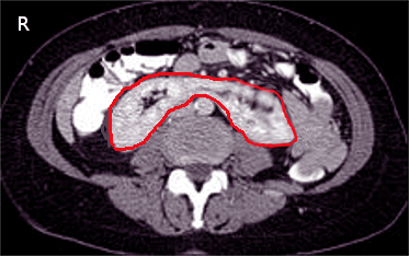
<p>CT Scan, Horseshoe Kidney</p>

CT Scan, Horseshoe Kidney

Contributed by S Bhimji, MD

Details

Updated:

5/30/2023 3:57:44 PM

Looking for an easier read?

Click here for a simplified version

References


[1]

Schiappacasse G,Aguirre J,Soffia P,Silva CS,Zilleruelo N, CT findings of the main pathological conditions associated with horseshoe kidneys. The British journal of radiology. 2015 Jan;     [PubMed PMID: 25375751]


[2]

Natsis K,Piagkou M,Skotsimara A,Protogerou V,Tsitouridis I,Skandalakis P, Horseshoe kidney: a review of anatomy and pathology. Surgical and radiologic anatomy : SRA. 2014 Aug     [PubMed PMID: 24178305]


[3]

Cook WA,Stephens FD, Fused kidneys: morphologic study and theory of embryogenesis. Birth defects original article series. 1977     [PubMed PMID: 588702]


[4]

Glodny B,Petersen J,Hofmann KJ,Schenk C,Herwig R,Trieb T,Koppelstaetter C,Steingruber I,Rehder P, Kidney fusion anomalies revisited: clinical and radiological analysis of 209 cases of crossed fused ectopia and horseshoe kidney. BJU international. 2009 Jan     [PubMed PMID: 18710445]

Level 3 (low-level) evidence

[5]

Stroosma OB,Scheltinga MR,Stubenitsky BM,Kootstra G, Horseshoe kidney transplantation: an overview. Clinical transplantation. 2000 Dec     [PubMed PMID: 11127302]

Level 3 (low-level) evidence

[6]

Crawford ES,Coselli JS,Safi HJ,Martin TD,Pool JL, The impact of renal fusion and ectopia on aortic surgery. Journal of vascular surgery. 1988 Oct     [PubMed PMID: 3172375]


[7]

Khougali HS,Alawad OAMA,Farkas N,Ahmed MMM,Abuagla AM, Bilateral pelvic kidneys with upper pole fusion and malrotation: a  case report and  review of the literature. Journal of medical case reports. 2021 Apr 5     [PubMed PMID: 33814014]

Level 3 (low-level) evidence

[8]

David RS, Horseshoe kidney: a report of one family. British medical journal. 1974 Dec 7     [PubMed PMID: 4434143]


[9]

Solhaug MJ,Bolger PM,Jose PA, The developing kidney and environmental toxins. Pediatrics. 2004 Apr     [PubMed PMID: 15060203]


[10]

Friedland GW,de Vries P, Renal ectopia and fusion. Embryologic Basis. Urology. 1975 May     [PubMed PMID: 1129903]


[11]

Mandell GA,Maloney K,Sherman NH,Filmer B, The renal axes in spina bifida: issues of confusion and fusion. Abdominal imaging. 1996 Nov-Dec     [PubMed PMID: 8875880]


[12]

Cereda A,Carey JC, The trisomy 18 syndrome. Orphanet journal of rare diseases. 2012 Oct 23     [PubMed PMID: 23088440]


[13]

Ranke MB,Saenger P, Turner's syndrome. Lancet (London, England). 2001 Jul 28     [PubMed PMID: 11498234]


[14]

Bhattarai B,Kulkarni AH,Rao ST,Mairpadi A, Anesthetic consideration in downs syndrome--a review. Nepal Medical College journal : NMCJ. 2008 Sep     [PubMed PMID: 19253867]


[15]

Cascio S,Sweeney B,Granata C,Piaggio G,Jasonni V,Puri P, Vesicoureteral reflux and ureteropelvic junction obstruction in children with horseshoe kidney: treatment and outcome. The Journal of urology. 2002 Jun     [PubMed PMID: 11992090]


[16]

Lallas CD,Pak RW,Pagnani C,Hubosky SG,Yanke BV,Keeley FX,Bagley DH, The minimally invasive management of ureteropelvic junction obstruction in horseshoe kidneys. World journal of urology. 2011 Feb     [PubMed PMID: 20204377]


[17]

GLENN JF, Analysis of 51 patients with horseshoe kidney. The New England journal of medicine. 1959 Oct 1     [PubMed PMID: 13828436]


[18]

O'Hara PJ,Hakaim AG,Hertzer NR,Krajewski LP,Cox GS,Beven EG, Surgical management of aortic aneurysm and coexistent horseshoe kidney: review of a 31-year experience. Journal of vascular surgery. 1993 May     [PubMed PMID: 8487363]


[19]

O'Brien J,Buckley O,Doody O,Ward E,Persaud T,Torreggiani W, Imaging of horseshoe kidneys and their complications. Journal of medical imaging and radiation oncology. 2008 Jun     [PubMed PMID: 18477115]


[20]

Lee CT,Hilton S,Russo P, Renal mass within a horseshoe kidney: preoperative evaluation with three-dimensional helical computed tomography. Urology. 2001 Jan     [PubMed PMID: 11164171]


[21]

Stein RJ,Desai MM, Management of urolithiasis in the congenitally abnormal kidney (horseshoe and ectopic). Current opinion in urology. 2007 Mar     [PubMed PMID: 17285023]

Level 3 (low-level) evidence

[22]

Rais-Bahrami S,Friedlander JI,Duty BD,Okeke Z,Smith AD, Difficulties with access in percutaneous renal surgery. Therapeutic advances in urology. 2011 Apr     [PubMed PMID: 21869906]

Level 3 (low-level) evidence

[23]

Sadiq AS,Atallah W,Khusid J,Gupta M, The Surgical Technique of Mini Percutaneous Nephrolithotomy. Journal of endourology. 2021 Sep;     [PubMed PMID: 34499550]


[24]

Skoog SJ,Reed MD,Gaudier FA Jr,Dunn NP, The posterolateral and the retrorenal colon: implication in percutaneous stone extraction. The Journal of urology. 1985 Jul     [PubMed PMID: 4009801]


[25]

Al Muzrakchi A,Aker LJA,Barah A,Alsherbini A,Omar A, Alternating Biplanar Fluoroscopy in Percutaneous Nephrostomy to Approach Stones in Patients With Horseshoe Kidney: An Institutional Experience. Cureus. 2021 Jul;     [PubMed PMID: 34430150]


[26]

Quintana Álvarez R,Herranz Amo F,Bueno Chomón G,Subirá Ríos D,Bataller Monfort V,Hernández Cavieres J,Hernández Fernández C, Surgical management of horseshoe kidney tumors. Literature review and analysis of two cases. Actas urologicas espanolas. 2021 Sep     [PubMed PMID: 34326031]

Level 3 (low-level) evidence

[27]

Quintana Álvarez R,Herranz Amo F,Bueno Chomón G,Subirá Ríos D,Bataller Monfort V,Hernández Cavieres J,Hernández Fernández C, Surgical management of horseshoe kidney tumors. Literature review and analysis of two cases. Actas urologicas espanolas. 2021 Sep     [PubMed PMID: 33958222]

Level 3 (low-level) evidence

[28]

Campi R,Sessa F,Rivetti A,Pecoraro A,Barzaghi P,Morselli S,Polverino P,Nicoletti R,Li Marzi V,Spatafora P,Sebastianelli A,Gacci M,Vignolini G,Serni S, Case Report: Optimizing Pre- and Intraoperative Planning With Hyperaccuracy Three-Dimensional Virtual Models for a Challenging Case of Robotic Partial Nephrectomy for Two Complex Renal Masses in a Horseshoe Kidney. Frontiers in surgery. 2021;     [PubMed PMID: 34136528]

Level 3 (low-level) evidence

[29]

Sobrinho ULGP,Albero JRP,Becalli MLP,Sampaio FJB,Favorito LA, Three-dimensional printing models of horseshoe kidney and duplicated pelvicalyceal collecting system for flexible ureteroscopy training: a pilot study. International braz j urol : official journal of the Brazilian Society of Urology. 2021 Jul-Aug;     [PubMed PMID: 33848082]

Level 3 (low-level) evidence

[30]

Decter RM, Renal duplication and fusion anomalies. Pediatric clinics of North America. 1997 Oct     [PubMed PMID: 9326964]


[31]

Neville H,Ritchey ML,Shamberger RC,Haase G,Perlman S,Yoshioka T, The occurrence of Wilms tumor in horseshoe kidneys: a report from the National Wilms Tumor Study Group (NWTSG). Journal of pediatric surgery. 2002 Aug     [PubMed PMID: 12149688]


[32]

Rubio Briones J,Regalado Pareja R,Sánchez Martín F,Chéchile Toniolo G,Huguet Pérez J,Villavicencio Mavrich H, Incidence of tumoural pathology in horseshoe kidneys. European urology. 1998     [PubMed PMID: 9519360]


[33]

Huang EY,Mascarenhas L,Mahour GH, Wilms' tumor and horseshoe kidneys: a case report and review of the literature. Journal of pediatric surgery. 2004 Feb     [PubMed PMID: 14966742]

Level 3 (low-level) evidence

[34]

Kölln CP,Boatman DL,Schmidt JD,Flocks RH, Horseshoe kidney: a review of 105 patients. The Journal of urology. 1972 Feb     [PubMed PMID: 5061443]


[35]

Pitts WR Jr,Muecke EC, Horseshoe kidneys: a 40-year experience. The Journal of urology. 1975 Jun     [PubMed PMID: 1152146]


[36]

Pawar AS,Thongprayoon C,Cheungpasitporn W,Sakhuja A,Mao MA,Erickson SB, Incidence and characteristics of kidney stones in patients with horseshoe kidney: A systematic review and meta-analysis. Urology annals. 2018 Jan-Mar     [PubMed PMID: 29416282]

Level 1 (high-level) evidence

[37]

Chopra P,St-Vil D,Yazbeck S, Blunt renal trauma-blessing in disguise? Journal of pediatric surgery. 2002 May     [PubMed PMID: 11987100]


[38]

Boatman DL,Kölln CP,Flocks RH, Congenital anomalies associated with horseshoe kidney. The Journal of urology. 1972 Feb     [PubMed PMID: 5061444]


[39]

Kumata H,Takayama T,Asami K,Haga I, Living-Donor Kidney Transplantation With Laparoscopic Nephrectomy From a Donor With Horseshoe Kidney: A Case Report. Transplantation proceedings. 2021 May     [PubMed PMID: 33892929]

Level 3 (low-level) evidence

[40]

De Pablos-Rodríguez P,Suárez JF,Riera Canals L,Sanz-Serra P,Vigués F, Horseshoe kidney splitting technique for transplantation. Urology case reports. 2021 Jul;     [PubMed PMID: 33665125]

Level 3 (low-level) evidence