Hip Fracture Overview

Earn CME/CE in your profession:


Continuing Education Activity

Hip fractures are fractures of the proximal femur and are commonly seen in the elderly population following a fall. This type of injury has a high rate of morbidity and mortality. To reduce this, surgical intervention is often required. This activity reviews the assessment and management of hip fractures and highlights the importance of the interprofessional team.

Objectives:

  • Summarize the etiology of hip fractures.
  • Outline the key aspects of the evaluation in hip fractures.
  • Review the management options available for hip fractures.
  • Explain some interprofessional team strategies for improving care coordination and communication to advance hip fractures and improve outcomes.

Introduction

Hip fractures are one of the most frequent fractures presenting to the emergency department and orthopedic trauma teams. The terms hip fracture and neck of femur fracture are used synonymously. Both terms describe a fracture of the proximal femur between the femoral head and 5 cm distal to the lesser trochanter.

Etiology

The majority of hip fractures are the result of a fall in the elderly population. Risk factors for falls in the elderly population are numerous, but those with a strong independent association are a previous history of falls, gait abnormalities, the use of walking aids, vertigo, Parkinson disease, and antiepileptic medications.[1] Many patients have multiple risk factors, and this, along with age-associated reduced bone quality, is the underpinning cause of most hip fractures.

Hip fractures that occur in younger adults are often the result of high-energy trauma. These patients are likely to have incurred multiple injuries and should be assessed and managed appropriately as per local trauma guidelines.

Around 5% of hip fractures have no history of trauma, and in these cases, an alternative cause should be suspected.[2] A pathological fracture is defined as a fracture caused by a disease process and not related to trauma. The two most frequent causes in relation to hip fractures are malignancy and bisphosphonate use. Arguably many more hip fractures could be described as pathological due to underlying osteoporosis, but this group is rarely labeled in this way.

Epidemiology

In 1990 the annual incidence of hip fractures globally was 1.3 million, and this is predicted to rise to 7 to 21 million by 2050.[3] In the U.S., the yearly incidence per 100,000 is thought to be between 197 to 201 for men and 511 to 553 for women.[4] Incidence increases with age, and the average age of patients presenting with a hip fracture is 80 years old.[2][4]

It is estimated that a patient spends $40,000 in the first year following a hip fracture, and the cost of hip fracture care annually in the U.S. is in excess of $17 billion.[5][6]

History and Physical

Most hip fractures can be diagnosed, or at least suspected, from history alone. Classically a fall leads to a painful hip with an associated inability to walk. Clinicians should explore potentially sinister causes of the fall, such as syncope, stroke, or myocardial infarction. As these patients are often elderly with a complex medical background, a full medical history is vital and should include both a history of the presentation as well as a full assessment of the patient's medical background. A thorough social history that provides baseline mobility and the patient's home circumstances is also of great value and is likely to guide postoperative rehabilitation and discharge planning.

It is recommended that a cognitive assessment be performed in all patients presenting with hip fractures. Ideally, this should be done both on admission and pos-operatively. The aim of this is to recognize patients with underlying dementia or those who are developing an acute delirium, both of which are associated with a poorer prognosis.[7]

The physical examination will demonstrate pain, immobility, and potentially a deformed limb. The degree of deformity seen is dependent on both the anatomical configuration of the fracture and the degree of displacement. The classically described presentation is a shortened and externally rotated limb due to the unopposed pull of the iliopsoas muscle that attaches to the lesser trochanter. Recognizing this, deformity immediately makes one suspicious of a hip fracture. Further examination often reveals pain on any, or all, of the following: palpation in the groin or greater trochanter, axial loading of the hip, and 'pin-rolling' of the leg.

A full primary trauma and secondary trauma assessment should be performed to assess the patient for other injuries. It is always useful to assess the patient's cardiovascular and respiratory status prior to undergoing surgery. Specific examinations to identify the cause of the fall should also be considered.

Evaluation

The majority of hip fractures can be diagnosed on plain film radiographs. An anteroposterior view of the pelvis should be obtained along with a lateral view of the affected hip. Occult fractures are those that are not visible on x-ray and account for between 2% to 10% of hip fractures.[8] MRI has demonstrated 100% sensitivity and 93% to 100% specificity at diagnosing occult hip fractures and is, therefore, the gold standard.[9] If MRI is not available, then CT is a valid alternative; however, it can miss fractures, particularly those that occur in the axial plane.

It is important to recognize the fracture pattern on radiographic images as this will dictate the surgical management. Generally speaking, hip fractures are described by comparing their location relative to the insertion of the joint capsule on to the femoral neck. The joint capsule of the hip originates from the acetabulum and encompasses the entirety of the femoral neck. Distally the capsule inserts into the femur at the intertrochanteric line anteriorly and the intertrochanteric crest posteriorly. The capsule is reinforced by three ligaments: the iliofemoral and pubofemoral ligaments anteriorly and the ischiofemoral ligament posteriorly. Fractures proximal to the capsular insertion are described as intracapsular, while fractures distal to this are extracapsular.

Intracapsular hip fractures may be classified using the Pauwel classification. This classification divides fractures into three groups based on the angle of the fracture from the horizontal plane[10]:

  • Type 1: <30 degree
  • Type 2: 31-50 degrees
  • Type 3: >50 degrees

An increased angle is associated with increasing shear forces and is, therefore, a more unstable fracture with reduced healing potential. This classification demonstrates marked inter-observer variation, particularly in displaced fractures.[11][12]

The Garden classification is a more commonly recognized classification system for intracapsular hip fractures. It describes four fracture patterns and defines them based on the completeness and displacement of the fracture.

  • Type 1: incomplete fracture and no displacement
  • Type 2: complete fracture and no displacement
  • Type 3: complete fracture and partial displacement
  • Type 4: complete fracture and complete displacement

This classification is more reproducible than the Pauwel classification but again demonstrates intra-observer variation.[12] Many clinicians simplify this classification to displaced or undisplaced, as this is what ultimately guides management. These fractures can also be described based on their position along the femoral neck. Sub-capital fractures are the most proximal intracapsular fractures followed by transcervical and finally, basicervical fractures at the base of the femoral neck.

Extracapsular fractures can be divided into trochanteric and subtrochanteric fractures. Trochanteric fractures are fractures that occur between the greater and lesser trochanter. These fractures are historically classified with Evan’s classification, which assesses the stability of the fracture. In modern practice extracapsular fractures are typically described using the AO classification:

  • A1: two-part, stable fracture
  • A2: comminuted, unstable fracture
  • A3: reverse or transverse, unstable fracture

Recognizing the different fracture types is important as it influences the surgical management.

Subtrochanteric fractures are fractures that occur between the lesser trochanter and 5 cm distal to the lesser trochanter. These were historically classified using the Russell-Taylor classification but now have a modernized AO classification system. Both of these classification methods are often academic and rarely influence management.

As part of the clinical evaluation, all patients should have blood tests on admission to assess for anemia, renal function, and coagulation profile. A bone screen is also of use and may aid the management of underlying osteoporosis or calcium abnormalities. Finally, a cross-match should be performed as operative management for fractured hips is associated with significant blood loss.

Patient evaluation should be approached in an interprofessional format with the involvement of the surgical, medical, and anesthetic teams, along with physiotherapists, pharmacists, and dieticians. This interprofessional approach ensures that the patient is comprehensively assessed and optimized prior to undergoing surgery. Hospitals are recommended to adopt this approach and formalize a hip fracture program to improve postoperative outcomes and reduce mortality.[13]

Treatment / Management

Initial management begins in the emergency department. Patients can lose up to 1 liter of blood from proximal femoral fractures, and thus fluid replacement and blood transfusion should be early considerations. 

Prolonged periods of preoperative fasting should be minimized, and nutritional supplementation should be made available until a likely time of operative intervention is known. Prolonged fasting is associated with increased catabolism, hypoglycemia, immunosuppression, and dehydration.[14] Hip fracture patients are particularly susceptible to complications of dehydration, and adequate pre and peri-operative hydration should be ensured.[15] Preoperative fasting times vary by hospital. The European Society of Anaesthesiology recommends fasting from fluids for 2 hours and from food for 6 hours prior to surgery.[16]

Oral or intravenous analgesia should be administered, but achieving adequate pain control can be challenging. It is now recommended that a fascia-iliaca nerve block be used in the preoperative period to reduce analgesia requirements and associated morbidity.[17] It is not advisable to apply limb traction, or attempt closed reduction in the emergency department.

The definitive management is largely dependent on the fracture location and configuration. The pre-morbid function and medical background of the patient are also considered, as well as their personal preferences. The approach, as with assessment, should involve the wider interprofessional team. The primary aim is to restore the patient’s mobilization status as swiftly as possible, and therefore operative intervention is normally preferred. Conservative management for fractured hips is associated with a higher 30 day and 1-year mortality and is generally reserved for patients who are not fit for surgery.[18] Early surgery in hip fracture patients is linked with improved outcomes, and therefore operating within 48 hours of admission is recommended.[19] Hyper-acute surgery, represented as surgery within 6 hours, does not reduce mortality or the rate of major complications but nor does it increase patient risk.[20] Hyper-acute surgery does reduce the rate of delirium and decreases the length of hospital stay by 1 day.[20]

Intracapsular Fractures

The blood supply to the femoral head plays an important role when deciding the management of hip fractures. The main arterial supply to the femoral head is from the medial and lateral circumflex femoral arteries. These vessels give branches that pass proximally through the joint capsule to supply the femoral head. In intracapsular fractures, these vessels can be damaged, which results in avascular necrosis of the femoral head.

Arthroplasty is shown to be superior to fixation in elderly patients with displaced intracapsular hip fractures with regards to pain, postoperative function, and complications.[21][22][23][24][25][26] Fixation is associated with a re-operation rate of around 30% over 24 months.[21][25] Early failure of fixation is often due to non-union or re-displacement of the fracture, while late failure is commonly the result of avascular necrosis.

The arthroplasty options for displaced intracapsular hip fractures are either a total hip replacement (THR) or a hemiarthroplasty. Both modalities demonstrate similar mortality rates, but there is evidence that suggests that THR improves postoperative pain and reduces acetabular wear.[22][27][27][28] A review of the UK national data registry has suggested a reduced rate of revision in the THR patient group but a higher rate of dislocation.[29] Conversely, a recent large multicentre randomized control trial demonstrated no difference in mortality, adverse events, or the incidence of secondary procedures between the hemiarthroplasty and THR patient groups.[30] The American Academy of Orthopaedic Surgeons (AAOS) and the National Institute of Clinical Excellence, UK (NICE), both currently recommend THR for all patients who can ambulate independently.

For patients with low functional demand, hemiarthroplasty is generally recommended due to the simplicity and speed of the procedure while still providing a good functional outcome.[30] When there is preexisting hip arthritis, a THR may be appropriate in the low functional demand patient group, but this should be decided on a case-by-case basis. Only young, highly active patients should be considered for open reduction and internal fixation of displaced intracapsular hip fractures due to the high risk of displacement, non-union, avascular necrosis, and, ultimately, revision surgery.

With regards to a hemiarthroplasty, there is thought to be little difference in functional outcome between unipolar and bipolar prostheses.[31][32] Unipolar hemiarthroplasties are associated with an increased rate of acetabular erosion, but they are also more cost-effective.[31] The use of cemented stems is favored due to improved postoperative hip function and lower rates of iatrogenic fracture.[33]

Undisplaced intracapsular fractures can be managed conservatively or surgically. Conservative management is associated with poor function and fracture displacement.[34] It is, therefore, only recommended in high-risk surgical candidates or patients who are pain-free and mobilizing. Surgical options are, in broad terms, fixation or arthroplasty. Fixation is thought to be preferable to arthroplasty as it preserves the native joint and therefore provides better long-term function and mobility.[35] A sliding hip screw (SHS) or cannulated hip screws (CHS) are both well-recognized fixation techniques. CHS is associated with reduced intraoperative blood loss and a lower rate of avascular necrosis.[36][37] Both fixation modalities have similar re-operation rates; however, patients with displaced or base of neck fractures along with current smokers demonstrate reduced re-operation rates with the use of an SHS.[36] When inserting an SHS, the risk of rotating the femoral head during implant insertion must be considered, and a de-rotation screw can be used to prevent this.

Following fixation of an intracapsular hip fracture, many surgeons will opt for protected weight-bearing in the immediate postoperative period to prevent fracture displacement. Avascular necrosis can still occur following fixation of undisplaced intracapsular fractures. Should this occur, further surgery in the form of core decompression or arthroplasty may be necessary. Patients must be counseled about this prior to undergoing fixation.

In patients with low functional demand or those who are unlikely to be able to comply with protected weight-bearing postoperatively, arthroplasty may be preferred as a primary operation for undisplaced intracapsular hip fractures. Arthroplasty removes the risk of avascular necrosis, thus reducing the rate of re-operation. The risks and benefits of both options should be discussed with the patient or their next-of-kin. Arthroplasty may also be preferred in patients with preexisting arthritis: there is little point attempting to preserve an already damaged articular surface.

Extracapsular Fractures

In extracapsular fractures, the blood supply to the femoral head is rarely compromised, and therefore fixation is the management of choice. The method of fixation is dependent on the fracture pattern. Sometimes the fracture pattern is not clear until it is screened on the operating table, and the surgical plan may need to be changed accordingly.

For stable trochanteric fractures (A1), intramedullary nails (IMN) and sliding hip screws (SHS) both demonstrate favorable postoperative outcomes.[38] SHS has a lower blood loss and operative time compared to IMN while also being a cheaper implant.[38]

IMNs are SHSs are both good options for fixing unstable trochanteric fractures (A2). IMNs are associated with better postoperative functional scores and are therefore recommended by the AAOS.[39] Short IMNs are a quicker procedure with reduced blood loss compared to long IMNs, but with regards to function and mortality, the two are similar.[39][40] Some surgeons still choose to use an SHS due to their operative simplicity and low cost, and in the UK NICE recommends the use of an SHS over an IM nail for both A1 and A2 fractures. When using an SHS or IMN, care should be taken to ensure the tip-apex distance is < 25 mm to reduce the risk of implant cut out.[41]

Subtrochanteric and reverse oblique trochanteric (A3) fractures are particularly challenging fractures to manage due to their instability. These fracture patterns are associated with a higher rate of non-union and failure of fixation when compared to other hip fractures.[42] Fixation of subtrochanteric fractures with an intramedullary device is recommended as IMNs demonstrate a lower rate of non-union when compared with extramedullary fixation techniques.[43][44] IMN fixation is also recommended for reverse oblique fractures as the technique demonstrates adequate fixation as well as shorter operative times and reduced length of hospital admission when compared with other techniques.[45] Open reduction techniques should be performed if an adequate closed reduction cannot be achieved.

Differential Diagnosis

Other injuries and disease processes that cause hip pain must be considered as a differential diagnosis. Pain caused by a chronically arthritic hip can be exacerbated by trauma in the absence of an acute fracture. Acute hip dislocation may also present with similar symptoms of pain, deformity, and reduced mobility. Pelvic fractures must be considered, and an x-ray of the pelvis, as well as the hip, is recommended. Injuries to the spine, femoral shaft, and knee can all present with hip pain, and a thorough examination is required to identify the site of injury. In the absence of trauma or fracture, then other causes for hip pain must be considered, including malignancy and infection.

Surgical Oncology

Pathological hip fractures can be as a result of either primary bone tumors or metastatic disease. If malignancy is suspected, then a full-length femoral X-ray and MRI of the femur should be obtained to assess the extent of the malignancy. Surgical management is then dependent on the disease prognosis following discussion with the oncology physicians. A curative resection and reconstruction is an option with curable disease. For patients with an incurable malignancy, then palliative procedures to reduce pain and restore mobility are indicated. Intra-operative tissue sampling can be useful to determine the origin of the primary tumor.

Prognosis

Mortality rates are reported to be between 18% to 31% within 1 year of sustaining a hip fracture.[46] Factors that increase mortality rates are age over 85, dependent functional status, ASA grade of 3 or more, male sex, previous history of cancer, and the development of a postoperative complication.[46][47] Multiple outcome predication models exist and are validated for predicting both 30 day and 1-year mortality.[47][48] In the UK, these risk stratification tools are used to identify high-risk patients whose operation should be performed or directly supervised by a senior surgeon.

Only 40% to 60% of patients regain their baseline mobility after a hip fracture, and 20% to 60% of previously independent people require assistance with at least one activity of daily living.[49] Patients who live in a residential care home prior to their injury are less likely to recover their pre-injury function.[49]

Complications

The complications following a hip fracture are vast, diverse, and multi-factorial. Infection rates following surgical management of fractured hips are between 0.6-3.6%, but these vary depending on the operation type.[50] Other generic surgical complications include postoperative pain, bleeding, neurovascular injury, and wound problems.

Specific complications for arthroplasty include dislocation, loosening, wear, leg length discrepancy, and peri-prosthetic fractures. For fixation devices, the recognized complications are failure of fixation or metalwork, avascular necrosis, and non-union.

Medical complications following a hip fracture present a significant problem. Early recognition and intervention of medical complications are imperative to reduce associated mortality. Below is a list of common medical complications following hip fracture along with their estimated prevalence[51]:

  • Delirium: 13.5% to 33%
  • Pulmonary embolism: 1.4% to 7.5%
  • Deep venous thrombosis: 27%
  • Pneumonia: 7%
  • Myocardial infarction or heart failure: 35% to 42%
  • Urinary retention or infection: 12% to 61%
  • Acute kidney injury: 11%
  • Anemia: 24% to 44%
  • Skin pressure damage: 7% to 9%

Postoperative and Rehabilitation Care

Pharmacological venous thromboembolism prophylaxis is recommended during the postoperative period providing there are no contraindications. Postoperative blood transfusion is only recommended if the hemoglobin is below 8 g/dL or the patient is symptomatic with their anemia.[52]

The postoperative rehabilitation is as important as the initial surgery. The aim of the surgery is to restore mobility, and therefore early mobilization should be commenced. Patients who have undergone arthroplasty or fixation of an extracapsular fracture can usually mobilize immediately after surgery without weight restrictions. Following fixation of an intracapsular fracture protected weight bearing is often recommended to reduce the risk of subsequent fracture displacement.

Regular intensive physiotherapy is required to encourage the rapid progression of mobility to restore the patient’s original mobility status. Medical management must also be optimized to reduce the risk of associated complications. Unfortunately, many patients do not regain their previous level of mobility or independence and therefore require social care input.

Deterrence and Patient Education

Deterrence of hip fractures has two main strands. Firstly preventing falls in the elderly population will, in turn, reduce the rate of fractures. This is by no means an easy undertaking as falls are often multifactorial, and some of the precipitating factors cannot be modified. Clinicians, and indeed the wider interprofessional team, should be aware of the risk factors for falls, and every attempt should be made to identify and manage these risks.

The second approach to reducing hip fractures is the diagnosis and management of osteoporosis. A fracture risk assessment tool is used to assess the risk of osteoporotic fracture over 10 years.[53] Both a personal fracture and a parental hip fracture increase a patient's score.[53] All patients over the age of 50, presenting with any fracture, should be assessed for osteoporosis and managed appropriately to reduce the risk of subsequent fracture.

Enhancing Healthcare Team Outcomes

As previously discussed, an interprofessional approach should be taken to manage this complex patient group. Physiotherapists play a crucial role in the post-operative care of patients with hip fractures. Dietician input is required to optimize nutrition to meet metabolic demands as well as to promote wound healing. Patients often require a lot of nursing care, both pre and postoperatively, and therefore nursing staff should be adequately trained and supported to manage this high demand patient group.

Input from elderly medicine physicians is crucial both pre and postoperatively to optimize patients for surgery, manage acute medical pathologies, assess falls risks, investigate for osteoporosis, and aid with patient’s on-going medical needs while rehabilitating. The orthopedic surgeon really only plays a small roll in the management of patients with an acute hip fracture.


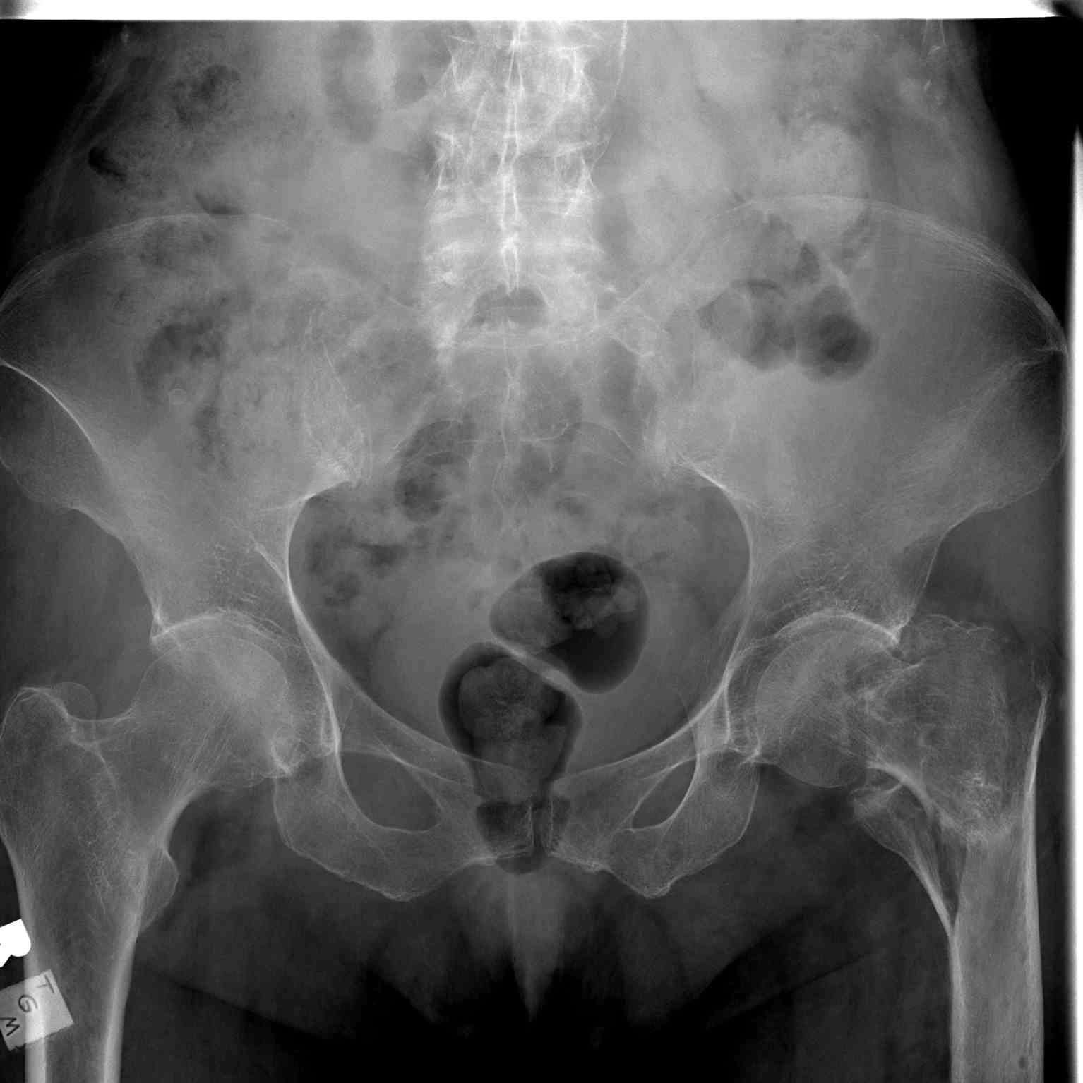
(Click Image to Enlarge)
<p>Pelvic Radiograph, Left Hip Fracture</p>

Pelvic Radiograph, Left Hip Fracture


Contributed by Scott Dulebohn, MD


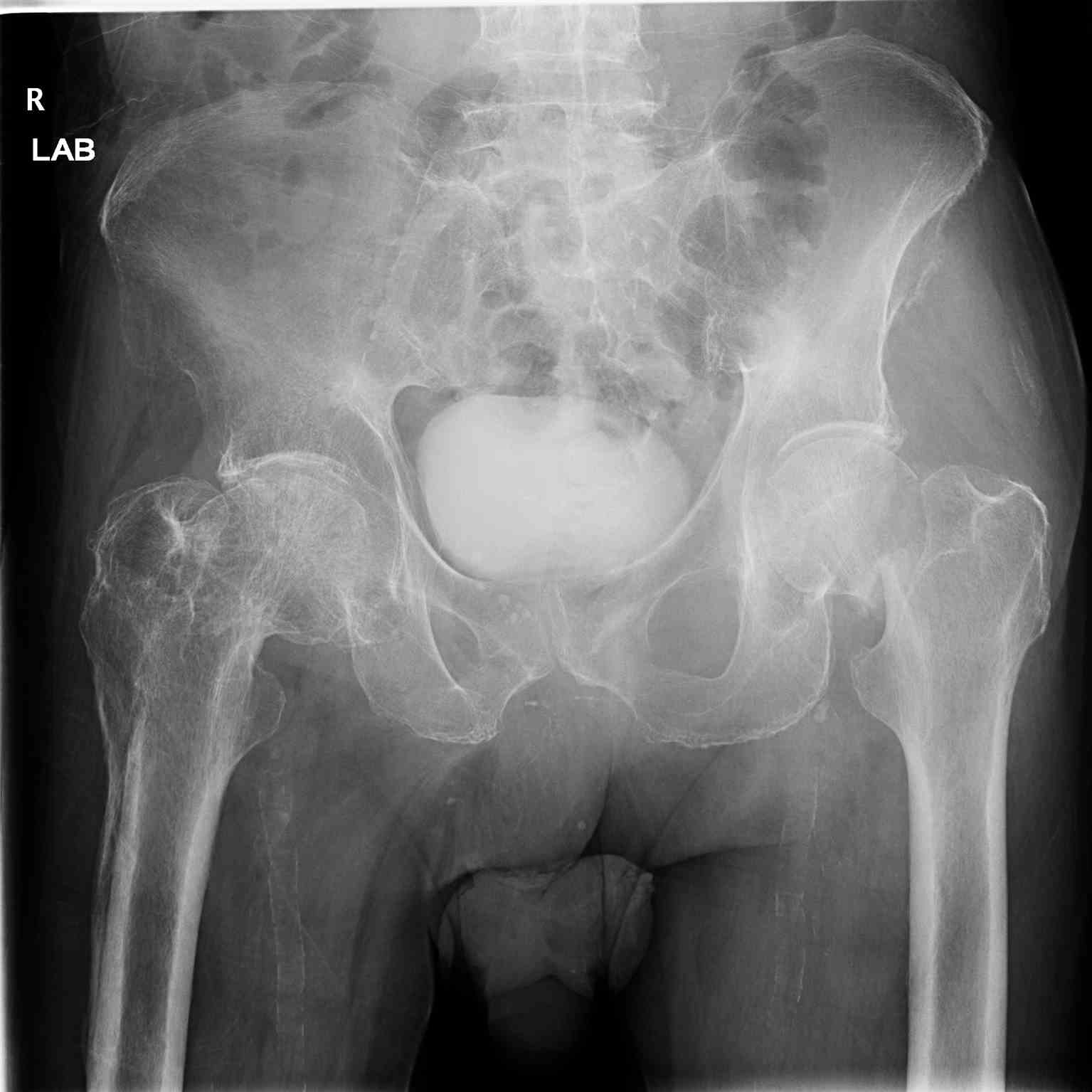
(Click Image to Enlarge)
<p>Pelvic Radiograph, Right Hip Fracture</p>

Pelvic Radiograph, Right Hip Fracture


Contributed by Scott Dulebohn, MD

Details

Editor:

Dominic Inman

Updated:

8/8/2023 12:16:56 AM

Looking for an easier read?

Click here for a simplified version

References


[1]

Deandrea S, Lucenteforte E, Bravi F, Foschi R, La Vecchia C, Negri E. Risk factors for falls in community-dwelling older people: a systematic review and meta-analysis. Epidemiology (Cambridge, Mass.). 2010 Sep:21(5):658-68. doi: 10.1097/EDE.0b013e3181e89905. Epub     [PubMed PMID: 20585256]

Level 1 (high-level) evidence

[2]

Parker M, Johansen A. Hip fracture. BMJ (Clinical research ed.). 2006 Jul 1:333(7557):27-30     [PubMed PMID: 16809710]


[3]

Gullberg B, Johnell O, Kanis JA. World-wide projections for hip fracture. Osteoporosis international : a journal established as result of cooperation between the European Foundation for Osteoporosis and the National Osteoporosis Foundation of the USA. 1997:7(5):407-13     [PubMed PMID: 9425497]


[4]

Dhanwal DK,Dennison EM,Harvey NC,Cooper C, Epidemiology of hip fracture: Worldwide geographic variation. Indian journal of orthopaedics. 2011 Jan;     [PubMed PMID: 21221218]


[5]

Brauer CA, Coca-Perraillon M, Cutler DM, Rosen AB. Incidence and mortality of hip fractures in the United States. JAMA. 2009 Oct 14:302(14):1573-9. doi: 10.1001/jama.2009.1462. Epub     [PubMed PMID: 19826027]


[6]

Youm T, Koval KJ, Zuckerman JD. The economic impact of geriatric hip fractures. American journal of orthopedics (Belle Mead, N.J.). 1999 Jul:28(7):423-8     [PubMed PMID: 10426442]


[7]

Mosk CA, Mus M, Vroemen JP, van der Ploeg T, Vos DI, Elmans LH, van der Laan L. Dementia and delirium, the outcomes in elderly hip fracture patients. Clinical interventions in aging. 2017:12():421-430. doi: 10.2147/CIA.S115945. Epub 2017 Mar 10     [PubMed PMID: 28331300]


[8]

Deleanu B,Prejbeanu R,Tsiridis E,Vermesan D,Crisan D,Haragus H,Predescu V,Birsasteanu F, Occult fractures of the proximal femur: imaging diagnosis and management of 82 cases in a regional trauma center. World journal of emergency surgery : WJES. 2015     [PubMed PMID: 26587053]

Level 3 (low-level) evidence

[9]

Foex BA, Russell A. BET 2: CT versus MRI for occult hip fractures. Emergency medicine journal : EMJ. 2018 Oct:35(10):645-647. doi: 10.1136/emermed-2018-208093.3. Epub     [PubMed PMID: 30249714]


[10]

Bartonícek J. Pauwels' classification of femoral neck fractures: correct interpretation of the original. Journal of orthopaedic trauma. 2001 Jun-Jul:15(5):358-60     [PubMed PMID: 11433141]


[11]

van Embden D, Roukema GR, Rhemrev SJ, Genelin F, Meylaerts SA. The Pauwels classification for intracapsular hip fractures: is it reliable? Injury. 2011 Nov:42(11):1238-40. doi: 10.1016/j.injury.2010.11.053. Epub 2010 Dec 13     [PubMed PMID: 21146815]


[12]

Kazley JM,Banerjee S,Abousayed MM,Rosenbaum AJ, Classifications in Brief: Garden Classification of Femoral Neck Fractures. Clinical orthopaedics and related research. 2018 Feb     [PubMed PMID: 29389800]


[13]

Pedersen SJ, Borgbjerg FM, Schousboe B, Pedersen BD, Jørgensen HL, Duus BR, Lauritzen JB, Hip Fracture Group of Bispebjerg Hospital. A comprehensive hip fracture program reduces complication rates and mortality. Journal of the American Geriatrics Society. 2008 Oct:56(10):1831-8. doi: 10.1111/j.1532-5415.2008.01945.x. Epub     [PubMed PMID: 19054201]


[14]

Downie S, Joss J, Sripada S. A prospective cohort study investigating the use of a surgical planning tool to improve patient fasting times in orthopaedic trauma. The surgeon : journal of the Royal Colleges of Surgeons of Edinburgh and Ireland. 2019 Apr:17(2):80-87. doi: 10.1016/j.surge.2018.05.003. Epub 2018 Jun 18     [PubMed PMID: 29929769]


[15]

Ylinenvaara SI, Elisson O, Berg K, Zdolsek JH, Krook H, Hahn RG. Preoperative urine-specific gravity and the incidence of complications after hip fracture surgery: A prospective, observational study. European journal of anaesthesiology. 2014 Feb:31(2):85-90. doi: 10.1097/01.EJA.0000435057.72303.0e. Epub     [PubMed PMID: 24145802]

Level 2 (mid-level) evidence

[16]

Smith I,Kranke P,Murat I,Smith A,O'Sullivan G,Søreide E,Spies C,in't Veld B, Perioperative fasting in adults and children: guidelines from the European Society of Anaesthesiology. European journal of anaesthesiology. 2011 Aug     [PubMed PMID: 21712716]


[17]

Callear J, Shah K. Analgesia in hip fractures. Do fascia-iliac blocks make any difference? BMJ quality improvement reports. 2016:5(1):. doi: 10.1136/bmjquality.u210130.w4147. Epub 2016 Jan 14     [PubMed PMID: 26893899]

Level 2 (mid-level) evidence

[18]

van de Ree CLP, De Jongh MAC, Peeters CMM, de Munter L, Roukema JA, Gosens T. Hip Fractures in Elderly People: Surgery or No Surgery? A Systematic Review and Meta-Analysis. Geriatric orthopaedic surgery & rehabilitation. 2017 Sep:8(3):173-180. doi: 10.1177/2151458517713821. Epub 2017 Jul 7     [PubMed PMID: 28835875]

Level 1 (high-level) evidence

[19]

Khan SK, Kalra S, Khanna A, Thiruvengada MM, Parker MJ. Timing of surgery for hip fractures: a systematic review of 52 published studies involving 291,413 patients. Injury. 2009 Jul:40(7):692-7. doi: 10.1016/j.injury.2009.01.010. Epub 2009 May 18     [PubMed PMID: 19450802]

Level 1 (high-level) evidence

[20]

HIP ATTACK Investigators. Accelerated surgery versus standard care in hip fracture (HIP ATTACK): an international, randomised, controlled trial. Lancet (London, England). 2020 Feb 29:395(10225):698-708. doi: 10.1016/S0140-6736(20)30058-1. Epub 2020 Feb 9     [PubMed PMID: 32050090]

Level 1 (high-level) evidence

[21]

Davison JN, Calder SJ, Anderson GH, Ward G, Jagger C, Harper WM, Gregg PJ. Treatment for displaced intracapsular fracture of the proximal femur. A prospective, randomised trial in patients aged 65 to 79 years. The Journal of bone and joint surgery. British volume. 2001 Mar:83(2):206-12     [PubMed PMID: 11284567]

Level 1 (high-level) evidence

[22]

Keating JF, Grant A, Masson M, Scott NW, Forbes JF. Displaced intracapsular hip fractures in fit, older people: a randomised comparison of reduction and fixation, bipolar hemiarthroplasty and total hip arthroplasty. Health technology assessment (Winchester, England). 2005 Oct:9(41):iii-iv, ix-x, 1-65     [PubMed PMID: 16202351]

Level 1 (high-level) evidence

[23]

Johansson T, Jacobsson SA, Ivarsson I, Knutsson A, Wahlström O. Internal fixation versus total hip arthroplasty in the treatment of displaced femoral neck fractures: a prospective randomized study of 100 hips. Acta orthopaedica Scandinavica. 2000 Dec:71(6):597-602     [PubMed PMID: 11145387]

Level 1 (high-level) evidence

[24]

Bray TJ, Smith-Hoefer E, Hooper A, Timmerman L. The displaced femoral neck fracture. Internal fixation versus bipolar endoprosthesis. Results of a prospective, randomized comparison. Clinical orthopaedics and related research. 1988 May:(230):127-40     [PubMed PMID: 3365885]

Level 1 (high-level) evidence

[25]

Frihagen F, Nordsletten L, Madsen JE. Hemiarthroplasty or internal fixation for intracapsular displaced femoral neck fractures: randomised controlled trial. BMJ (Clinical research ed.). 2007 Dec 15:335(7632):1251-4     [PubMed PMID: 18056740]

Level 1 (high-level) evidence

[26]

Sikorski JM, Barrington R. Internal fixation versus hemiarthroplasty for the displaced subcapital fracture of the femur. A prospective randomised study. The Journal of bone and joint surgery. British volume. 1981:63-B(3):357-61     [PubMed PMID: 7263746]

Level 1 (high-level) evidence

[27]

Macaulay W, Nellans KW, Iorio R, Garvin KL, Healy WL, Rosenwasser MP, DFACTO Consortium. Total hip arthroplasty is less painful at 12 months compared with hemiarthroplasty in treatment of displaced femoral neck fracture. HSS journal : the musculoskeletal journal of Hospital for Special Surgery. 2008 Feb:4(1):48-54. doi: 10.1007/s11420-007-9061-4. Epub     [PubMed PMID: 18751862]


[28]

Hedbeck CJ, Enocson A, Lapidus G, Blomfeldt R, Törnkvist H, Ponzer S, Tidermark J. Comparison of bipolar hemiarthroplasty with total hip arthroplasty for displaced femoral neck fractures: a concise four-year follow-up of a randomized trial. The Journal of bone and joint surgery. American volume. 2011 Mar 2:93(5):445-50. doi: 10.2106/JBJS.J.00474. Epub     [PubMed PMID: 21368076]

Level 1 (high-level) evidence

[29]

Metcalfe D, Judge A, Perry DC, Gabbe B, Zogg CK, Costa ML. Total hip arthroplasty versus hemiarthroplasty for independently mobile older adults with intracapsular hip fractures. BMC musculoskeletal disorders. 2019 May 17:20(1):226. doi: 10.1186/s12891-019-2590-4. Epub 2019 May 17     [PubMed PMID: 31101041]


[30]

HEALTH Investigators, Bhandari M, Einhorn TA, Guyatt G, Schemitsch EH, Zura RD, Sprague S, Frihagen F, Guerra-Farfán E, Kleinlugtenbelt YV, Poolman RW, Rangan A, Bzovsky S, Heels-Ansdell D, Thabane L, Walter SD, Devereaux PJ. Total Hip Arthroplasty or Hemiarthroplasty for Hip Fracture. The New England journal of medicine. 2019 Dec 5:381(23):2199-2208. doi: 10.1056/NEJMoa1906190. Epub 2019 Sep 26     [PubMed PMID: 31557429]


[31]

Yang B, Lin X, Yin XM, Wen XZ. Bipolar versus unipolar hemiarthroplasty for displaced femoral neck fractures in the elder patient: a systematic review and meta-analysis of randomized trials. European journal of orthopaedic surgery & traumatology : orthopedie traumatologie. 2015 Apr:25(3):425-33. doi: 10.1007/s00590-014-1565-2. Epub 2014 Dec 5     [PubMed PMID: 25476243]

Level 1 (high-level) evidence

[32]

Hedbeck CJ, Blomfeldt R, Lapidus G, Törnkvist H, Ponzer S, Tidermark J. Unipolar hemiarthroplasty versus bipolar hemiarthroplasty in the most elderly patients with displaced femoral neck fractures: a randomised, controlled trial. International orthopaedics. 2011 Nov:35(11):1703-11. doi: 10.1007/s00264-011-1213-y. Epub 2011 Feb 8     [PubMed PMID: 21301830]

Level 1 (high-level) evidence

[33]

Lin FF, Chen YF, Chen B, Lin CH, Zheng K. Cemented versus uncemented hemiarthroplasty for displaced femoral neck fractures: A meta-analysis of randomized controlled trails. Medicine. 2019 Feb:98(8):e14634. doi: 10.1097/MD.0000000000014634. Epub     [PubMed PMID: 30813202]

Level 1 (high-level) evidence

[34]

Cserháti P, Kazár G, Manninger J, Fekete K, Frenyó S. Non-operative or operative treatment for undisplaced femoral neck fractures: a comparative study of 122 non-operative and 125 operatively treated cases. Injury. 1996 Oct:27(8):583-8     [PubMed PMID: 8994566]

Level 2 (mid-level) evidence

[35]

Parker MJ, White A, Boyle A. Fixation versus hemiarthroplasty for undisplaced intracapsular hip fractures. Injury. 2008 Jul:39(7):791-5. doi: 10.1016/j.injury.2008.01.011. Epub 2008 Apr 14     [PubMed PMID: 18407277]


[36]

Fixation using Alternative Implants for the Treatment of Hip fractures (FAITH) Investigators. Fracture fixation in the operative management of hip fractures (FAITH): an international, multicentre, randomised controlled trial. Lancet (London, England). 2017 Apr 15:389(10078):1519-1527. doi: 10.1016/S0140-6736(17)30066-1. Epub 2017 Mar 3     [PubMed PMID: 28262269]

Level 1 (high-level) evidence

[37]

Shehata MSA, Aboelnas MM, Abdulkarim AN, Abdallah AR, Ahmed H, Holton J, Consigliere P, Narvani AA, Sallam AA, Wimhurst JA, Imam MA. Sliding hip screws versus cancellous screws for femoral neck fractures: a systematic review and meta-analysis. European journal of orthopaedic surgery & traumatology : orthopedie traumatologie. 2019 Oct:29(7):1383-1393. doi: 10.1007/s00590-019-02460-0. Epub 2019 Jun 5     [PubMed PMID: 31165917]

Level 1 (high-level) evidence

[38]

Ahrengart L, Törnkvist H, Fornander P, Thorngren KG, Pasanen L, Wahlström P, Honkonen S, Lindgren U. A randomized study of the compression hip screw and Gamma nail in 426 fractures. Clinical orthopaedics and related research. 2002 Aug:(401):209-22     [PubMed PMID: 12151898]

Level 1 (high-level) evidence

[39]

Li AB, Zhang WJ, Wang J, Guo WJ, Wang XH, Zhao YM. Intramedullary and extramedullary fixations for the treatment of unstable femoral intertrochanteric fractures: a meta-analysis of prospective randomized controlled trials. International orthopaedics. 2017 Feb:41(2):403-413. doi: 10.1007/s00264-016-3308-y. Epub 2016 Oct 8     [PubMed PMID: 27722824]

Level 1 (high-level) evidence

[40]

Zhang Y, Zhang S, Wang S, Zhang H, Zhang W, Liu P, Ma J, Pervaiz N, Wang J. Long and short intramedullary nails for fixation of intertrochanteric femur fractures (OTA 31-A1, A2 and A3): A systematic review and meta-analysis. Orthopaedics & traumatology, surgery & research : OTSR. 2017 Sep:103(5):685-690. doi: 10.1016/j.otsr.2017.04.003. Epub 2017 May 22     [PubMed PMID: 28546048]

Level 1 (high-level) evidence

[41]

Baumgaertner MR, Curtin SL, Lindskog DM, Keggi JM. The value of the tip-apex distance in predicting failure of fixation of peritrochanteric fractures of the hip. The Journal of bone and joint surgery. American volume. 1995 Jul:77(7):1058-64     [PubMed PMID: 7608228]


[42]

Park SY, Yang KH, Yoo JH, Yoon HK, Park HW. The treatment of reverse obliquity intertrochanteric fractures with the intramedullary hip nail. The Journal of trauma. 2008 Oct:65(4):852-7. doi: 10.1097/TA.0b013e31802b9559. Epub     [PubMed PMID: 18849802]


[43]

Ekström W, Karlsson-Thur C, Larsson S, Ragnarsson B, Alberts KA. Functional outcome in treatment of unstable trochanteric and subtrochanteric fractures with the proximal femoral nail and the Medoff sliding plate. Journal of orthopaedic trauma. 2007 Jan:21(1):18-25     [PubMed PMID: 17211264]


[44]

Rahme DM, Harris IA. Intramedullary nailing versus fixed angle blade plating for subtrochanteric femoral fractures: a prospective randomised controlled trial. Journal of orthopaedic surgery (Hong Kong). 2007 Dec:15(3):278-81     [PubMed PMID: 18162669]

Level 1 (high-level) evidence

[45]

Sadowski C, Lübbeke A, Saudan M, Riand N, Stern R, Hoffmeyer P. Treatment of reverse oblique and transverse intertrochanteric fractures with use of an intramedullary nail or a 95 degrees screw-plate: a prospective, randomized study. The Journal of bone and joint surgery. American volume. 2002 Mar:84(3):372-81     [PubMed PMID: 11886906]

Level 1 (high-level) evidence

[46]

Cheng SY, Levy AR, Lefaivre KA, Guy P, Kuramoto L, Sobolev B. Geographic trends in incidence of hip fractures: a comprehensive literature review. Osteoporosis international : a journal established as result of cooperation between the European Foundation for Osteoporosis and the National Osteoporosis Foundation of the USA. 2011 Oct:22(10):2575-86. doi: 10.1007/s00198-011-1596-z. Epub 2011 Apr 12     [PubMed PMID: 21484361]


[47]

Tsang C, Boulton C, Burgon V, Johansen A, Wakeman R, Cromwell DA. Predicting 30-day mortality after hip fracture surgery: Evaluation of the National Hip Fracture Database case-mix adjustment model. Bone & joint research. 2017 Sep:6(9):550-556. doi: 10.1302/2046-3758.69.BJR-2017-0020.R1. Epub     [PubMed PMID: 28947603]

Level 3 (low-level) evidence

[48]

Nijmeijer WS, Folbert EC, Vermeer M, Slaets JP, Hegeman JH. Prediction of early mortality following hip fracture surgery in frail elderly: The Almelo Hip Fracture Score (AHFS). Injury. 2016 Oct:47(10):2138-2143. doi: 10.1016/j.injury.2016.07.022. Epub 2016 Jul 20     [PubMed PMID: 27469403]


[49]

Dyer SM,Crotty M,Fairhall N,Magaziner J,Beaupre LA,Cameron ID,Sherrington C, A critical review of the long-term disability outcomes following hip fracture. BMC geriatrics. 2016 Sep 2;     [PubMed PMID: 27590604]


[50]

Mackay DC, Harrison WJ, Bates JH, Dickenson D. Audit of deep wound infection following hip fracture surgery. Journal of the Royal College of Surgeons of Edinburgh. 2000 Feb:45(1):56-9     [PubMed PMID: 10815382]


[51]

Carpintero P, Caeiro JR, Carpintero R, Morales A, Silva S, Mesa M. Complications of hip fractures: A review. World journal of orthopedics. 2014 Sep 18:5(4):402-11. doi: 10.5312/wjo.v5.i4.402. Epub 2014 Sep 18     [PubMed PMID: 25232517]


[52]

Carson JL, Terrin ML, Noveck H, Sanders DW, Chaitman BR, Rhoads GG, Nemo G, Dragert K, Beaupre L, Hildebrand K, Macaulay W, Lewis C, Cook DR, Dobbin G, Zakriya KJ, Apple FS, Horney RA, Magaziner J, FOCUS Investigators. Liberal or restrictive transfusion in high-risk patients after hip surgery. The New England journal of medicine. 2011 Dec 29:365(26):2453-62. doi: 10.1056/NEJMoa1012452. Epub 2011 Dec 14     [PubMed PMID: 22168590]


[53]

Kanis JA, Johansson H, Oden A, Dawson-Hughes B, Melton LJ 3rd, McCloskey EV. The effects of a FRAX revision for the USA. Osteoporosis international : a journal established as result of cooperation between the European Foundation for Osteoporosis and the National Osteoporosis Foundation of the USA. 2010 Jan:21(1):35-40. doi: 10.1007/s00198-009-1033-8. Epub 2009 Aug 25     [PubMed PMID: 19705047]