Continuing Education Activity
Hammertoes are among the most common deformities of the forefoot. This activity outlines the evaluation and management of hammertoe deformity and explains the role of the interprofessional team in evaluating and treating patients with this condition.
Objectives:
- Identify the etiology of hammertoe deformity.
- Summarize the evaluation of hammertoe deformity.
- Outline the management options available for hammertoe deformity.
- Describe how an interprofessional team can coordinate the care of patients with hammertoe deformity to improve outcomes.
Introduction
Hammertoes are among the most common deformities of the forefoot.[1] It results from an imbalance between the weak intrinsic muscles and the stronger extrinsic muscles surrounding the metatarsophalangeal joints (MTPJ) of the lesser digits. Hammertoe is a deformity that involves flexion at the interphalangeal joints (IPJ) and can be distinguished into categories including the classic hammertoe, mallet toe or claw toe. With the lesser digits being an important component in the balance of the foot, as well as in pressure distribution, deformities may lead to compensatory gait changes, distortions in cosmetics, callous formations, and pain. It is, therefore, important to know that there is a multitude of viable treatment options to consider. Treatments should first and foremost be centered around conservative measures such as wearing shoes with a wider toe box, toe pads, and the proper utilization of orthotics. If conservative management fails and pain persists with worsening deformity, the patient may benefit from surgical intervention. There are different types of characteristics of the deformities, and depending on its rigidity, the surgical approach will differ. A proper clinical evaluation of the patient is, therefore, of the utmost importance when aiming for long-term reduction of the deformity.
Anatomy
Deformities of the lesser digits result from an imbalance between the weak intrinsic muscles and the stronger extrinsic muscles. Any imbalance in these forces will favor the stronger extrinsic muscles and thus will result in an extended proximal phalanx and possible MTPJ hyperextension, as well as with a PIPJ and/or DIPJ flexion due to the long unopposed flexor.[2]
- Extensor digitorum longus (EDL) primary function is in the swing phase of gait and functions to dorsiflex the foot. EDL tendon splits into four separate tendon slips as it courses across the ankle, with each one going to each of the lesser digits. Over the proximal phalanx, the tendon again divides into 3 slips, with the middle slip inserting into base of the middle phalanx, and the 2 lateral slips converting into a terminal tendon and inserting into the base of the distal phalanx.
- Extensor digitorum brevis (EDB) only has three slips and inserts into the fibrous expansion of EDL at the level of MTPJ of digits 2, 3, 4 forming what is called the extensor hood apparatus. Because of this unique anatomical construct, the pull of the EDL and EDB creates significant dorsiflexion of the MTPJ and minimal dorsiflexion power at the IPJ.
- Flexor digitorum longus (FDL) divides into 4 separate slips that insert onto the lesser digits distal phalanx and flex at the DIPJ, while FDB inserts onto the middle phalanx and flexes the PIPJ. With no flexor inserting into the proximal phalanx and working as an antagonist to the MPJ in an extended position, the force results in flexion in the PIP and DIP joints.
MTPJ instability is commonly found in patients with digital deformities. The structures providing stability to this joint include the plantar plate along with the accessory and proper collateral ligaments at the lateral and medial aspects of the joint. If these key ligamentous structures attenuate or rupture, it will lead to instability at the level of the MTPJ. Plantar plate ruptures may also lead to subluxation, and a collateral ligament injury may result in a medial or lateral drift with a potential valgus or varus rotation of the affected digit; this pathology leads to a 'cross-over deformity.'[3]
Etiology
The causes of hammertoe deformity are many and multifactorial, including congenital and acquired, with the most accepted factor and component being a biomechanical dysfunction. The etiology of deformities of the lesser digits includes:[4][5]
- Neuromuscular conditions
- Diabetes
- Inflammatory arthropathies
- Ill-fitting shoes and high heels
- Intrinsic muscle imbalance
- Hallux valgus
- Long metatarsals
- Pes planus
Epidemiology
Deformities of the lesser digits are one of the most common problems to affect the foot and ankle, with up to 20% of reported incidences. Lesser toe problems increase with advancing age, occurring more frequently in women and have high heritability.[6] The condition also has a strong correlation to the presence of a hallux abductovalgus deformity, increased length of the involved toe, as well as pes planus foot posture.[7][8]
Pathophysiology
There are different types of characteristics of the digital deformities; they may be static or dynamic, flexible or rigid and may occur in conjunction with other pathologies such as Charcot-Marie-Tooth disease, cavus deformity and rheumatoid arthritis (RA). RA is distinctive in the way that it causes hammertoe formation by progressive joint destruction at the MTPJ leading to subluxation and dislocation. Whether due to neuromuscular, anatomic abnormalities such as the second ray being longer than the first or improperly fitted shoes, there is an imbalance in the extrinsic or intrinsic forces that are exerted on the digit causing a deformity.[2]
There are three major categories explaining the cause of the deformity and the loss of intrinsic and extrinsic muscle balance at the MTPJ; flexor stabilization, extensor substitution, and flexor substitution. These biomechanical mechanisms will induce deformity at different levels of the digits.
- Flexor Stabilization - the most common cause of digital deformities and occurs with excessive pronation. With the pronation of the subtalar joint, it unlocks the midtarsal joint as well as causing hypermobility of the forefoot. In an attempt to stabilize the forefoot, the flexors now fire longer and earlier and overpower the interosseous muscles. It is easily recognizable by adductovarus rotation of the fifth digit, as well as hammering or clawing of the lesser digits in stance position.
- Flexor substitution - occurs when there is a weak triceps surae muscle, and the deep and lateral leg muscles try to compensate for inadequate plantarflexion. This leads to the flexors gaining a mechanical advantage over the interossei muscles.
- Extensor substitution - clinically recognizable by bowstringing of the extensor tendons. In this mechanism, the extensors gain a mechanical advantage over the lumbricals and cause contractions at the MTPJ.
History and Physical
History
The hammertoe is commonly described as a chronic progressive deformity with flexion noted to the proximal interphalangeal joint of the affected digit. It is not uncommon that the affected toe will become red and painful. Patients typically present with chronic pain that is exacerbated by ambulation and shoewear. As the deformity progresses, the severity of symptoms gradually increases. By evaluating the skin, there may be blisters, callosities, ulcerations, and irritated skin dorsally over the PIPJ, plantar to the head of the metatarsals, and/or at the distal tip of the toe which is formed from the increased pressure to these areas. Patients will occasionally complain of pain to the plantar aspect of the head of the metatarsal, and this usually occurs when the MTPJ is hyperextended, subluxated, or dislocated.[9]
Physical
During the physical examination, the trained examiner should evaluate the biomechanics of the patient's feet to look for possible causes of the hammertoe deformity and for accompanying deformities such as hallux valgus. It is important to evaluate the patient's feet in standing as well as in a sitting position as many deformities cannot be correctly appreciated solely during the seated examinations. Therefore, the evaluation and assessment of the pathology are often divided into weight-bearing and non-weight-bearing exams. A Lachman test should be performed to evaluate the MTPJ instability along with recordings of the flexibility of all deformities. The physical examination should also include neurovascular evaluation, including palpation of pulses.
Flexible hammertoes are generally present upon weight-bearing and are corrected when the ankle is passively placed in a neutral position, whereas the rigid deformity is not. Attempting passive correction at the IPJ is important since it will assist in determining the treatment options. The MTPJ should undergo a range of motion and quality assessments as well. If palpating the articular portions of the metatarsal head reveals an increase in tenderness and instability, it may require a different treatment than if the patient presented with an isolated hammertoe deformity.[10]
Evaluation
To properly evaluate hammertoes, it is beneficial to retrieve diagnostic imaging.
Radiograph
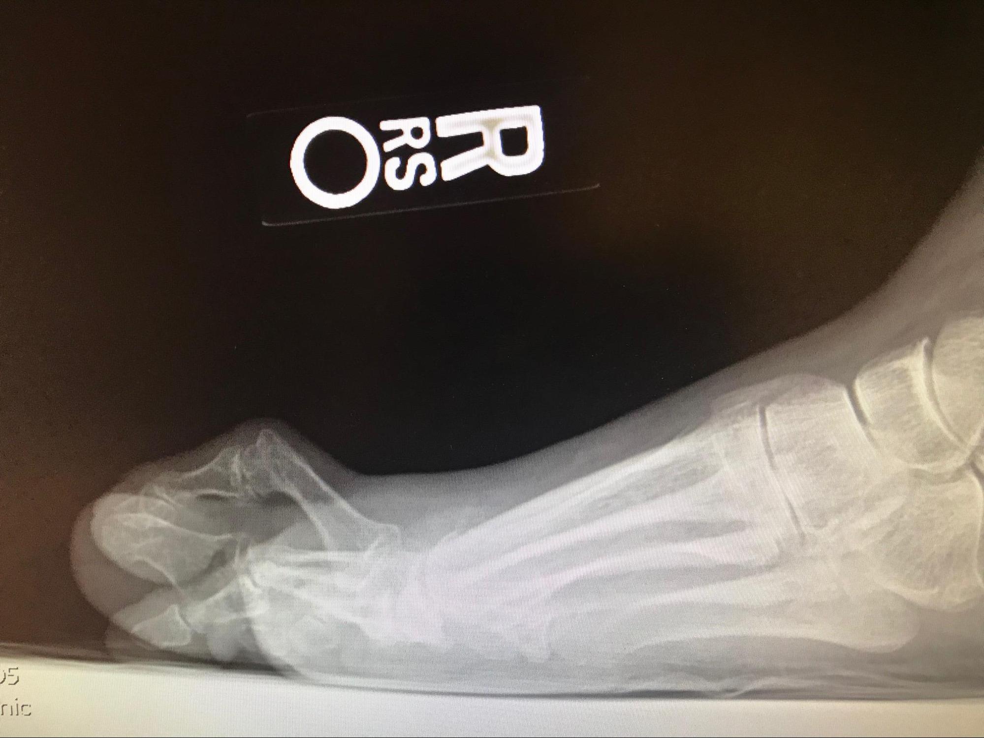
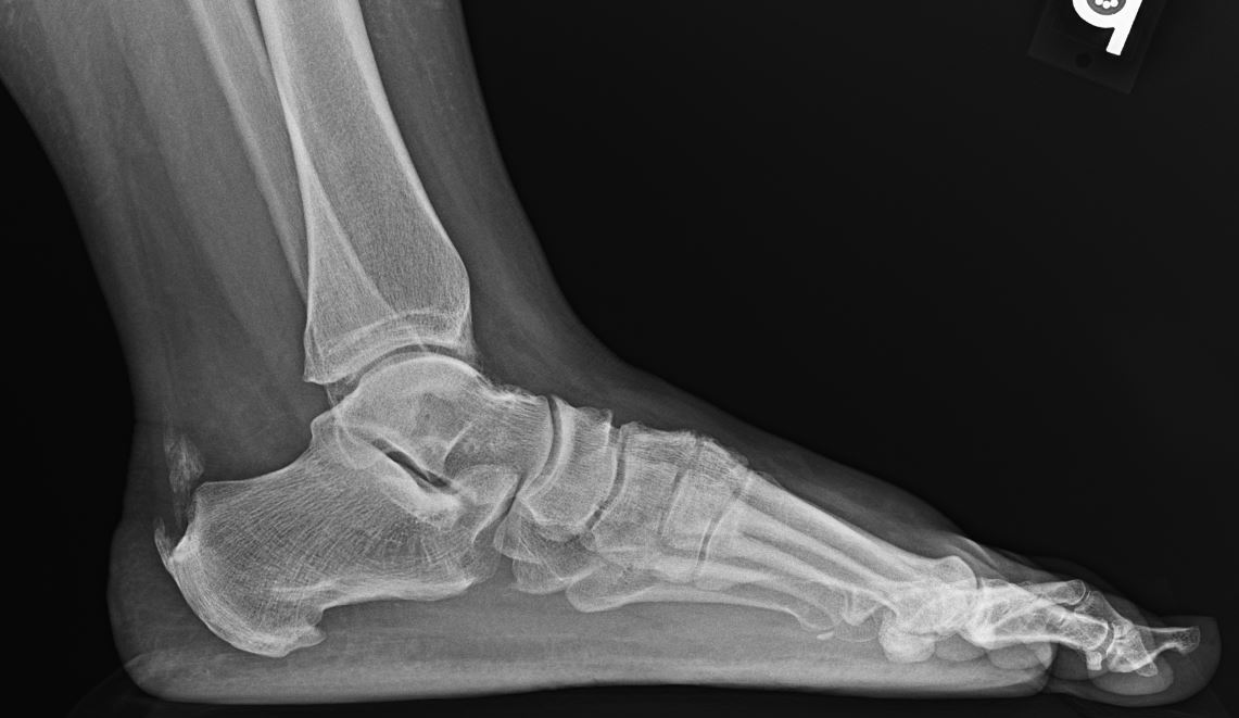
The anterior-posterior, oblique, and lateral plain film radiographs are helpful in the evaluation of hammertoe deformities and should be taken with the patient being weight-bearing. It may be used to assess contractures, seeing into the medullary canal of the proximal phalanx is associated with hammertoe deformities, and this sign is called the "gun barrel." Imaging makes it further possible to evaluate the relative metatarsal lengths, identifying hallux valgus as well as metatarsus adductus by examining the overall forefoot alignment. This is particularly helpful during a pre-operative assessment. To determine the length of the hammertoe pre-operatively, a transverse line may be drawn on the radiograph from the tips of the distal phalanges of the adjacent digits on either side; a long toe would overlap this line.[8]
Magnetic Resonance Imaging
Magnetic resonance imaging series may be helpful in the event that there is a suspicion for a plantar plate rupture. This kind of advanced diagnostic imaging may further help detect soft tissue or osseous pathologies such as avascular necrosis of the metatarsal head, with the second metatarsal being the most commonly affected, or defects of the cartilage. If the patient has a medical history with comorbidities such as diabetes, distal peripheral neuropathy, or peripheral vascular disease, noninvasive arterial studies may be warranted to assure healing assessment.
Treatment / Management
Hammertoe deformities can be extremely painful and can have a significant impact on a person’s quality of life. Surgical correction of hammertoe deformities is therefore among the most commonly performed surgical procedures performed on the forefoot.[8]
Non-surgical Treatment
Before the surgical route is considered, conservative treatments are usually attempted. These treatments are concerned with relieving pressure dorsally over the involved PIPJ, plantarly to its metatarsal head as well as relieving the pressure to the tip of the involved toe. With lesser digit deformities, it is recommended that the patient begins using insoles or orthotics and shoes with a wide toe box to accommodate the deformities as well as to alleviate the pain that may present with the impingement of the digits.[4] High heel shoes are not recommended due to the continued increased transfer of pressure to the forefoot. Padding or periodic shaving of the painful calluses may alleviate some of the patient’s discomfort, and strapping or taping the flexible deformities may improve some alignment. These modifications can be beneficial for managing forefoot disorders however none of these techniques are permanent solutions to the deformity.[5]
Surgical Treatment
Once the flexion contractures start forming and becoming constant along with pain, surgical intervention may be indicated. Historically, this has been based on balancing out the forces from the extensors and the flexors by altering the relative lengths of the toe, including its osseous structures and its tendons. The distinction between flexible and rigid hammertoes, as well as the absence or presence of associated MTPJ deformity, will help to guide you to conservative care or to the best surgical intervention. Recent findings have shown that lesser toe surgery makes up 48% of forefoot surgeries with hammertoe surgery being the most common procedure.[1][2]
Treatments must address and evaluate the deformity at all joints of the affected digit including DIPJ, PIPJ, and MTPJ. The most common surgical techniques employed to address the rigid or so-called fixed hammertoe deformities are PIPJ resection arthroplasty or PIPJ arthrodesis. For the correction of flexible hammertoes, soft tissue release is often used and this will maintain the structural stability of the toe.
If digital surgery is warranted and instability is noted at the MTPJ, this should be addressed as a concomitant procedure such as an osteotomy to address the affected metatarsal, and/or plantar plate repair. Plantar plate rupture may be diagnosed on physical examination by the Lachman test and is considered the most accurate clinical test for diagnosing MTPJ pathology. During the test, the metatarsal head should be stabilized while the proximal phalanx is displaced dorsally. If displacement is over 2 mm or 50% of the MTPJ, the test is positive indicating a plantar plate rupture or insufficiency. A dorsal medial deviation of the digit, often affecting the second toe, is commonly seen as an objective finding of MTPJ instability along with exquisite point tenderness just distal to MTPJ at the insertion of the plantar plate apparatus into the base of the proximal phalanx.
- PIPJ resection arthroplasty - Resection of the head of the proximal phalanx will shorten the distance from the origin to the insertion of the flexor digitorum longus and flexor digitorum brevis causing weakness. However, the extensors will not be weakened since their insertions are into the MTPJ via the extensor hood apparatus. This procedure is therefore most effective in treating flexor induced hammertoe deformities that are semirigid or rigid, as well as for elongated digits. Due to the loss of structural integrity, multiple arthroplasties should be avoided.
- PIPJ arthrodesis - The fusion of the PIPJ is an effective procedure for most deformities related to the digits. It is particularly useful and preferred when there are significant deforming forces, as well as when all or multiple digits are involved since it maintains the structural stability to the toes. The fusion converts the toe to a rigid lever arm, leading to FDL and FDB tendons now augmenting the intrinsic muscles providing plantarflexion stability at the MTPJ.
- Tendon transfers - Transferring the FDL and FDB tendons to the dorsal aspect of the proximal phalanx convert these tendons to plantar flexors of the MTPJ during weight-bearing in the same fashion as PIPJ arthrodesis. This procedure is indicated for flexible deformities. For the extensor tendon transfer, the EDL may be transferred to the metatarsal neck to eliminate the deforming force.
- Tenotomy - Another procedure indicated for flexible deformity is a flexor tenotomy performed at the PIPJ which effectively releases both the FDL and FDB. For mild, flexible extensor hammertoes the extensor tenotomy is indicated. The tenotomy must be performed proximal to the extensor hood apparatus to effectively release the pull. If the incision is made at the MTPJ, it requires a full capsulotomy.
- Weil Osteotomy - This is a distal oblique osteotomy resulting in a shortening of the metatarsal and is a procedure most commonly seen when addressing hammertoes or claw toes. It is often used to correct dislocations of the digits and angular deformities.
- Plantar Plate Repair - Commonly performed in conjunction with a Weil osteotomy. It is typically carried out through a dorsal approach where the injured or ruptured plantar plate is repaired using suture.
Differential Diagnosis
- Claw toe - Over the years, there has been some confusion regarding the exact definition of hammertoes and claw toes in the literature. This is probably in part because its treatment path is usually the same, so the exact definition may not be of any clinical importance. Coughlin and Mann defined the distinguishing deformity to occur at the MTPJ.[11]
- Most authors would agree with defining the claw toe as primarily a flexion deformity at the PIPJ and DIPJ with a simultaneous hyperextension at the MTPJ. Meanwhile, the hammertoe can be defined as a primary flexion deformity at the PIPJ but with a neutral or hyperextended DIPJ, with or without a hyperextended MTPJ.[4] Claw toes are more severe and usually involve multiple toes, both feet, and is frequently associated with neuromuscular conditions. The most typical foot type occurring in association with the claw toes is a cavus foot. Hammertoes, on the other hand, can occur in isolation and are most commonly affecting the second toe.
- Mallet toe - isolated flexion deformity at the DIPJ
- Turf toe
- Sesamoiditis
- Gout
- Osteochondrotic lesion of the first metatarsal head
- Osteochondritis dissecans
- Metatarsalgia
- Metatarsal stress fracture
Prognosis
Hammertoe deformity is a chronic progressive deformity. However, the overall prognosis of a hammertoe deformity is good. Once the patients have been evaluated, conservative management should be initiated. If functionality and pain do not improve, surgical intervention should be considered. Depending on the type of procedure, the technique used, and the surgeon’s preference, the postoperative recovery period differs. The standard post-op protocol usually includes partial weight-bearing with heel touch in a postoperative shoe for 2-6 weeks with the transition into a rigid athletic shoe. If the patient is a smoker or diabetic, the healing may take longer. Postoperative complications may arise depending on the surgical technique being used. Some of the more common post-surgical complications include infection, non-union, hematoma, numbness, and recurrence.[12]
Rates of recurrence and revision surgery of digital deformities are relatively high and has been showing with rates ranging up to 10%. It has been found that the 2nd digit is more likely to fail relative to the third and the fourth. Patients that showed a greater transverse deformity pre-operatively is shown to experience a greater failure rate than if the deformity is mainly in the sagittal plane. Furthermore, performing concomitant surgery to address a deviated 1st MTPJ at the first ray has shown a decrease in hammertoe recurrence with nearly 50%.[13] Recurrence may develop due to tight flexor tendon or due to inadequate bone resection, however care should be taken since excessive resection of bone may lead to a flail toe.
Complications
Hammertoes usually start out as mild deformities that progressively worsen over time, which can result in complications such as pain, gait imbalance, decreased quality of life, and skin changes like callouses, corns, and blisters. Surgical correction of the hammertoe deformity may lead to complications including but not limited to:[5][8]
- Nonunion
- Malunion
- Avascular necrosis
- Metatarsalgia
- Malalignment
- Infection
- Numbness
- PIP joint instability (flail toe)
- Pain
- Recurrent deformity
- Stiffness
- Vascular impairment
- Chronic edema
- Mallet toe
Deterrence and Patient Education
Hammertoes are one of the most common deformities of the forefoot. Hammertoe is a deformity that is involving flexion of the interphalangeal joints (IPJ) Deformities may lead to compensatory gait changes, callous formations, and pain. Management includes the use of shoes with a wider toe box, toe pads, taping, and the use of orthotics. Depending on if the deformity is flexible or rigid in nature or showing MTPJ instability, the surgical approach will differ. Most patients can return to their normal activity levels following clearance from their physician.
Enhancing Healthcare Team Outcomes
An interprofessional team that includes primary care physicians, podiatric surgeons, nurses, and physical therapists is the best approach for the management of hammertoe deformity. It may start as an initial visit to the primary care physician due to pain, swelling, or aesthetic concern. The diagnosis is made through a clinical examination, and radiographic imaging is helpful in evaluating the underlying structures and necessity for potential future surgical planning. However, conservative management should be considered as the primary approach. If proven ineffective, then the patient should get a referral to a surgeon and undergo a thorough surgical evaluation. If the patient undergoes surgery, post-operative protocol, including rehabilitation, should follow to maximize functionality; post-operative pain management with a focus on minimizing the opioid use should also be initiated. It is important for the patient to continue long term follow up to ensure the proper recovery timeline and milestones are being met.


