Continuing Education Activity
Freiberg disease is an uncommon yet clinically significant condition characterized by osteonecrosis of the metatarsal heads, prominently the second metatarsal. Osteonecrosis leads to flattening and eventual collapse of the metatarsal head, precipitating degenerative changes in the metatarsophalangeal joint and evolving into arthritic manifestations.
This course elucidates the nuances of Freiberg disease, highlighting its rarity and positioning avascular necrosis of the second metatarsal as the fourth most common osteochondrosis. Through historical insights and contemporary understanding, clinicians gain a comprehensive understanding of disease patterns, imaging findings, clinical presentations, and evolving management strategies for improved diagnosis and tailored interventions in Freiberg disease.
Objectives:
Identify clinical presentations and imaging findings indicative of Freiberg disease, considering osteonecrosis affecting the metatarsal heads, particularly the second metatarsal.
Conduct thorough foot examinations and imaging studies to screen for osteonecrosis in patients presenting with metatarsal head pain or joint abnormalities, particularly focusing on the second metatarsal.
Implement evidence-based treatment strategies, including conservative measures or surgical interventions tailored to the stage and severity of Freiberg disease.
Collaborate with orthopedic specialists, radiologists, and physical therapists to optimize comprehensive care, especially in cases requiring surgical interventions or advanced rehabilitative strategies.
Introduction
Freiberg disease is a rare condition in which the metatarsal head undergoes osteonecrosis.[1] In 1914, Dr Alfred Freiberg initially described it after 6 patients presented with a similar infraction pattern affecting the second metatarsal head.[1] Although the second metatarsal head is most commonly affected, Freiberg disease can involve any of the 5 metatarsal heads.[2][3][4][5]
As observed by Dr Freiberg, this pattern results in flattening and collapse of the head, leading to degenerative changes of the metatarsophalangeal joint and progressing to arthritis. Considered an uncommon process, avascular necrosis of the second metatarsal is the fourth most common osteochondrosis.[1][6]
Etiology
Freiberg disease is osteochondrosis affecting the metatarsal heads. Osteochondroses are a family of disorders resulting from an epiphysis injury that alters enchondral ossification and produces irregularity at the joint surface. Several potential explanations have been presented for Freiberg disease, but the most popular are microtrauma, vascular compromise, and systemic disorders.[1][7] Some systemic conditions associated with the development of Freiberg disease include diabetes mellitus, systemic lupus erythematosus, and hypercoagulability.[1] A genetic component is also thought to play a role since the condition has been reported in identical twins.[8]
A recent study by Samanta and Cobb described Freiberg infraction as the first clinical sign of an 18-year-old woman with Sneddon syndrome, a rare vasculopathy associated with livedo racemosa and frequent ischemic strokes.[9]
Epidemiology
Freiberg disease is the only osteochondrosis more common in females at a rate of 5:1 relative to males.[2][7] The dominant foot is involved 36% of the time.[7] Bilateral involvement is reported in less than 10% of cases. The condition affects the second metatarsal in 68% of cases, the third metatarsal in 27%, and the fourth metatarsal in 3%, with the fifth rarely affected.[1] The peak age of presentation is between 11 and 17 years, but can affect women in their seventh decade.[3]
Pathophysiology
The pathologic origin of articular osteochondroses occurs in 3 stages, as described by Omer.[4] The intra-articular and periarticular soft tissues swell and engorge in the first stage. In the second stage, there is an irregularity of the epiphyseal contour. In the last stage, the necrotic tissue is replaced.[5]
History and Physical
Patients present with pain and swelling localized to the involved metatarsal head region of the forefoot. They describe the sensation of walking on something hard, such as a stone. Symptom onset is typically gradual, with no specific acute event. Patients describe their symptoms worsening with walking, especially when barefoot or wearing shoes with elevated heels.[10][7][10] A short course of steroids following foot trauma can also be associated with an atypical presentation of acute Freiberg disease.[11]
The affected toe may appear swollen on physical exams, especially near the metatarsophalangeal (MTP) joint.[12] Elevation (dorsiflexion) of the toe may be present. In the more chronic or advanced stages, sagittal or coronal plane malalignment may develop, such as hammertoes or crossover deformities. The range of motion at the MTP joint is reduced, and crepitation may be palpated. A callus may develop under the involved metatarsal head at the plantar fat pad. Digital Lachman testing can be performed, which evaluates joint instability and is graded based on the amount of dorsal translation of the proximal phalanx relative to the metatarsal head and compared to the contralateral foot. The test is abnormal when the joint subluxes dorsally, which will typically reproduce the patient’s pain.[13]
Evaluation
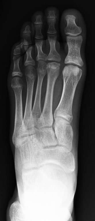
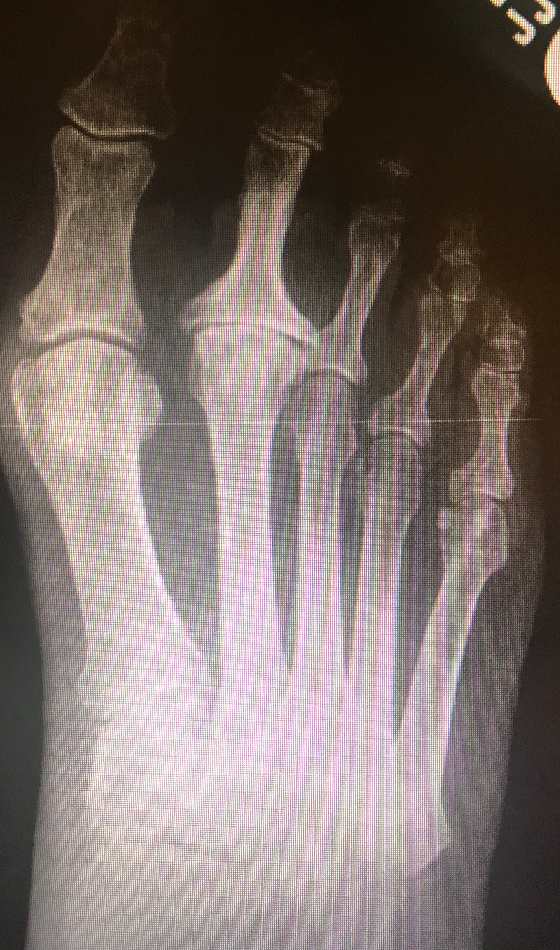
The diagnosis of Freiberg disease can be confirmed following the clinical exam with radiographs. On weight-bearing foot radiographs, there may be subtle changes early in the disease presentation (characterized by joint space widening due to effusion) that may be present for 3 to 6 weeks following the onset of symptoms. As the disease process progresses, there is increased bone density at the subchondral region and flattening of the metatarsal head. Oblique radiographs assist in the evaluation of the dorsal aspect of the metatarsal head, allowing for a full evaluation in the identification of flattening of the metatarsal head in subtle cases. As the disease progresses, later findings include central joint depression, loose bodies, and sclerosis of the metatarsal head. There may be reactive thickening of the metatarsal shaft as a late response due to abnormal stress. The final stages of this disease include joint space narrowing and arthrosis.[12]
Although classically described intraoperatively through observed structural changes to the metatarsal head by Smillie, these findings are evident radiographically and have been adapted nonoperatively.[14] This staging system includes:
- Stage 1: A fissure fracture in the ischemic epiphysis. The cancellous bone at the fracture appears sclerotic. Compared with the adjacent metaphysis, the epiphysis shows the absence of blood supply.
- Stage 2: Absorption of cancellous bone occurs proximally. The central cartilage sinks into the head while the margins and plantar cartilage remain intact. This process results in an altered contour of the articular surface.
- Stage 3: Further absorption occurs, and the central portion sinks deeper, creating larger projections on either side. The plantar cartilage remains intact.
- Stage 4: The central portion continues to sink, so the plantar hinge gives way. The peripheral projections fracture and fold over the central portion. Restoration of the anatomy is no longer possible.
- Stage 5: The final stage shows arthrosis with flattening and deformity of the metatarsal head. Only the plantar portion of the metatarsal cartilage retains the original contour of the head. Loose bodies have reduced in size, and the metatarsal shaft is thickened and dense.
MRI can also be used to evaluate these patients and may assist in the early detection of Freiberg disease when radiographs are normal. The MRI will reflect changes in the marrow signal with an edema-like signal localized to the affected metatarsal head. As the process progresses, changes similar to osteonecrosis seen in other parts of the body occur. These changes include a hypointense signal on T1-weighted images and mixed hypointense and hyperintense signals on T2-weighted images with flattening of the affected metatarsal head, best appreciated on the sagittal images.[12]
Nuclear medicine bone scans can also be used to evaluate these patients in the setting of early presentation or if there are no appreciable changes on radiographs. Early changes on bone scans include a photopenic area surrounded by increased radiotracer uptake, the typical pattern for early avascular necrosis. In later stages, these will be diffuse hyperactivity secondary to revascularization, osseous repair, and progression to arthritic involvement of the MTP joint.[12]
Treatment / Management
Initially, nonoperative management is attempted to alleviate symptoms and minimize epiphyseal deformity to limit the progression to arthritis regardless of the severity of the disease.[12] Activity modification, protected weight-bearing (stiff-soled shoe, fracture boot, or cast), shoe wear modifications, and oral anti-inflammatory medications are utilized in this early treatment. Shoe wear modifications may include orthoses with metatarsal bars designed to offload the painful metatarsal head, which has been shown to help patients respond without long-term disability.[12]
The use of bisphosphonate is a new method that shows promising results. A single injection of 5 mg of intravenous zoledronic acid followed by 70 mg weekly of oral alendronate for 1 year has eliminated symptoms and slowed the progression of early-stage avascular necrosis of the second metatarsal head.[15]
Most patients with Smillie stages 1 through 3 respond to conservative treatment and obtain long-term success.[6] However, there is a large number of surgical procedures proposed for the treatment of Freiberg disease when conservative measures have failed, primarily reserved for patients with Smillie stage 4 and stage 5 disease.[5]
Surgeons have little consensus about which procedure should be primarily performed. In a review by Carmont et al, surgical options were divided into two categories: either altering the abnormal physiology and biomechanics or restoring articular congruency/arthritic sequelae encountered in the later stages of the disease. Those procedures aimed at altering abnormal physiology include core decompression and corrective osteotomies. The procedures intended to restore articular congruency include debridement, osteotomy, grafting, and arthroplasty.[8]
Among different joint-sparing procedures, the Gauthier osteotomy is perhaps the most popular with the most extended follow-up reported. It consists of an intra-articular dorsal closing-wedge osteotomy, during which the diseased cartilage is removed, and the healthy plantar cartilage is reoriented into the central joint.[16] This procedure showed no complications and a high satisfaction rate in all patients with a follow-up of 23.4 years.[17]
When the disease advances, treatment may require a joint-destructive procedure. As with other procedures, an effort should be made to remove the avascular portion. However, this may lead to significant shortening of the metatarsal. Due to this reason, an interpositional arthroplasty is among the favorite procedures for late-stage disease.[12][18][19][1] This procedure consists of the interposition of soft tissue autograft (dorsal metatarsophalangeal joint capsule) [20], extensor digitorum longus tendon [21], extensor digitorum brevis tendon [19], or allograft [22] in the affected joint. The technique decreases the need for artificial implants while preserving the length of the toe, with a success rate of up to 90%.[12]
Differential Diagnosis
The differential diagnosis for these patients based on clinical presentation includes a stress fracture, neuroma, plantar plate tear, or other inflammatory arthritis such as rheumatoid arthritis or gout. Radiographs assist in narrowing the differential diagnosis and exclude these entities. The classic finding of flattening of the metatarsal heads on the radiograph will confirm the clinical suspicion.
Prognosis
Most patients with Smillie stages 1 through 3 respond to conservative treatment and obtain long-term success. Patients presenting at high grades typically undergo surgery to restore articular congruency and limit the progression to arthritis.
Complications
Complications include progression to advanced arthritis with the associated pain and limited range of motion.
Postoperative and Rehabilitation Care
Postoperative protocols vary according to the wide variety of available surgical procedures. Patients undergoing joint-sparing procedures such as the Gauthier dorsiflexion wedge osteotomy are usually allowed partial weight bearing to the heel or a forefoot offloading shoe for 3 weeks after surgery.[23] However, patients undergoing joint-destructive procedures will likely be required to be non-weight-bearing for a short period.
Physical therapy is indicated after patients transition to full weight bearing. A course of rehabilitation that includes a metatarsal phalangeal joint range of motion and walking retraining is advised.[23]
Consultations
The patient should seek medical attention from a podiatrist or orthopedic surgeon once symptoms arise. A physical therapist may be involved to relieve symptoms conservatively or following surgery as part of a rehabilitation program. A primary care physician may be consulted if bisphosphonate medications are employed.
Deterrence and Patient Education
Freiberg disease is a rare condition affecting the metatarsal bones located between the arch of the foot and the toes. The disease usually occurs in teenage girls that are growing. Several potential explanations for the cause of Freiberg disease have been proposed, but the most popular are microtrauma, vascular compromise, and systemic disorders.[1][7]
Patients initially complain of swelling and discomfort localized to the metatarsal head. They then describe the sensation of walking on something hard, such as a stone. Walking barefoot will be painful. Symptom onset is typically gradual, with no specific acute event.
If symptoms arise, the patient must seek medical attention. History, physical examination, and imaging studies will confirm the diagnosis.
Shoe accommodations and NSAIDs should suffice if the patient's condition is determined to be early-stage Freiberg disease. If the condition is found to be in later stages, surgery may be necessary.
Pearls and Other Issues
Key facts regarding Freiberg disease are as follows:
- Freiberg disease is caused by osseous infraction at the head of a metatarsal; the exact etiology is unknown
- The disease is more common in females and athletes
- The goals of treatment are early identification to place the patient in conservative therapy to allow healing and prevent progression to advanced arthritis.
Enhancing Healthcare Team Outcomes
Freiberg disease is an uncommon condition that may severely affect patients regarding the quality of life and their level of activities, primarily due to the young age of onset during the second or third decades of life. The etiology exact etiology is unknown but felt to be related to a multitude of factors. The diagnosis is made on clinical exam and confirmed with imaging, most commonly radiographs.
An interprofessional team approach can be helpful for accurate and timely diagnosis, especially in the early stages. Most patients will initially seek care from primary care and be referred to a podiatrist or an orthopedic surgeon. Foot and nail care specialists and orthopedic nurses can provide education and assist in coordinating care. They report changes in status to the team. Radiologists can assist with interpreting radiographs, MRIs, or bone scans. Conservative therapy is the first line of treatment, and if the patient does not respond, then operative intervention may be considered. In the operative setting, anesthesiologists and registered nurses will also play a role in the patient's care. Ultimately, therapy goals are to prevent or slow the progression of arthritis and clinical disability.