Continuing Education Activity
Erythema multiforme is an immune-mediated hypersensitivity reaction affecting both the skin and mucous membranes, characterized by the appearance of macular, papular, or bullous lesions that often form distinct "target lesions," primarily found on the extremities. Mucosal involvement, including the eyes, mouth, or genitals, is seen more frequently in erythema multiforme major compared to the minor form. Erythema multiforme is most often triggered by infections, particularly herpes simplex virus, but can also be caused by medications, autoimmune disorders, and occasionally vaccines. In more severe cases, erythema multiforme can cause significant pain and require hospitalization, especially when mucosal lesions or dehydration occur.
Erythema multiforme is a self-limiting condition that usually resolves without significant complications; however, a limited number of cases become persistent. Treatment focuses on managing symptoms and preventing recurrence, often through antiviral therapy for recurrent herpes-related cases. This activity for healthcare professionals is designed to enhance the learner's competence in recognizing erythema multiforme, performing the recommended evaluation, and implementing an appropriate interprofessional management approach to improve patient outcomes.
Objectives:
Differentiate erythema multiforme from conditions with similar presentations, such as Stevens-Johnson syndrome and toxic epidermal necrolysis, to ensure accurate diagnosis and treatment.
Screen for common triggers, including herpes simplex virus and medication use, to guide management and prevention strategies.
Implement appropriate symptom management protocols, including antiviral therapy for herpes-related cases and supportive care for mucosal involvement.
Apply interprofessional team strategies to improve care coordination and outcomes for a patient with erythema multiforme.
Introduction
Erythema multiforme is an immune-mediated hypersensitivity reaction with cutaneous and mucosal involvement. Clinically, erythema multiforme is characterized by macular, papular, bullous, or urticated lesions with a distinctive pattern of "target lesions" distributed primarily across extremities. Mucosal surfaces, including ocular, oral, and genital mucosa, may also be involved. Significant mucosal involvement differentiates erythema multiforme major from multiforme minor. Erythema multiforme is a self-limiting condition, usually resolving without significant complications, and is now classed as separate from Stevens-Johnson syndrome and toxic epidermal necrolysis. Only a limited number of cases become persistent. Erythema multiforme's etiologies are variable and numerous, and its clinical course is generally favorable.[1][2][3][4]
Most lesions appear in 48 to 72 hours, most frequently in the extremities. Generally, the lesions remain localized to 1 site and heal within 7 to 21 days. Common precipitating factors include infections such as herpes simplex virus, histoplasmosis, and Epstein Barr virus. Recurrences are not uncommon if the trigger is herpes simplex. While most erythema multiforme cases are mild, severe cases can be life-threatening. The mucous membranes are involved in 2% to 10% of individuals. Overall, the majority of cases of erythema multiforme are linked to medications. Management of acute erythema multiforme focuses on improving symptoms, managing pain, and supporting recovery, which usually happens within 2 weeks. Interventions for recurrent erythema multiforme aim to reduce or eliminate repeated disease episodes.
Etiology
The etiology of erythema multiforme is multifactorial, including infections, medications, autoimmune disease, malignancy, immunizations, radiation exposure, allergic contact dermatitis, sarcoidosis, and menstruation.[5][6] Approximately 90% of cases of erythema multiforme are caused by infections (viral, bacterial, or fungal), with herpes simplex virus being the most common culprit.[7] Moreover, a strong association between the COVID-19 vaccine and erythema multiforme has recently been found.[8] Other viral causes include adenovirus, coxsackievirus B5, cytomegalovirus, enterovirus, echoviruses, Epstein-Barr virus, hepatitis A, B, and C viruses, influenza, measles, mumps, parvovirus B19, poliomyelitis, and varicella-zoster virus.[9] Autoimmune conditions (eg, systemic lupus erythematosus) are also associated with erythema multiforme, particularly causing a typical but rare syndrome called Rowell syndrome.[10]
Another common cause of erythema multiforme, particularly in children, is Mycoplasma pneumoniae infection.[11] Other bacterial causes include borreliosis, catscratch disease, diphtheria, legionellosis, hemolytic streptococci, staphylococcus, leprosy, salmonellosis, Neisseria meningitidis, Mycobacterium avium complex, pneumococci, tuberculosis, Treponema avium complex, pneumococci, tuberculosis, Treponema pallidum, tularemia, and rickettsial infections.[12] Although herpes simplex virus (HSV) types 1 and 2 and Mycoplasma pneumonia are the common causes of erythema multiforme, many other viral, fungal, and bacterial infections have been implicated. Human immunodeficiency virus (HIV) infection and HIV treatment medications have been found to have a direct or indirect effect on the development of erythema multiforme.[13]
Also, various vaccines leading to erythema multiforme have been reported, including Bacille Calmette-Guérin, oral polio, vaccinia, and tetanus/diphtheria vaccines.[14][15][16][17] Drugs associated with erythema multiforme include antibiotics (eg, penicillins, cephalosporins, macrolides, and sulfonamides), antituberculosis agents, and antipyretics.[18] In some patients, contact with heavy metals, herbal agents, topical therapies, and poison ivy can trigger erythema multiforme.
Epidemiology
Erythema multiforme is reported worldwide without any ethnic preference. Although erythema multiforme occurs at any age, the condition presents more frequently in young adults. The average age of onset is between 20 and 40 years, with 20% of cases occurring in children. Erythema multiforme is more common in men than women, with a ratio of 1 in 5. The prevalence is not known but appears to be well below 1%.[19] As the classification is not always clear, cases of Stevens-Johnson syndrome have frequently been included in studies on erythema multiforme.
Pathophysiology
The exact pathophysiology behind erythema multiforme is unknown. Most data has been collected to study the underlying pathogenesis behind HSV-associated erythema multiforme. The mechanism by which HSV causes erythema multiforme is thought to involve a cell-mediated immune response against viral antigens in skin lesions.[20] Viral deoxyribonucleic acid (DNA) in biopsy specimens of individuals with affected skin supports this theory.[21]
In the development of mucocutaneous lesions of HSV-associated erythema multiforme, an interplay between cluster of differentiation (CD)34+ Langerhans cell precursors, viral DNA fragments, epidermal keratinocytes, HSV-specific CD4 TH1 cells, interferon (IFN)-γ, and autoreactive T cells is observed. These interactions lead to epidermal damage and the characteristic inflammatory infiltration of cutaneous lesions of erythema multiforme.[22] Similar mechanisms may be involved in other causes of erythema multiforme, but inadequate evidence exists. Damage to the epithelial cells is caused by cell-mediated immunity. During the early phase of the disease, an influx of macrophages and CD8 T lymphocytes occurs, which release a wide range of cytokines that mediate the inflammation and resultant cell death.
A slight difference in drug-induced erythema multiforme is the presence of tumor necrosis factor (TNF)-alpha instead of IFN-gamma, making the earliest pathological feature the necrosis of keratinocytes.[23] Genetic susceptibility also plays a role in the development of erythema multiforme. Recurrent erythema multiforme has been found to have an association with human leukocyte antigen (HLA) types A33, B35, DR4, DQB1*03:01, DQ3, and DR53.[24]
Study results have demonstrated the presence of HSV DNA by a polymerase chain reaction in acute or sequellae erythema multiforme lesions. The predisposing factors are unknown. HLA-DQ3 is reported to be associated with postherpetic erythema multiforme and has been suggested as an additional diagnostic marker. Other human leukocyte antigen groups have also been reported as markers of recurrent erythema multiforme.
Histopathology
A punch biopsy can confirm the diagnosis of erythema multiforme. However, findings may vary slightly based on the site of the biopsy. The classic lesion will reveal vacuolar interface dermatitis with marked infiltration of lymphocytes along the dermo-epidermal junction.[25] In addition, dyskeratosis of basal keratinocytes and hydropic changes may be noted. With advanced lesions, epidermal necrosis, subepidermal blisters, and vesiculation is often observed. A predominance of CD8 T lymphocytes and macrophages is also present.
History and Physical
Erythema multiforme can present with both mucosal and cutaneous lesions; however, it may mostly spare mucosal surfaces. A detailed history should focus on the triggers, including medications, infections, and constitutional symptoms.
Prodrome Features
In patients with erythema multiforme minor, prodromal symptoms are usually nonspecific, absent, or mild.[19] Patients may report fatigue, malaise, or upper respiratory tract infection. The onset of a rash usually occurs within 3 days, with centripetal spreading. In erythema multiforme major, patients have more marked symptoms, such as moderate fever, generalized aches, cough, sore throat, chest pain, vomiting, and diarrhea. These symptoms may classically persist for 2 weeks before the cutaneous lesions appear. Cough and respiratory symptoms can be seen in patients with erythema multiforme related to Mycoplasma pneumoniae infection.
Cutaneous Features
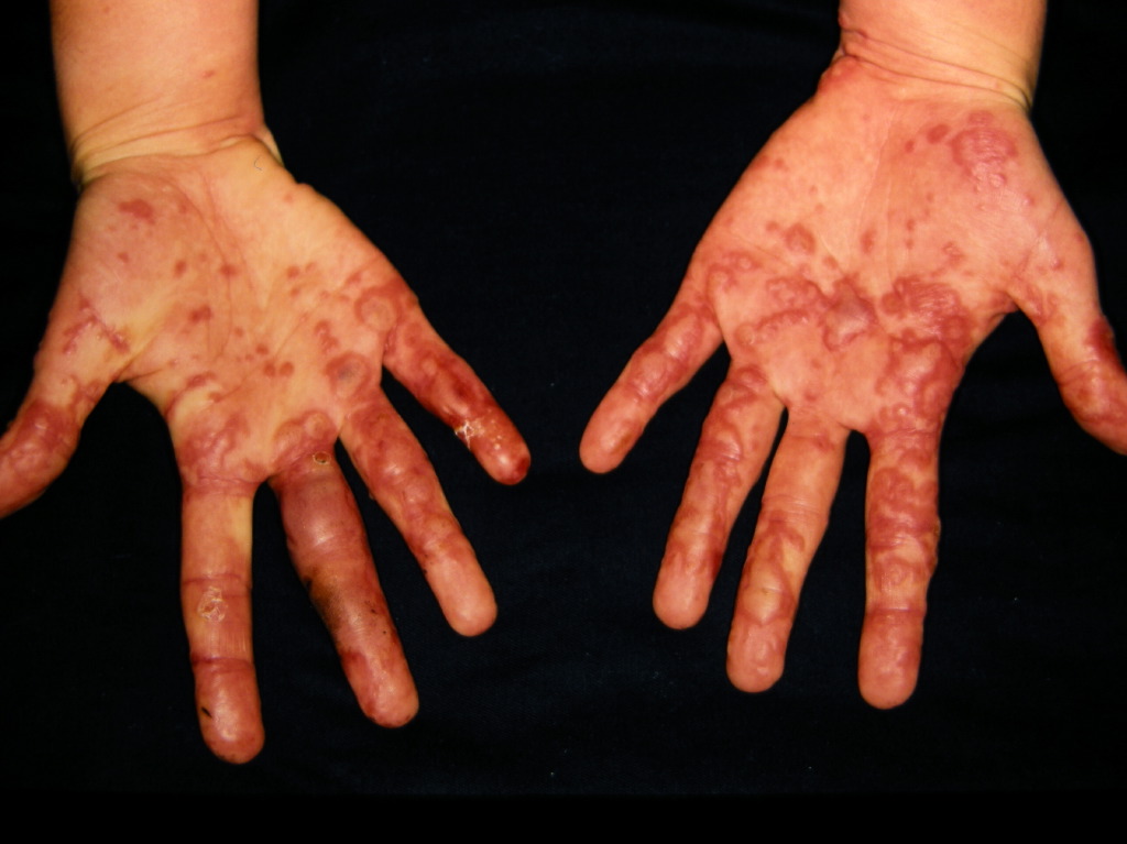
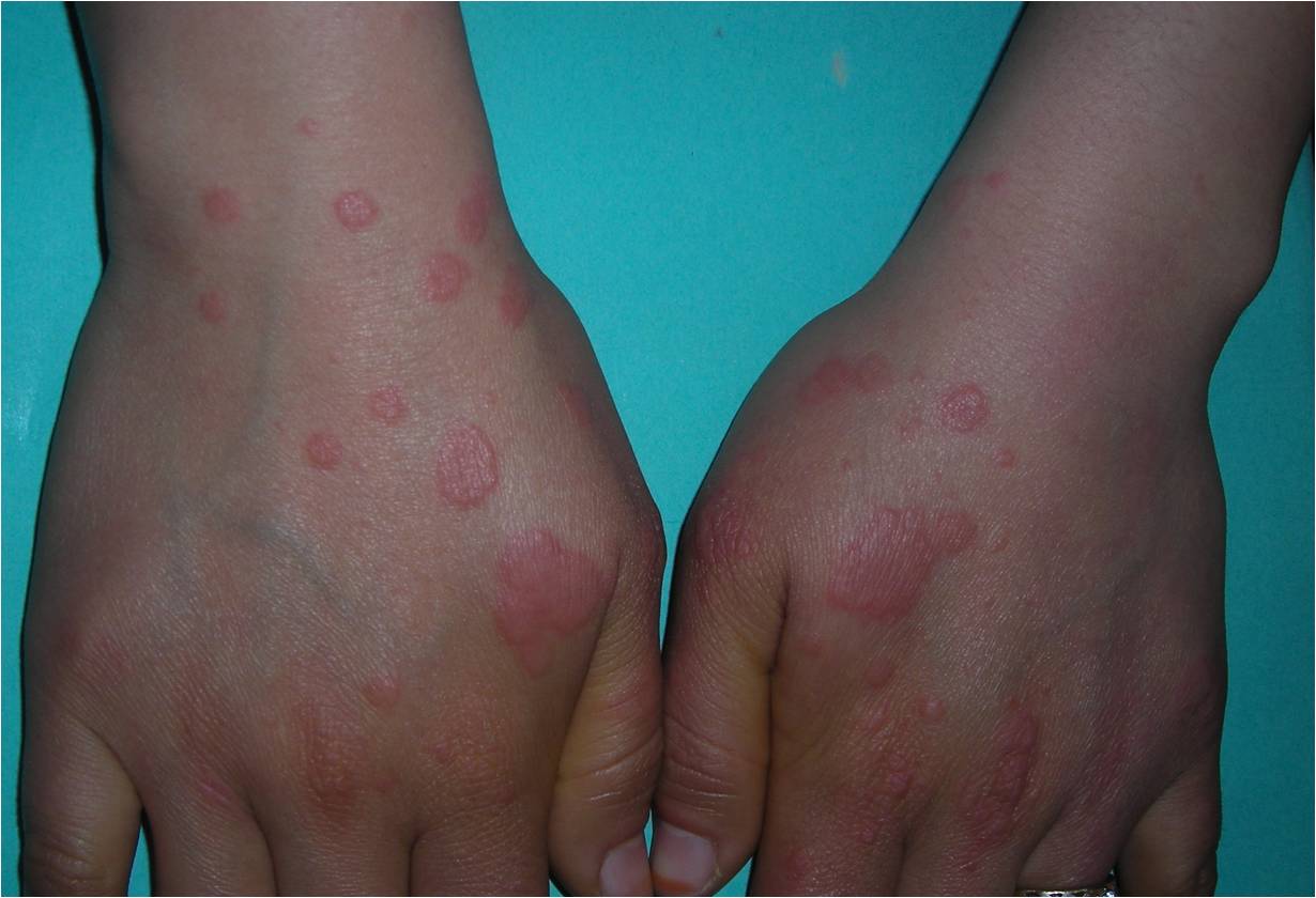
Clinically, the typical lesion of the erythema multiforme is the target lesion; however, they may not always be present. Lesions are rounded, regular lesions with 3 concentric circles and a well-defined border. The peripheral ring is erythematous, sometimes microvesicular; the middle zone is often more precise, oedematous, and palpable, and the center is erythematous, covered by a blister. These different aspects evoke different stages of the evolving lesion. The distribution of these lesions is usually symmetrical, with a predilection for extensor surfaces (see Images. Erythema Multiforme and Erythema Multiforme Lesions). Atypical lesions may be mixed with typical lesions or raised with poorly defined borders and fewer zones of color variation.[26]
The lesions generally measure less than 3 cm, and their location is mainly acral. They are symmetrical in the palms and backs of the hands, the feet, and the extensor surfaces of the limbs. The trunk is often spared, but the face and ears can get involved; typical lesions do not have pruritus.
Mucosal Features
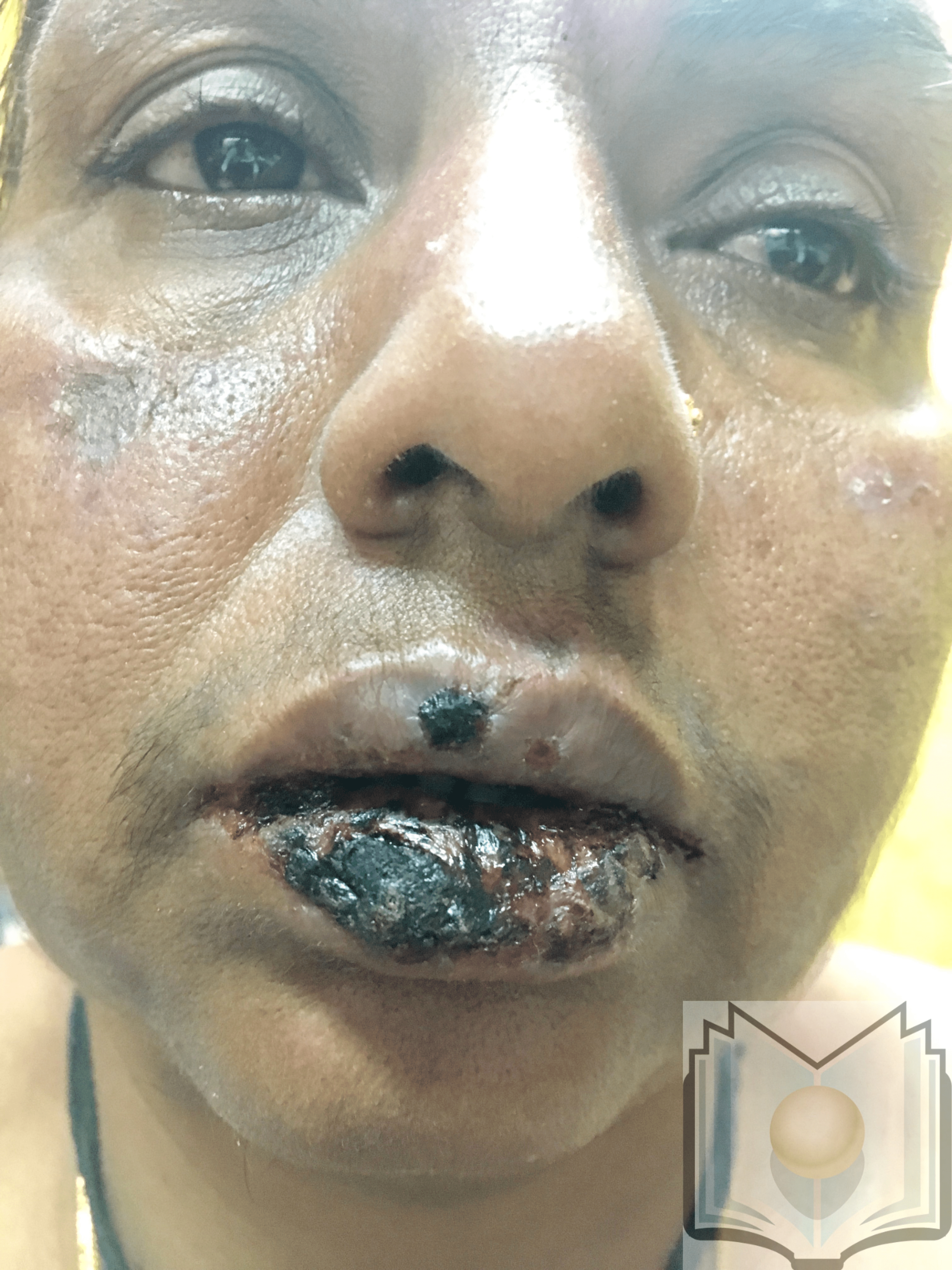
Mucosal lesions are common, mainly in the mouth, and less frequently in the urogenital and ocular mucous membranes.[27] The eruptions start with blisters, eroding to show a white overlying pseudo-membrane. Thick hemorrhagic crusts may cover the labial lesions, and a fibrin-whitish coating may line the mucosal erosions of the cheeks, palate, and genitalia (see Image. Erythema Multiforme Oral Lesions).
These mucosal lesions often coincide with skin lesions but may precede or follow cutaneous lesions. While skin lesions are nonpainful, mucosal lesions are frequently painful. When extensive skin involvement occurs, some patients may show signs of dehydration. Others with mucosal involvement may lose weight because of difficulty eating.
Evaluation
The diagnosis of erythema multiforme is based on history and clinical examination. Certain investigations, including skin biopsy, may help establish the diagnosis in cases with clinical diagnostic uncertainty, including:
- Complete blood cell count: May show moderate leukocytosis, eosinophilia, neutropenia, mild anemia, and thrombocytopenia
- Erythrocyte sedimentation: May be elevated in severe cases
- Immunofluorescence study: Can detect specific HSV antigens within keratinocytes
- Polymerase chain reaction: Can detect HSV DNA primarily within the keratinocytes
- Chest x-ray: May show interstitial radiological infiltrate (mainly in erythema multiforme due to Mycoplasma pneumoniae)
A skin biopsy of the lesion center can be performed for histological study with immunofluorescence, as it shows epithelial intercellular edema with keratinocyte necrosis, which is responsible for an intraepidermal or subepidermal blister covered with a necrotic epidermis. A perivascular lymphohistiocytic infiltrate is present in the superficial dermis without necrotic vascular lesions. Direct immunofluorescence is negative. The biological assessment provides no argument for the diagnosis of erythema multiforme. However, it is helpful to appreciate the severity of the disease. Renal, hepatic, or hematologic lesions have also been described and are not systematically sought after in mild forms.[28]
A characteristic lymphocytic infiltrate can also be seen around the superficial vascular plexuses. In advancing disease, epidermal necrosis, subepidermal blisters, or intraepidermal vesiculation may appear due to spongiosis and damage to the basal epidermal layer. Occasionally, severe papillary edema may be present. The lymphocytic inflammatory infiltrate in the dermis is rich in T cells and macrophages.
Treatment / Management
The treatment approaches of erythema multiforme can be divided into strategies for acute episodes and suppressing recurrent disease. Supportive measures can treat acute episodes, aiming to reduce the patient's burden of symptoms until the natural resolution occurs, usually in 2 weeks. In recurrent cases, the focus is on managing acute symptoms and eliminating the triggering factor to prevent future episodes.
Acute Phase Treatment
The first step in managing erythema multiforme is to remove the underlying cause, eg, discontinuing the medication that could have triggered the reaction. Etiological treatment must be instituted when a cause is identified. Mycoplasma pneumoniae infection should be treated with antibiotics without waiting for the results of the bacteriological examinations, especially if a cough or radiological evidence is present. Some studies suggest treating herpes with acyclovir or valacyclovir if it is suspected.
Topical treatment for erythema multiforme includes antiseptics for bullous lesions, antiseptic mouthwashes, and anesthetic agents for pain relief. Vaseline for the lips and vitamin A ointment for the eyes support healing, particularly for mucosal involvement. Ophthalmologists play a key role in managing any ocular complications. Hospitalization may be required for severe pain, dehydration, or difficulty eating. While systemic corticosteroids and intravenous immunoglobulins have been explored, limited evidence supports their effectiveness. Daily monitoring is essential for patients with extensive lesions to track progress and promptly address any complications.
Prevention of Recurrence
Herpes is the most common cause of recurrent erythema multiforme. Even if specimens have not established the evidence, long-term treatment with acyclovir or valacyclovir should be proposed. Typical regimens would include acyclovir 400 mg twice daily, valacyclovir 500 mg twice daily, or famcyclovir 500 mg twice daily for at least 6 months.[29] Preventive therapy is indicated, in theory, for patients with more than 5 erythema multiforme outbreaks per year or fewer in the case of severe forms.
Valacyclovir treatment prevents HSV-induced erythema multiforme outbreaks but appears to have no impact on an erythema multiforme outbreak if it starts after the beginning of the eruption. If no etiology is identified, other therapeutics may be proposed in the long term, eg, hydroxychloroquine, dapsone, or early treatment with systemic corticosteroids.[30][31][32] In nonresponsive cases, immunomodulation may be required with medications including mycophenolate mofetil, dapsone, or azathioprine.[33][34][35]
Inpatient Admission
Severe cases of erythema multiforme will require hospital admission to manage complications, dehydration, and infection, which are best handled in an intensive care facility. However, debridement should be avoided while the lesions progress. Instead, the eroded lesions should be bathed in Burrow solution or saline with nonadherent dressings. All offending drugs must be immediately discontinued.
While one may apply silver nitrate, silver sulfadiazine should be avoided because it may worsen the injury. Reepithelialization can take 7 to 21 days. Nutritional support is vital, and total parenteral nutrition is an option if the patient has diarrhea. Central lines should be avoided to lower the risk of infection, and strict asepsis should be practiced. Hypothermic individuals may require a warming blanket, warmed intravenous solutions, or a heating lamp.
Differential Diagnosis
Although erythema multiforme is not very common, erythema multiforme major is a severe and life-threatening condition, and clinicians should keep a strong index of suspicion to note signs of erythema multiforme. As the etiology of erythema multiforme is so diverse, the list of differential diagnoses is also extensive. The following differential diagnoses should also be considered when a patient presents with typical or atypical lesions that may or may not suggest erythema multiforme:
- Herpes simplex virus infection
- Mycoplasma pneumoniae infection
- Urticaria
- Viral exanthem
- Fixed drug eruptions
- Acute generalized exanthematous pustulosis
- Collagen vascular disease
- Erythroderma
- Lupus erythematosus [36]
- Serum sickness
- Bullous pemphigoid
- Dermatological aspects of Behçet disease
Stevens-Johnson syndrome affects up to 10% of the body surface area, a larger area than in erythema multiforme. The mucosal involvement is similar. The cutaneous involvement differs from erythema multiforme by the absence of typical targets and the predominantly axial disposition. The target-like lesions are asymmetrical and made of 2 concentric zones and purpuric evolution. A drug origin is most often involved, and the outcome is more severe since it can progress into Lyell syndrome, unlike erythema multiforme.
Staging
Erythema multiforme minor presents with typical lesions and a symmetrical acral disposition. Mucosal involvement is rare, and when it is present, mucosal involvement is minimal, affecting a single mucosal area, often the mouth. In erythema multiforme major, the skin lesions are more extensive, though not exceeding 10% of the body surface area. Characteristic target lesions are present. The mucosal involvement is severe and affects at least 2 different mucosal sites, with the oral mucosa typically affected.
Prognosis
The prognosis of erythema multiforme is mainly related to the body surface area affected; however, most cases are self-limited, and the lesions eventually subside within 2 to 3 weeks without scarring. In contrast, resolving erythema multiforme major takes 4 to 6 weeks. The mortality rate in erythema multiforme major is less than 5% and is directly proportional to the extent of sloughed epithelium. The mucosal lesions always take longer to heal. The healing of the mucocutaneous lesions is without scarring but with frequent dyschromia. Recurrences are seen in less than 5% of cases, mainly in forms due to herpes infection.
The main long-term risk is the development of synechias or adhesions in case of mucosal involvement. Ocular sequelae can be severe, leading to blindness. Synechiae can also produce functional sequelae at the genital level. Poor prognostic factors include renal dysfunction, prior bone marrow transplant, visceral involvement, immunosuppression, and advanced age. Rarely, some patients may develop recurrent or persistent forms, which are recalcitrant to treatment. This may occur in patients with HSV infection, reactivation of Epstein-Barr virus, inflammatory bowel disease, and occult renal cell cancer.
Complications
While mucosal lesions heal completely, the skin lesions may result in scars. In addition, strictures of the urethra, esophagus, vagina, and anus are not uncommon. Due to strictures, urinary retention, phimosis, and hematocolpos have all been reported. Eye complications occur in up to 20% of patients and can result in uveitis, conjunctivitis, scarring, panophthalmitis, and permanent blindness. Epiphora can result if the nasolacrimal duct is narrowed. Many patients develop dry eye syndrome and corneal scarring.
Consultations
The role of primary care clinicians is crucial in managing erythema multiforme, as in most instances, they would be the initial healthcare professional to see patients with lesions. The following specialists may be consulted based on the presentation of the patient:
- Dermatologists can take the lead in diagnosing and managing patients. They may also conduct tissue biopsies if necessary.
- An internal medicine specialist or a pediatric specialist should coordinate the patient's care and diagnose any underlying trigger for erythema multiforme.
- An ophthalmologist can help treat the ocular disease to prevent future complications, eg, synechiae formation.
- Respiratory therapists can help manage patients with tracheobronchial involvement.
- Psychologists, psychiatrists, and social workers may help address mental health and environmental issues.
- Infectious disease specialists should provide a guide to the treatment of any viral, fungal, or bacterial infection.
Deterrence and Patient Education
Patient education is essential as it allows patients to manage their symptoms appropriately and to have a shared care plan where they understand when to reach out for help. Another vital role of clinicians is to reassure patients of erythema multiforme that the condition is usually self-limited. Additionally, clinicians advise patients regarding the risk of recurrence and emphasize the role of antiviral therapy in suppressing recurrent HSV.
Enhancing Healthcare Team Outcomes
The effective management of erythema multiforme requires a coordinated, interprofessional approach to ensure optimal patient-centered care, safety, and outcomes. Clinicians, particularly dermatologists, play a central role in diagnosis, but primary care clinicians and advanced clinicians are essential for follow-up and ongoing care. Nurses contribute to patient education on general skin care, ensuring treatment adherence, and monitoring symptom progression. Pharmacists must carefully review medications to discontinue any potential triggers or exacerbating drugs.
In cases of diagnostic uncertainty, dermatologists may perform skin biopsies while ophthalmologists manage ocular involvement. Severe cases with mucosal involvement may require specialized care from burn surgeons and respiratory therapists. Physical therapists help maintain mobility and restore joint function, particularly in more critical cases. Interprofessional communication and care coordination is crucial for managing acute episodes, preventing complications, and addressing recurrences, ensuring a comprehensive strategy that supports patient recovery and safety.


