Definition/Introduction
Computed Tomography (CT) instrumentation and physics encompasses the equipment, devices, and tangible components integral to CT imaging technology. CT is a medical imaging modality that employs x-rays and computer processing to generate detailed internal body images. Instrumentation refers to the specialized machinery and apparatuses used for conducting CT scans. This includes the x-ray machine, detectors, the circular gantry that houses both the x-ray machine and detectors while revolving around the patient, the patient table, and various electronic components responsible for overseeing the imaging procedure. This term also encompasses the properties of CT imaging, such as the fundamental principles of x-ray physics, radiation exposure considerations, and image acquisition mechanics.
Basic Principles of CT
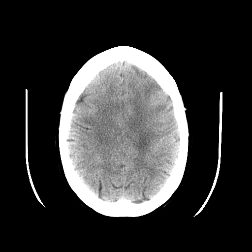
CT uses x-ray beams and a computer to create cross-sectional images of the body. CT slices reveal specific anatomy levels, with slice thickness chosen to minimize scatter radiation and superimposition using collimators. CT data are divided into pixels, forming a matrix, each representing different image details. Structures in CT images are depicted in varying shades of gray based on x-ray beam attenuation principles. The linear attenuation coefficient quantifies photon interaction with matter and varies with density, atomic number, and energy.[1] Higher density and atomic number lead to more significant attenuation, affecting image contrast.[1] CT images reflect these coefficients, showing denser materials as white and less dense ones as black. Contrast agents, like iodine, temporarily modify structure density differences for better visualization.[2] Hounsfield units (HU) quantify attenuation, aiding tissue characterization. However, HU values may be slightly inaccurate due to various factors. Polychromatic x-rays in CT can cause artifacts, like beam hardening, when low-energy photons are preferentially absorbed.[2] Filtering the x-ray beam with materials like aluminum can reduce these artifacts and improve image quality while lowering patient radiation dose.[2] CT exams involve selecting slice thickness based on anatomy and pathology. Thinner slices are ideal for detecting minor details and minimizing volume averaging effects, where normal and pathological tissue blend.[3] X-ray photons are created in the gantry-mounted tube, with voltage and tube current controlling intensity.[3] Detectors convert x-rays to electric current, which is processed by the data acquisition system (DAS) and converted into images by the central processing unit (CPU).[4] Often shown in HU, these images rely on pixel values to depict anatomical structures.
While designed differently, CT scanners share typical phases: data acquisition, image reconstruction, and image display. Data collection occurs in the first phase, processing in the second assigns pixel values, and in the final stage, data are displayed as shades of gray for viewing.
Data Acquisition & Image Reconstruction
CT utilizes detectors, which can be single elements or part of a larger detector array, and includes reference detectors to aid in calibration and artifact reduction. The size of the fan beam and the number of detector elements collecting data depend on the selected scan field of view.[4] Ideal detectors exhibit high efficiency in capturing transmitted photons, minimal afterglow (persistent scintillation), effective scatter suppression, and stability that eliminates frequent calibration interruptions.[5][6] Overall detector efficiency results from multiple factors, including the stopping power of the detector material, scintillator efficiency (for solid-state detectors), charge collection efficiency (for xenon detectors), geometric efficiency (comparing detector collimator plates to surface area), and scatter rejection.[7][4] Other terms related to detector efficiency include capture efficiency (the ability to acquire photons), absorption efficiency (the number of absorbed photons), response time (the time for the signal to return to zero after x-ray stimulation), and dynamic range (the ratio of the maximum to minimum measurable signals).[3][4]
Modern CT scanners primarily employ solid-state crystal detectors. At the same time, older models may utilize xenon gas detectors, which are becoming less prevalent due to limitations in multidetector row CT (MDCT) systems. Third-generation CT scanners feature a detector array and an x-ray tube that generates a fan-shaped beam, eliminating the need for beam and detector translation.[8] This design reduces scan times and motion artifacts, improving image quality, although it may lead to ring artifacts.[2][8] Fourth-generation scanners have a fixed detector array within the gantry while the tube rotates. Despite having more detectors, this design poses challenges related to motion artifacts. To address this issue, over-scans are utilized, but they elevate patient radiation exposure.[9] Another CT design, electron beam imaging (EBCT), employs a fixed electron gun and anode target. While it offers high speed, its clinical utility is limited due to spatial resolution concerns, cost factors, and difficulty obtaining insurance reimbursement.[10] The emergence of newer multidetector-row technology has reduced the relevance of EBCT.
The gantry's DAS converts analog signals from detectors into digital signals.[2] Continuous x-rays create rays read by the DAS. The system correlates ray attenuation with position, creating profiles for each view. These are projected onto a matrix, potentially causing streak artifacts. To reduce them, data undergoes filtered mathematical operations. Iterative reconstruction, a newer method, updates the image by comparing computed projections with original data, reducing noise and radiation dose by up to 50%.[11][12]
Image Quality & Quality Assurance
The quality of the image is influenced by numerous factors, some of which are within the operator's control, while others, like the patient's size, are not. The operator can manipulate variables, including milliampere (mA) level, scan duration, slice thickness, field of view, reconstruction method, and kilovolt peak (kVp).[1] Additionally, the operator can select a pitch value when employing helical scanning techniques. Collectively, these factors are typically referred to as scanning parameters. Image quality relies on two key elements: spatial resolution, which discerns fine, high-contrast details, and contrast resolution, which distinguishes objects with similar densities.[12][13] These aspects together enhance diagnostic precision in medical imaging. Spatial resolution is commonly assessed with the modulation transfer function (MTF), which ranges from 0 to 1, indicating how faithfully object details are reproduced.[14] In-plane resolution depends on matrix size and display field of view (DFOV). Matrix size determines pixel size, and adjusting DFOV changes to image and pixel dimensions. Larger DFOV leads to larger pixels and lower spatial detail. Pixel size affects accuracy, with smaller pixels reducing volume averaging and enhancing spatial resolution.[15] Voxel size, influenced by slice thickness and matrix dimensions, is also crucial in spatial resolution.[15] Thinner slices and consistently sized voxels lead to better results. The operator's choice of slice thickness determines the voxel shape, and all tissue data within a voxel are averaged to produce a single user-selected CT number.
CT systems offer different reconstruction algorithms that operators can choose or build into scan protocols to enhance or suppress specific data aspects for optimal diagnosis.[13] Some algorithms emphasize data smoothing by reducing pixel differences and reducing artifacts at the expense of spatial resolution.[16] Conversely, certain filters accentuate pixel differences to optimize spatial resolution but sacrifice low-contrast resolution.[16] These filters are suitable when extreme tissue density variations are present and low-contrast resolution is less critical. Contrast resolution distinguishes subtle density variations (low-contrast detectability).[3] Subject contrast depends on object size, making smaller objects harder to discern.[3] Organ contrast stems from physical traits, eg, the lung's air content. Temporal resolution is crucial for capturing moving structures and dynamic contrast studies influenced by gantry speed, detector channels, and signal recording.[17]
Quality control programs aim to optimize CT image quality while minimizing patient radiation exposure.[18] These programs systematically monitor the CT system's performance to detect specific issues or malfunctions. CT technologists and medical physicists share responsibility for conducting and documenting quality control tests. Technologists typically handle routine tests, while physicists perform annual or semi-annual tests, including acquiring necessary dosimetric data.[19] Quality assurance programs must adhere to three key principles: 1) regular performance of program tests, 2) consistent documentation of results in a standardized format, and 3) indication of whether the tested parameters meet specified guidelines.[19]
Summary and additional points to consider
A CT scanner is composed of a gantry, which constitutes the external apparatus of the scanner, an x-ray tube often composed of tungsten targets, a filter that attempts to create a monochromatic beam, a collimator which removes scatter, and a detector (most often a solid-state detector). Compared to typical diagnostic x-ray tubes, CT tubes function with higher amperage and a similar voltage to x-ray tubes and have similar focal spot sizes. Filters serve to reduce patient dose by removing low-energy x-rays and create a monochromatic beam as a result. Filters are typically composed of low-Z materials. While filters attenuate low-energy x-rays, collimators determine the thickness of the x-ray beam and section thickness. On the detector end, septa are also employed to reduce scatter and improve image quality by decreasing noise.
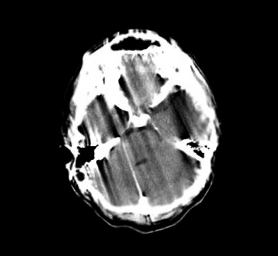
- CT scans use a kVp of around 120, although this can range from 70-140. An increased kVp increases the dose in CT imaging. Increasing kVp also increases noise, although this might be necessary for troubleshooting metal artifacts and imaging osseous structures. Iodinated contrast, whose K edge is around 33 keV, is better imaged at a lower kVp (typically about 80).
- The application of mathematical filters called kernels serves to smoothen or sharpen data. Kernels serve to produce images with varying levels of noise and spatial resolution. A smooth kernel can help visualize soft tissue anatomy, while a sharp kernel has superior spatial resolution and is used to evaluate osseous structures.[20]
- Helical/spiral CT obtains images with an x-ray tube by enabling a table to move continuously at a constant speed, thereby having the advantage of less motion artifact.[21]
- Pitch is the ratio of the distance the table moved in a single x-ray tube rotation divided by the beam width. A beam width of 1 will have no slice overlap. A pitch greater than 1 will have slice gaps, while a less than 1 will overlap.[22]
- Iterative reconstruction is now the more commonly used method of newer algorithms of image reconstruction, often uses a lower dose and can decrease noise to a better degree than older image reconstruction algorithms such as back projection and filtered back projection.[23]
- Housfield units constitute density and attenuation data of tissues with the standard of water used as the point of zero.
- Window width and level constitute a way to manipulate an image better to interrogate anatomy and pathology. The "level" constitutes the grey scale midpoint and is often established by the tissue attenuation being interrogated. The window "width" constitutes the desired range of densities used to compare tissues. Comparing bone with soft tissue will have a wide window width, while comparing tissues of similar densities will have a narrow window width.
- Automatic exposure control will alter mA but not kVp. Increasing mA by a factor of 4 increases your signal-to-noise ratio by a factor of 2 while increasing dose by a factor of 2.[24]