Learning Outcome
- Know the causes of ascites
- Describe the clinical features of ascites
- Understand the treatment of ascites
- Recall the nursing management plans for ascites

Ascites is the pathologic accumulation of fluid within the peritoneal cavity. It is the most common complication of cirrhosis and occurs in about 50% of patient with decompensated cirrhosis in 10 years. The development of ascites denotes the transition from compensated to decompensated cirrhosis. Mortality increases from complications such as spontaneous bacterial peritonitis and hepatorenal syndrome. Mortality ranges from 15% in a year to 44% in 5 years.[1][2][3]
In the United States, the most common disease that causes patients to get ascites is cirrhosis, which accounts for approximately 80% of cases. Other causes of ascites include cancer, 10%; heart failure, 3%; tuberculosis, 2%; dialysis, 1%; pancreatic disease, 1%; and others, 2%. Up to 19% of patients with cirrhosis will have hemorrhagic ascites; this may develop spontaneously with 72% of the cases most likely due to bloody lymph and 13% due to hepatocellular carcinoma. It can also develop following paracentesis.[4][5]
Patients with cirrhotic ascites have a 3-year mortality rate of approximately 50%. Refractory ascites carries a poor prognosis, with a 1-year survival rate of less than 50%. Males have little intraperitoneal fluid, females have approximately 20 mL, depending on the phase of their menstrual cycle.
Patients typically report progressive abdominal distension that may be painless or associated with abdominal discomfort, weight gain, early satiety, shortness of breath, and dyspnea resulting from fluid accumulation and increased abdominal pressure. Symptoms such as fever, abdominal tenderness, and confusion can be seen in spontaneous bacterial peritonitis.
Patients with malignant ascites can have symptoms related to malignancy, which may include weight loss. On the other hand, patients with ascites due to heart failure may report dyspnea, orthopnea, and peripheral edema, and those with chylous ascites report diarrhea, steatorrhea, malnutrition, edema, nausea, enlarged lymph nodes, early satiety, fevers, and night sweats.
Patients with ascites typically will have flank dullness on examination, shifting dullness, a fluid wave, evidence of pleural effusions, and findings related to the underlying cause of the ascites, such as stigmata of cirrhosis (cirrhosis includes spider angioma, palmar erythema, and abdominal wall collaterals.
Spider angiomata, jaundice, muscle wasting, gynecomastia, and leukonychia are present in patients with advanced liver disease.
An umbilical nodule that is not bowel or omental, such as a Sister Mary Joseph nodule, provides evidence for cancer as the cause of ascites. In heart failure, physical examination findings may include jugular venous distension, pulmonary congestion, or peripheral edema.
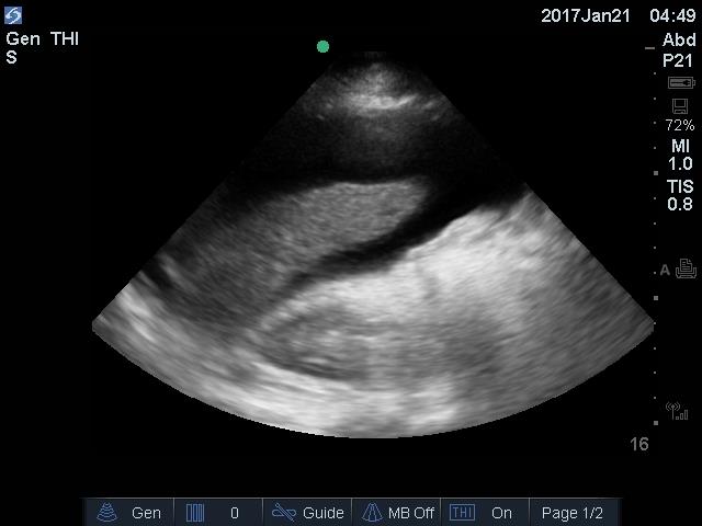
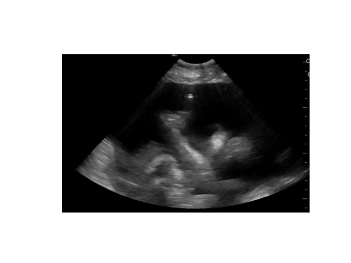
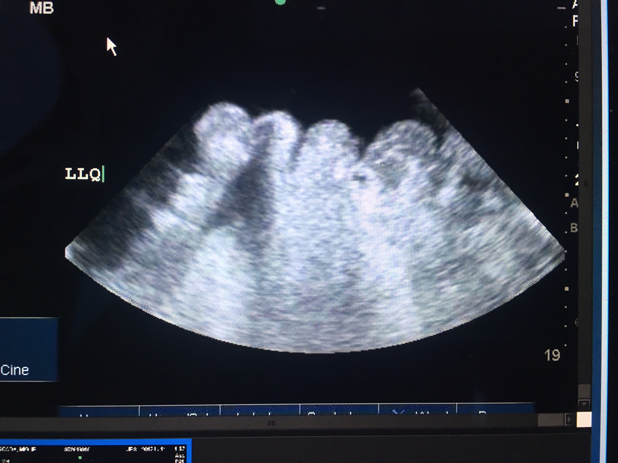
Diagnostic abdominal paracentesis with the appropriate ascitic fluid analysis is probably the most rapid and cost-effective method of diagnosing the cause of ascites.[6][7]
The initial tests that should be performed on the ascitic fluid include a blood cell count, with both a total nucleated cell count and polymorphonuclear neutrophils (PMN) count, and a bacterial culture by bedside inoculation of blood culture bottles.
Ascitic fluid protein and albumin are measured simultaneously with the serum albumin level to calculate the serum-ascites albumin gradient (SAAG).
The presence of a gradient greater or equal to 1.1 g/dL (greater or equal to 11 g/L) predicts that the patient has portal hypertension with 97% accuracy. This is seen in cirrhosis, alcoholic hepatitis, heart failure, massive hepatic metastases, heart failure/pericarditis, Budd-Chiari syndrome, portal vein thrombosis, and idiopathic portal fibrosis. A gradient less than 1.1 g/dL (less than 11 g/L) indicates that the patient does not have portal hypertension and occurs in peritoneal carcinomatosis, peritoneal tuberculosis, pancreatitis, serositis, and nephrotic syndrome.
Additional tests may be performed only if a specific diagnosis is suspected clinically. LDH and glucose level should be determined in suspected cases of secondary peritonitis. Other tests to consider include amylase (greater than 1000 U/L suggests pancreatic ascites). Mycobacterial culture should be performed only if tuberculosis is strongly suspected. Other ascitic fluid indices such as lactate and pH offer little to no additional information.
Appropriate treatment of ascites depends on the cause of fluid retention. The goals of therapy in patients with ascites are to minimize the ascitic fluid volume and decrease peripheral edema, without causing intravascular volume depletion.[8][9][10]
In cases of high-albumin-gradient ascites which occurs in cirrhosis, the treatment of ascites in these patients includes abstinence from alcohol, restricting dietary sodium to 88 mEq (2000 mg) per day, and treating with diuretics (spironolactone and furosemide in a ratio of 100:40 mg/day)
Patients with a treatable liver condition, such as autoimmune hepatitis, chronic hepatitis B with reactivation, hemochromatosis, or Wilson disease, should receive specific therapy for these diseases. Occasionally, cirrhosis due to causes other than alcohol or hepatitis B is reversible; however, these diseases are usually less reversible than in alcoholic liver disease, and by the time ascites is present, these patients may be better candidates for liver transplantation than for protracted medical therapy.
Low-albumin-gradient ascites commonly occurs in non-ovarian peritoneal carcinomatosis. These patients often benefit from an outpatient therapeutic paracentesis. Patients with ovarian malignancy may benefit from surgical debulking and chemotherapy.
TB peritonitis is treated with anti-tuberculous medications, while pancreatic ascites and postoperative lymphatic leak from a distal splenorenal shunt or radical lymphadenectomy may resolve spontaneously.
Chlamydia peritonitis is cured with antibiotics using doxycycline and ascites caused by lupus serositis may respond to glucocorticoids.
Abdomen girth normal
Can breath
Can ambulate and eat
No abdominal discomfort
No malaise
While it may appear that ascites is a GI related problem, the pathology affects almost all organ systems and has very high morbidity and mortality. Ascites is not a benign disorder and depending on the cause can have a mortality exceeding 20%. [11]To prevent complications and improve the quality of life, a streamlined protocol for management is vital.[3][12] (Level V)
Countless articles on a multidisciplinary approach have been published so that the morbidity and mortality can be improved. Besides the gastroenterologist the following interprofessional group of health professionals is highly recommended:
Outcomes and Evidence-based Medicine
Overall the prognosis is much worse for patients who have decompensated cirrhosis compared to those with compensated cirrhosis. Even patients who are ambulatory and have cirrhotic ascites have a 3-year mortality rate of 50%. Patients with refractory ascites have a 1-year survival of less than 50%. There are many evidence-based studies on managing patients with ascites.[13] (Level V). The bottom line is that aggressive care of these patients is critical if one wants to avoid the high mortality. Ascites patients are complex to manage and thus the need for a multidisciplinary team to ensure that the patient gets the right treatment, including a liver transplant.
Transfusion of blood products (fresh frozen plasma or platelets) routinely before paracentesis in patients with cirrhosis and coagulopathy, presumably to prevent hemorrhagic complications, is not supported by data
Contraindications to paracentesis include coagulopathy in the presence of DIC, massive ileus with bowel distension unless the procedure is image-guided to guarantee that the bowel is not entered.
Complications of ascites may include but are not limited to the following: Spontaneous bacterial peritonitis, cellulitis, tense ascites, pleural effusion, and abdominal wall hernias.
Lee JC, Kim JS, Kim HW, Cho IK, Lee J, Jang ES, Lee SH, Hwang JH, Kim JW, Jeong SH, Kim J. Outcome of endoscopic retrograde cholangiopancreatography in patients with clinically defined decompensated liver cirrhosis. Journal of digestive diseases. 2018 Oct:19(10):605-613. doi: 10.1111/1751-2980.12661. Epub 2018 Sep 23 [PubMed PMID: 30126061]
Privitera G, Figorilli F, Jalan R, Mehta G. Portosystemic Shunt Embolization and Recurrent Ascites: A Single-Center Case Series. Gastroenterology. 2018 Nov:155(5):1649-1650. doi: 10.1053/j.gastro.2018.06.092. Epub 2018 Aug 15 [PubMed PMID: 30118744]
Sarin SK, Choudhury A. Management of acute-on-chronic liver failure: an algorithmic approach. Hepatology international. 2018 Sep:12(5):402-416. doi: 10.1007/s12072-018-9887-5. Epub 2018 Aug 16 [PubMed PMID: 30116993]
Conangla-Planes M,Serres X,Persiva O,Augustín S, Imaging diagnosis of portal hypertension. Radiologia. 2018 Jul - Aug [PubMed PMID: 29472014]
Kibrit J, Khan R, Jung BH, Koppe S. Clinical Assessment and Management of Portal Hypertension. Seminars in interventional radiology. 2018 Aug:35(3):153-159. doi: 10.1055/s-0038-1660793. Epub 2018 Aug 6 [PubMed PMID: 30087517]
Fukui H, Kawaratani H, Kaji K, Takaya H, Yoshiji H. Management of refractory cirrhotic ascites: challenges and solutions. Hepatic medicine : evidence and research. 2018:10():55-71. doi: 10.2147/HMER.S136578. Epub 2018 Jul 3 [PubMed PMID: 30013405]
Szkodziak PR, Czuczwar P, Wrona W, Paszkowski T, Szkodziak F, Woźniak S. Ascites Index - a novel technique to evaluate ascites in ovarian hyperstimulation syndrome: a concept-proof study. Ginekologia polska. 2018:89(4):182-8. doi: 10.5603/GP.a2018.0031. Epub [PubMed PMID: 29781072]
Long B, Koyfman A. The emergency medicine evaluation and management of the patient with cirrhosis. The American journal of emergency medicine. 2018 Apr:36(4):689-698. doi: 10.1016/j.ajem.2017.12.047. Epub 2017 Dec 23 [PubMed PMID: 29290508]
Burgos AC, Thornburg B. Transjugular Intrahepatic Portosystemic Shunt Placement for Refractory Ascites: Review and Update of the Literature. Seminars in interventional radiology. 2018 Aug:35(3):165-168. doi: 10.1055/s-0038-1661347. Epub 2018 Aug 6 [PubMed PMID: 30087519]
Adebayo D, Neong SF, Wong F. Refractory Ascites in Liver Cirrhosis. The American journal of gastroenterology. 2019 Jan:114(1):40-47. doi: 10.1038/s41395-018-0185-6. Epub [PubMed PMID: 29973706]
Storni F, Stirnimann G, Banz V, De Gottardi A. Treatment of Malignant Ascites Using an Automated Pump Device. The American journal of gastroenterology. 2018 Jul:113(7):1060-1061. doi: 10.1038/s41395-018-0100-1. Epub 2018 Jun 5 [PubMed PMID: 29867179]
Strunk H,Marinova M, Transjugular Intrahepatic Portosystemic Shunt (TIPS): Pathophysiologic Basics, Actual Indications and Results with Review of the Literature. RoFo : Fortschritte auf dem Gebiete der Rontgenstrahlen und der Nuklearmedizin. 2018 Aug [PubMed PMID: 30045395]
Siddique SM, Lane-Fall M, McConnell MJ, Jakhete N, Crismale J, Porges S, Khungar V, Mehta SJ, Goldberg D, Li Z, Schiano T, Regan L, Orloski C, Shea JA. Exploring opportunities to prevent cirrhosis admissions in the emergency department: A multicenter multidisciplinary survey. Hepatology communications. 2018 Mar:2(3):237-244. doi: 10.1002/hep4.1141. Epub 2018 Jan 26 [PubMed PMID: 29507899]
Ede CJ, Nikolova D, Brand M. Surgical portosystemic shunts versus devascularisation procedures for prevention of variceal rebleeding in people with hepatosplenic schistosomiasis. The Cochrane database of systematic reviews. 2018 Aug 3:8(8):CD011717. doi: 10.1002/14651858.CD011717.pub2. Epub 2018 Aug 3 [PubMed PMID: 30073663]