Tonometry

Earn CME/CE in your profession:


Continuing Education Activity

Tonometry is a common procedure employed by healthcare professionals to measure intraocular pressure (IOP) using a calibrated instrument. IOP is important in diagnosing, screening, and managing ocular hypertension and glaucoma. Instruments measuring intraocular pressure assume the eye is a closed globe with uniform pressure distributed throughout the anterior chamber and vitreous cavity. The normal range of intraocular pressure is 10 to 21 millimeters of mercury. This activity discusses the numerous past and present methods of tonometry available to assess intraocular pressure and identify potential diseases causing abnormalities in eye pressure.

Objectives:

  • Identify the indications and contraindications of different types of tonometry.

  • Describe the equipment, personnel, preparation, and technique in regard to performing tonometry.

  • Summarize the appropriate evaluation of the potential complications of tonometry.

  • Explain interprofessional team strategies for improving care coordination and communication to advance the proper use of tonometry and optimize patient outcomes.

Introduction

Tonometry is a standard procedure employed by ophthalmologists to measure intraocular pressure (IOP) using a calibrated instrument. Devices used to measure IOP are based on the assumption that the eye is a closed globe with uniform pressure distributed throughout the anterior chamber and vitreous cavity. The normal range of intraocular pressure is 10 to 21 millimeters of mercury (mmHg), which is based on the average levels of IOP in normal individuals.[1] Glaucoma is a progressive optic neuropathy that leads to irreversible visual field damage, retinal nerve fiber layer thinning, and possible central visual acuity loss at the end stages of the disease. There are various risk factors in the development and progression of glaucoma,[2][3] the greatest being ocular hypertension (OHT), in which the IOP tends to be higher than 21 mmHg.[3] 

Treatment strategies, be it local IOP-lowering drops, laser, or surgery,[4] all aim to lower IOP to a target IOP level to limit, prevent, or slow glaucomatous progression.[5][6] Multiple methods of tonometry have been proposed since the 19th century to assess intraocular pressure.[7] The true IOP in the eyeball can only be obtained by manometry, in which a probe is inserted into the eye, which is invasive, risky, and not practical in a clinical setting.[8] 

Numerous methods have been proposed since the development of the first tonometer in 1863 by von Graefe.[9] These numerous methods are based on different principles of physics to obtain an estimate of the true IOP, which all aim to be accurate, specific, and representative of the actual IOP that is of utmost importance in the management of patients in a routine clinical setting. Instruments can be classified according to principles used in the device to measure IOP, which include applanation tonometry, indentation tonometry, rebound tonometry, Dynamic Contour tonometry, and continuous IOP monitoring.[10]

Anatomy and Physiology

IOP is determined by a delicate equilibrium between aqueous humor production and outflow. Elevated IOP can arise from increased production and/or decreased outflow mechanisms. The most common cause of elevated IOP in glaucoma is reduced aqueous humor outflow. Many forms of glaucoma are defined by elevated IOP, including primary open-angle glaucoma (the most frequent), closed-angle glaucoma, and secondary glaucoma (i.e., steroid-induced, pseudoexfoliation, traumatic, pigment dispersion glaucoma, etc.), and congenital glaucoma.[11][12][13][14][15][14][13][12][11][16][17] 

Most forms of open-angle glaucoma are silent sight-threatening diseases considering that central visual acuity tends not to be influenced, there is no associated pain or irritation, and peripheral visual field loss normally occurs gradually over numerous years in eyes with high IOP. Asymptomatic chronic elevated IOP leads to irreversible glaucomatous optic neuropathy, retinal nerve fiber layer thinning, and visual field damage. Tonometry is of great importance in the screening, diagnosis, and management of all patients, especially those with risk factors for developing glaucoma.[10] Tonometers must be properly calibrated, easy to use in routine clinics, and provide precise, accurate measurements with low inter-and intra- variability, preferably not influenced by external and ocular factors.[18]

Indications

Tonometry is utilized to measure IOP, which is essential in all forms of glaucoma. Tonometers are most commonly employed as part of a routine clinic visit by the clinician to screen for and monitor IOP elevation in glaucoma.[19] It is also used in the setting of ocular trauma without globe rupture, in cases of ocular inflammation and infection, and before and after ophthalmic surgical procedures.[7] It is crucial in managing patients at risk of developing the disease or those in therapy for glaucoma to prevent or slow down glaucomatous morphologic and functional defect progression. IOP is of clinical importance in numerous clinical situations, which include:

  • Screening for OHT and glaucoma in all adult patients, especially those with known risk factors.[20][21]
  • Diagnosis and management of patients with OHT or in therapy for glaucoma.[22][23]
  • Differential diagnosis and management of various ophthalmologic conditions can give rise to high levels of IOP, including pigment dispersion syndrome,[16] ocular infections, [24] pseudoexfoliation syndrome,[13] eye inflammation, Posner-Schlossman syndrome,[25] use of steroids in cortisone responders,[13] uveitis,[26] surgery,[27][28] trauma,[15] etc.

Contraindications

Information regarding IOP is of clinical importance during the ophthalmologic examination. There are situations, however, in which tonometry is challenging to obtain with certain instruments or can worsen the clinical conditions of the patient in specific conditions, which include:

Ruptured globe: Contact tonometry should be avoided or limited in the setting of a ruptured globe or suspicion of one.[15] Additional pressure on the globe with tonometry methods can further damage the globe and cause extrusion of the aqueous and vitreous humor. A ruptured globe is often apparent in an acute situation with the examination of the anterior ocular anatomy.

Infection: When an infection such as keratoconjunctivitis is suspected in one eye, the clinician should thoroughly disinfect or use a new cover before measuring the contralateral eye. This is also important when using the same tonometer between patients to prevent disease transmission.[19]

Unhealed corneal abrasions or ulcers, corneal scars, and elevated eyes with astigmatism: Manipulation with tonometry can further aggravate active corneal lesions.[29] The precision and accuracy of several tonometers that have direct contact with the cornea can be limited in the presence of scarring or high astigmatism; thus, proper instrument use in these select patients is of clinical importance in measuring IOP.[30]

Inability to administer or intolerance/allergies to topical anesthesia and/or fluorescein: This contraindication is applicable to methods of tonometry requiring topical anesthesia before measurement of IOP.[31] Alternative methods that do not require topical anesthesia can be considered in these patients, which include rebound (iCare) and non-contact "air puff" tonometry.[10]

Difficulties or inability to use the tonometer: Certain tonometry, like the gold standard Goldmann applanation, can only be used with a collaborative individual in an upright position. Other portable manual instruments, like iCare rebound tonometers, can be used in a supine position and require little patient collaboration, thus can be considered in bedridden and non-collaborative individuals and children.[10]

Equipment

Methods  

Applanation Tonometry

Goldmann Applanation Tonometer (GAT)

In 1948, Hans Goldmann proposed GAT to measure IOP.[32] To this date, GAT remains the gold standard method for measuring IOP and is most frequently used by ophthalmologists in a routine clinical setting.[33] It utilizes applanation, which measures the force required to compress the cornea over a given area. Applanation tonometry is based on the Imbert-Fick law from the 1800s, which states that the pressure inside the eye (P) can be approximated by the force (F) needed to flatten a fixed area of the cornea divided by the surface area (S) of the flattened region of the surface.[34][35] The higher the intraocular pressure, the greater the force needed to compress the cornea.[19] 

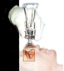
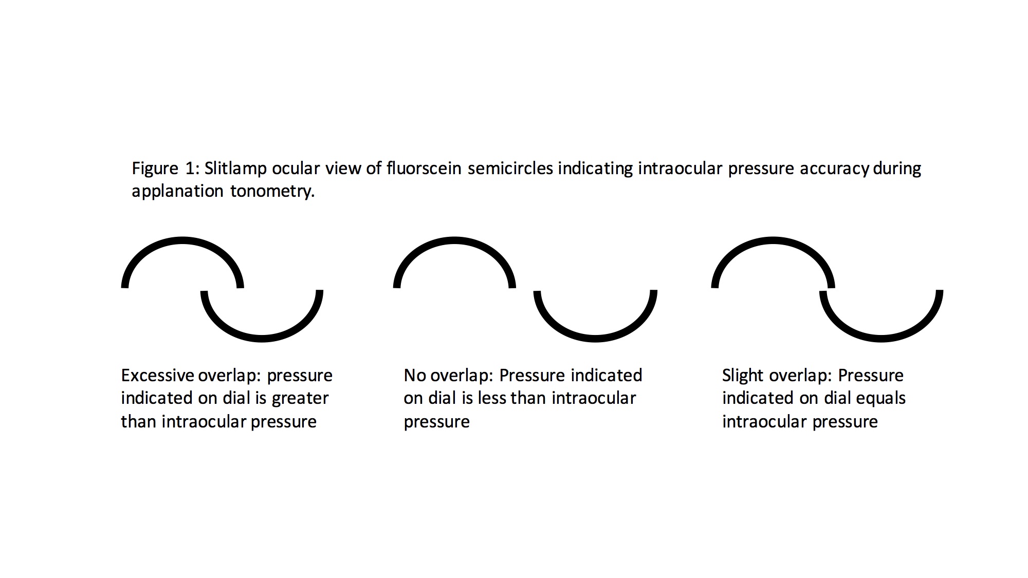
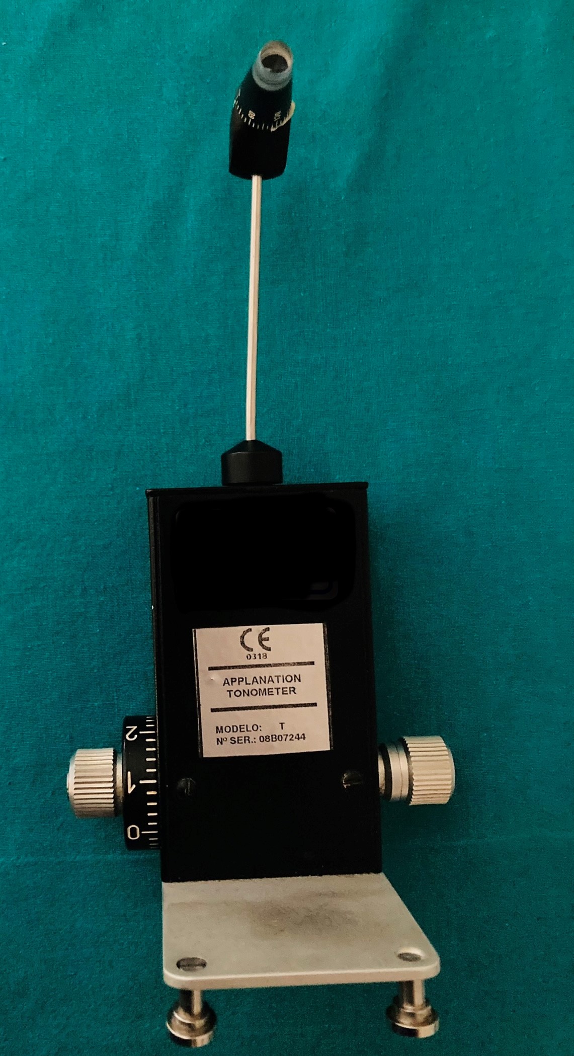
Eye clinicians use GAT as a standard component of a slit-lamp (See slit-lamp and GAT tonometer images) eye exam.[7] To perform GAT, topical anesthetic drops and a fluorescein dye are first applied to the eyes. Filtered cobalt blue light is then used to visualize the fluorescein dye while the tonometer tip compresses the center of the cornea with a diameter of 3.06 mm (See GAT tip images). The blue light and dye highlight the circular border of the tear film created by the applanator tip pressed against the cornea (See GAT tip images). A prism in the tip splits this circular film of fluorescein into two green semicircles when visualized from the slit lamp oculars (See images with arcs). The clinician then alters the tonometer force against the cornea by turning the dial on the side of the instrument (See tonometer with dial images) until the two semicircles overlap slightly (See images with arcs). This slight overlap of the two semicircles indicates that the cornea has been flattened by a calibrated amount to give an accurate IOP measurement in mmHg, which is read on the dial (see tonometer with dial images) of the tonometer.[29]

Several factors can influence the accuracy and precision of IOP measurements, which include central corneal thickness (CCT), elevated astigmatism, ocular rigidity, corneal hysteresis, tear film, corneal edema, the amount of fluorescein, previous refractive surgery, etc.[36][37][32][34][32][37][36] Although GAT is considered the gold standard method to measure IOP, several limits have enticed the proposal of new tonometers in the past 70 years.[7] The disadvantages of the Goldmann method include a high level of skill to operate, inability to measure in supine and non-collaborative patients, need for topical anesthesia and fluorescein, and decreased accuracy on an irregular or scarred cornea.[7][19]

Perkins and Draeger Applanation Tonometer

The Perkins and Drager handheld tonometers use the same mechanism of applanation as the Goldmann.[7] Considering the portability of these devices, these tonometers can be useful in settings where a slit lamp exam is not feasible, such as an emergency department or operating room, and can also be used in upright or supine positions. The tonometers have a built-in forehead rest, illuminating blue light source, a prism holder, a thumb-wheel dial with a scale to measure IOP, and a power source. The limits of these tonometers are similar to those in GAT, considering that these tonometers use the applanation principle to measure IOP. Other disadvantages of these portable devices include a high level of skill to operate and a decrease in stability with the handheld versions.[38]

Non-contact Tonometers

Non-contact tonometers (NCT),[7] which first came available in the 1970s, are also known as "air-puff" tonometers. Several desktop and portable handheld models are available from various manufacturers.[39][40] Numerous modern desktop instruments used as pachymeters and refractometers also have a built-in NCT to provide versatility and convenience for clinicians (See refractometer with pachymeter and NCT images). These instruments use a small puff of air to applanate the central cornea.[41] The returning air from the cornea's surface is measured by a membrane that records the force using sensors, detectors, and beams of light, which are then converted by an internal algorithm to give IOP measurements. The advantages of NCT include no requirement for topical anesthesia and its usefulness in screening, especially in children and adults who cannot tolerate contact methods.[19] NCT can be helpful in a clinical screening setting; however, it is considered a less accurate, precise, and sensitive method than GAT, in addition to being instrument-dependent and highly influenced by CCT, thus not advisable in the management of patients with glaucoma or individuals with OHT or other risk factors.[42][43][42][44][45]

Ocular-Response Analyzer (ORA) and Corvis ST Tonometers

ORA was developed in 2005 and represented a newer version of NCT that utilizes a column of air as the applanating force.[7] The flow of air is used to move the cornea inwardly, which slowly decreases when the cornea indents outwards and then moves to the flattened state.[46] Based on the force of airflow and the rate of recovery from deformation, the optical sensors can evaluate the elasticity of the cornea to provide a corneal hysteresis (CC) value, a corrected intraocular pressure (IOPg), and a corneal-compensated measurement (IOPcc).[47] 

Corneal hysteresis is the ability of the cornea to absorb and dissipate applied forces based on its viscoelastic and elasticity properties. ORA was designed to theoretically improve IOP accuracy, allowing clinicians to account for the variability in corneal biomechanical properties seen among patients.[47][48] Corneal hysteresis has been demonstrated to contribute to glaucoma progression risk, with lower values imparting a greater risk of glaucoma progression.[49][50] Like GAT, ORA is a desktop instrument and thus cannot be used on supine patients. Although the corneal compensation parameters have been developed to provide IOP measurements less affected by corneal factors, studies have shown overestimated ORA measurements in eyes with high levels of IOP.[51] Further clinical studies are needed to assess the clinical use of ORA in glaucoma management. 

Corvis ST is an NCT based on the indentation of the cornea by a jet of air, which was proposed in 2011 by Oculus in Germany.[52] A built-in Scheimpflug camera is used to provide precise measurements of the deformation of the cornea during the air-induced indentation process.[53] The instrument also has a pachymeter and provides several other corneal parameters.[54] Although the precision of the Corvis ST readings has been reported to be very good, and measurements appear to be less influenced by corneal factors, studies have shown that the Corvis ST tonometer tends to underestimate IOP when compared to GAT.[55][56][57][58]

Indentation Tonometry

Schiotz Tonometer

The principle of indentation is based on the fact that weight or force will sink more into a softer object than a harder one. The Norwegian physician, Hjalmar Schiotz, invented the first clinically useful indentation tonometer in 1905.[59] This portable tonometer (See Schiotz images) employs a rarely used, older technique. It consists of a weighted plunger attached to a footplate positioned on the cornea. Weights are stacked onto the probe to cause depression of the cornea. The number of weights stacked onto the probe correlates to a calibrated intraocular pressure using a conversion chart provided with the device.[7] The patient must be supine during measurement, and a topical corneal anesthetic is required.[19] This tonometer is relatively inexpensive and easy to use and is sometimes still used in children under general anesthesia and developing countries.[60] However, this tonometer is not routinely used in a clinical setting, considering that IOP measurements are subject to several errors, including defective instruments or calibration, high variability, bad positioning on the eyeball, and influences due to ocular rigidity and other ocular factors.[61]

Tono-Pen Indentation/Applanation Tonometer

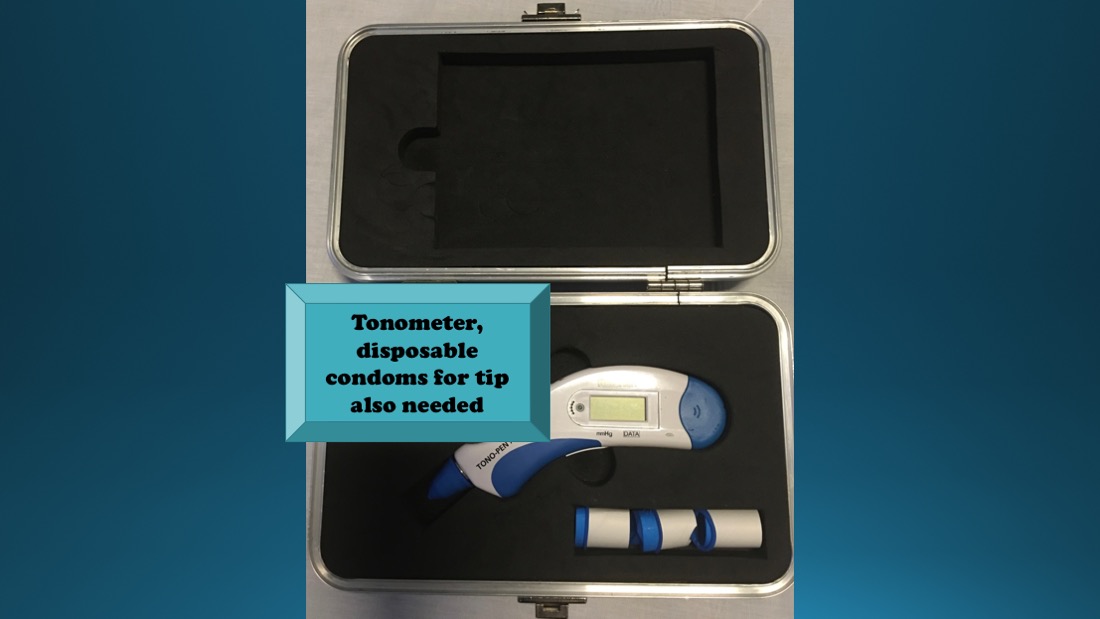
The Tono-pen is a battery-powered, lightweight, portable handheld device that uses a small plunger to record the force needed to applanate the cornea. This tonometer is based on both the indentation and applanation principles. An early version of this instrument was introduced in 1959 by McKay and Marg.[62] The Tono-pen averages multiple readings of this small force converted to IOP.[7] The newer versions of the Tono-Pen include XL and Avia, which give IOP measurements as an average of  4 and 10 measurements, respectively.[9] 

It requires daily recalibration and topical anesthesia and uses disposable covers (See Tono-pen image). It can prove useful in portable screenings, emergency rooms, bedridden patients, or operating rooms to measure intraocular pressure. Advantages of this method include the ability to measure over a soft contact lens, on an irregular corneal surface, averaging multiple readings, and the potential to measure at the peripheral cornea if a central corneal scar or ulcer exists. It can also measure intraocular pressure independent of patient position.[7] Studies have shown Tono-Pen to be useful in certain clinical situations; however, the intrasession variability has been shown to be rather high, and measurements with Tono-Pen appear to be significantly affected by CCT.[63][64]

Pneumatonometer

The pneumatonometer uses a stream of air to indent the cornea with a 5 mm diameter silicone tip on a piston that rides on the air.[7] This tonometry is based on both indentation and applanation principles.[65] The force of air that indents the cornea is recorded and converted to intraocular pressure. A built-in transducer is used to convert the force generated during corneal applanation into IOP in mmHg. Studies have shown that this method correlates well with GAT in normal pressure ranges.[66] 

The Pulsatile Ocular Blood Flow pneumotonometer is the newer version that provides pulse fluctuation and information regarding ocular blood pulse.[67] Similar to the Tono-pen, it has the advantage of measuring irregular cornea surfaces, over a soft contact lens, at the peripheral cornea. Accuracy is independent of patient position, although it requires topical anesthesia.[7] The pneumatonometer is accessible in a handheld probe attached to a table-mounted device. The device is also utilized for pneumatonography, which uses changes in IOP to measure the outflow resistance of aqueous humor from the anterior chamber.[68] If increased outflow resistance is found, it can cause increased IOP and contribute to glaucoma. However, the clinical usefulness of this tonometer remains debatable, considering that studies have shown IOP values with pneumatonometry tend to be overestimated and more influenced by corneal parameters compared to gold standard GAT.[69]

Rebound Tonometry

iCare tonometer

The iCare tonometer, based on the principles of rebound, is a portable battery-powered handheld device that is easy to use.[70] Several versions of this tonometer include the iCare TA01i, IC100, IC200, PRO, and HOME (See images with different iCare models). It utilizes a small disposable probe (composed of a tiny plastic round tip on a thin steel wire) that bounces off the cornea in the horizontal plane. The deceleration of the probe produces a level of voltage that is converted to IOP.[70] The faster the rate of deceleration against the cornea, the higher the pressure. The slower the rate of deceleration, the lower the pressure.[71] The advantages of this method include ease of use,[33]  no requirement for topical anesthetics, and its usefulness in children, non-collaborative individuals, those allergic to fluorescein and anesthetic drops, and dementia patients who are not tolerant of a slit-lamp exam or more involved contact methods.[47]

The HOME version can be used autonomously for at-home use in managing glaucoma patients, especially in suspect normal-tension glaucoma patients that tend to have normal IOP during clinical examinations.[72][73][74] Most versions need to be used while the patients are in an upright position. However, the iCare PRO, which became available in 2011, was designed with a shorter probe and can be used in a supine position.[75] The iCare tonometer can be of important clinical use in screening, routine examination of normal adults and children, and in patients with recent ocular surgeries, considering the facility, rapidness, limited pressure applied on the cornea, and no need for fluorescein or anesthetic drops.[70][27]

Several studies have shown that when compared to GAT and other tonometers, iCare showed good agreement, specificity, reliability, and reproducibility, especially for low-to-moderate IOP measurements.[76][77][78][77][76][79] Considering the facility, low cost, and good agreement with GAT, numerous clinicians currently find the iCare tonometer useful in a routine clinical setting, of course, keeping in mind that elevated IOP readings with rebound tonometry need to be confirmed with GAT. The gold standard in tonometry remains GAT, which to date cannot be replaced, especially when managing patients with glaucoma or individuals at risk of developing OHT.

Dynamic Contour Tonometry (DCT)

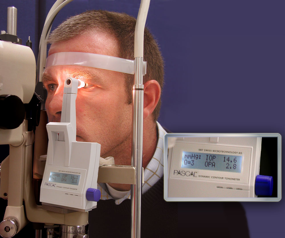
DCT was developed in 2003 and calculates IOP based on the Pascal principle, which deals with the pressure of a fluid within an enclosed space.[80][81] Similar to GAT, this tonometer needs to be mounted on a slit-lamp (See DCT Pascal image), with the patient in an upright position, and the use of anesthesia drops (but not fluorescein) considering the tip of the tonometer needs to be in contact with the cornea. This device utilizes a contour-matched, piezoelectric sensor to measure minuscule dynamic pulsations in intraocular pressure at the cornea.[82] Disposable plastic caps are used on the sensor tip to prevent cross-infection. This method allows the measurement of IOP without deforming the cornea; therefore, its readings are independent of corneal thickness, unlike other methods.[47][18][83] 

Studies have shown DCT to have good precision, reproducibility, and correlatability compared to GAT and other tonometers.[84][85][86][85][84][87] When using DCT, the tip needs to be in contact with the cornea for about 10 seconds to provide an accurate measurement of IOP; thus rather difficult to use and not ideal for all patients.[33] The other disadvantages include the high learning curve, the need for a slit-lamp and anesthetic drops, difficulties to use in a routine clinical setting, and IOP measurements influenced by several artifacts, measurement errors, CCT, and other corneal parameters.[88][89][90][91][92]

Continuous IOP Monitoring

The routine clinical setting normally provides the ophthalmologist with only a few spot readings of IOP for each patient. Patients can have ophthalmologic examinations up to 4 times yearly; however, in normal cases, this tends to be biannual or annual for the most part. In the ideal world of glaucoma management, IOP would be continuously measured 24 hours a day to provide the true fluctuations of IOP and IOP spikes during the waking and sleeping hours, which could have a great impact on understanding the true target pressure, progression rates, and the best individually tailored treatment strategies for each patient.[93][94][95] Unfortunately, current methods are not available for routine clinical use. However, several options for continuous IOP monitoring have been proposed using an externally applied contact lens or an intraocular surgically inserted lens or sensor.[96]

Regarding sensors applied to contact lenses on the cornea surface, a company from Switzerland launched the Sensimed Triggerfish system.[97] This 24-hour IOP-monitoring device is composed of a disposable silicone contact lens with an embedded electromechanical microprocessor that collects more than 280 repeated IOP measurements during the 24 hours of application.[98] The data is sent with a wireless antenna on the lens to a portable recorder that the patient carries in a handy shoulder purse. Studies have shown that the Triggerfish contact lens is well tolerated, provides reproducible readings of IOP, and can help detect IOP peaks throughout the day.[99][100][101] 

The main limits of this system, however, include no direct correlation between corneal changes and IOP readings and poor correlation with GAT readings regarding measurements taken several hours after the device has been activated.[102][103] The clinical use of Triggerfish and its application in routine use remains debatable. 

Implantable IOP-sensor devices, which can be positioned during cataract surgery in the anterior chamber, suprachoroidal space, or embedded in a foldable artificial intraocular lens, have been proposed; however, they remain experimental and not of widespread use yet.[104][105][106][105][107]   

Technique or Treatment

Technique Considerations

  • Care must be taken to not artificially raise the intraocular pressure by pressing on the eyelid during IOP measurement. Ensure the patient is comfortable and not attempting to squint, as this can raise IOP.
  • Ensure that the patient is appropriately positioned for the specific tonometry method (seated versus supine). Table-top methods such as GAT can only be used in upright and seated patients. Rebound (I-Care) tonometry can only be used in the horizontal plane.[7]
  • Clean and disinfect all contact tonometers between patients unless they utilize disposable covers. This assists in preventing disease transmission and possible corneal abrasions from residual debris.
  • Methods that rely on applanation are affected by the central corneal thickness and other biomechanical properties such as elasticity and hysteresis.[44] The calibration of these methods assumes uniform corneal thickness and biomechanical properties. Thinner and more elastic corneas will result in lower readings, while thicker and more rigid corneas will result in higher readings due to the variability in forces needed to deform the cornea. Additional factors that may affect tonometer accuracy are astigmatisms, irregular corneas, Valsalva maneuver, breath-holding, and natural variation of IOP during the cardiac cycle.[7]
  • GAT currently remains the gold standard technique in measuring IOP, especially in managing patients with OHT, risk factors, and in therapy for glaucoma. Elevated IOP measurements taken with alternative tonometers should be confirmed with GAT.

Complications

Corneal abrasion, aggravation of globe rupture, the transmission of infection, and reaction to ocular drugs are potential complications of tonometry. The risk of these complications is considerably low (less than 1%). Non-contact methods and rebound tonometry can be considered in special cases, even if GAT remains the gold standard approach to tonometry.

Clinical Significance

Measurement of intraocular pressure is important in the screening and monitoring of glaucoma, a progressive optic neuropathy that can be slowed with intraocular pressure reduction. Intraocular pressure is the only modifiable risk factor for glaucoma progression at this time.[7] Prescribing medications to reduce intraocular pressure and monitoring pressure over time using tonometry has been demonstrated to slow the progression of peripheral and central vision loss related to glaucoma. Tonometry is also used to evaluate acutely elevated intraocular pressure, as seen in acute-angle closure glaucoma and after ocular trauma. Acute angle-closure glaucoma is an ophthalmic emergency requiring immediate intervention to lower IOP and avoid vision loss.[19]

Enhancing Healthcare Team Outcomes

In addition to an ophthalmologist, an optometrist, ophthalmic technician, nurse, or emergency medicine physician may use tonometry during an ophthalmic office visit, emergency room visit, or ambulatory settings as needed. Communication among these professionals, operating as an interprofessional team, regarding the particular tonometry method is important so eye pressure measurements can be accurately compared throughout time.  

Portable techniques such as the Tono-pen and rebound tonometer are simple to use, while stationary methods such as GAT require more specialized training. Each medical professional mentioned above may use various tonometry methods based on comfort level and training. A point of emphasis among all professionals performing tonometry must be the proper use of topical anesthesia to minimize patient discomfort for methods including Tono-pen and GAT. Avoid any tangential movement across the cornea during tonometric measurement to prevent corneal abrasions. Additionally, ensure any disinfecting solution is rinsed from the tonometry surface that contacts the cornea to avoid potential toxic keratitis.  

Tonometry readings may need to be re-measured among professionals to ensure consistency of readings. If there is a question about the accuracy of a reading or device, an alternative tonometry method may be used to help confirm or refute the reading. In an acute setting where acute-angle closure glaucoma is suspected, an emergent intraocular pressure reading must be acquired. This is typically initially done in the emergency room by a physician or nurse, with a consulting ophthalmologist confirming the pressure on arrival and administering pressure-lowering medication or performing additional interventions to save vision.[108] [Level 1]


(Click Image to Enlarge)
Tonometer, disposable condoms for tip also provided in this example of one type
Contributed by Tammy J. Toney-Butler, RN, CEN, TCRN, CPEN

(Click Image to Enlarge)
The PASCAL Dynamic Contour Tonometer. A monitor for detection of increased intraocular pressure.
Contributed by Wikimedia Commons, Tony Wirthlin (Public Domain)

(Click Image to Enlarge)
tonometer for glaucome
Contributed by S Bhimji MD

(Click Image to Enlarge)
Fluorescein semicircles during Goldman Applanation Tonometery (GAT) to measure intraocular pressure (IOP)
Fluorescein semicircles during Goldman Applanation Tonometery (GAT) to measure intraocular pressure (IOP). Elevated IOP is a modifiable risk factor for Glaucoma
Created by John Bader, BS

(Click Image to Enlarge)
Goldmann Applanation Tonometer (GAT) used to measure intraocular pressure (IOP).
Contributed by Marco Zeppieri, MD, PhD.
Details

Author

John Bader

Editor:

Shane J. Havens

Updated:

12/12/2023 6:29:01 AM

References


[1]

ALIMUDDIN M. Normal intra-ocular pressure. The British journal of ophthalmology. 1956 Jun:40(6):366-72     [PubMed PMID: 13355942]


[2]

Salvetat ML, Zeppieri M, Tosoni C, Brusini P, Medscape. Baseline factors predicting the risk of conversion from ocular hypertension to primary open-angle glaucoma during a 10-year follow-up. Eye (London, England). 2016 Jun:30(6):784-95. doi: 10.1038/eye.2016.86. Epub 2016 May 13     [PubMed PMID: 27174381]


[3]

Popović-Suić S, Sikić J, Vukojević N, Cerovski B, Nasić M, Pokupec R. Target intraocular pressure in the management of glaucoma. Collegium antropologicum. 2005:29 Suppl 1():149-51     [PubMed PMID: 16193700]


[4]

Brusini P, Papa V, Zeppieri M. Canaloplasty in Pseudoexfoliation Glaucoma. Can It Still Be Considered a Good Choice? Journal of clinical medicine. 2022 Apr 30:11(9):. doi: 10.3390/jcm11092532. Epub 2022 Apr 30     [PubMed PMID: 35566656]


[5]

Toneatto G, Zeppieri M, Papa V, Rizzi L, Salati C, Gabai A, Brusini P. 360° Ab-Interno Schlemm's Canal Viscodilation with OMNI Viscosurgical Systems for Open-Angle Glaucoma-Midterm Results. Journal of clinical medicine. 2022 Jan 4:11(1):. doi: 10.3390/jcm11010259. Epub 2022 Jan 4     [PubMed PMID: 35012000]


[6]

Brusini P, Tosoni C, Zeppieri M. Canaloplasty in Corticosteroid-Induced Glaucoma. Preliminary Results. Journal of clinical medicine. 2018 Feb 11:7(2):. doi: 10.3390/jcm7020031. Epub 2018 Feb 11     [PubMed PMID: 29439499]


[7]

Aziz K, Friedman DS. Tonometers-which one should I use? Eye (London, England). 2018 May:32(5):931-937. doi: 10.1038/s41433-018-0040-4. Epub 2018 Feb 19     [PubMed PMID: 29456251]


[8]

McAllister F, Harwerth R, Patel N. Assessing the True Intraocular Pressure in the Non-human Primate. Optometry and vision science : official publication of the American Academy of Optometry. 2018 Feb:95(2):113-119. doi: 10.1097/OPX.0000000000001171. Epub     [PubMed PMID: 29370024]


[9]

Stamper RL. A history of intraocular pressure and its measurement. Optometry and vision science : official publication of the American Academy of Optometry. 2011 Jan:88(1):E16-28. doi: 10.1097/OPX.0b013e318205a4e7. Epub     [PubMed PMID: 21150677]


[10]

Brusini P, Salvetat ML, Zeppieri M. How to Measure Intraocular Pressure: An Updated Review of Various Tonometers. Journal of clinical medicine. 2021 Aug 27:10(17):. doi: 10.3390/jcm10173860. Epub 2021 Aug 27     [PubMed PMID: 34501306]


[11]

Reis TF, Paula JS, Furtado JM. Primary glaucomas in adults: Epidemiology and public health-A review. Clinical & experimental ophthalmology. 2022 Mar:50(2):128-142. doi: 10.1111/ceo.14040. Epub 2022 Jan 26     [PubMed PMID: 35037725]


[12]

Teo ZL, Soh ZD, Tham YC, Yu M, Chee ML, Thakur S, Nongpiur ME, Koh V, Wong TY, Aung T, Cheng CY. Six-Year Incidence and Risk Factors for Primary Angle-Closure Disease: The Singapore Epidemiology of Eye Diseases Study. Ophthalmology. 2022 Jul:129(7):792-802. doi: 10.1016/j.ophtha.2022.03.009. Epub 2022 Mar 16     [PubMed PMID: 35306094]


[13]

Wijnants D, Stalmans I, Vandewalle E. The Effects of Intranasal, Inhaled and Systemic Glucocorticoids on Intraocular Pressure: A Literature Review. Journal of clinical medicine. 2022 Apr 3:11(7):. doi: 10.3390/jcm11072007. Epub 2022 Apr 3     [PubMed PMID: 35407615]


[14]

Paulson C, Thomas SC, Gonzalez O, Taylor S, Swiston C, Herrick JS, McCoy L, Curtin K, Chaya CJ, Stagg BC, Wirostko BM. Exfoliation Syndrome in Baja Verapaz Guatemala: A Cross-Sectional Study and Review of the Literature. Journal of clinical medicine. 2022 Mar 24:11(7):. doi: 10.3390/jcm11071795. Epub 2022 Mar 24     [PubMed PMID: 35407402]

Level 2 (mid-level) evidence

[15]

Hou X, Guo X, Cui Z, Wang Y, Zhou J, Hu D. Value of ocular trauma score in predicting the incidence of secondary glaucoma after closed globe injury. European journal of ophthalmology. 2022 Sep:32(5):3005-3011. doi: 10.1177/11206721211055628. Epub 2021 Nov 29     [PubMed PMID: 34841932]


[16]

Zeppieri M, Tripathy K. Pigment Dispersion Glaucoma. StatPearls. 2024 Jan:():     [PubMed PMID: 35593820]


[17]

Panta Sitoula R, Gurung J, Anwar A. Primary Congenital Glaucoma among the Children Under 3 Years of Age in the Outpatient Department in a Tertiary Care Hospital: A Descriptive Cross-sectional Study. JNMA; journal of the Nepal Medical Association. 2021 Sep 11:59(241):867-870. doi: 10.31729/jnma.5889. Epub 2021 Sep 11     [PubMed PMID: 35199734]

Level 2 (mid-level) evidence

[18]

Fogagnolo P, Figus M, Frezzotti P, Iester M, Oddone F, Zeppieri M, Ferreras A, Brusini P, Rossetti L, Orzalesi N. Test-retest variability of intraocular pressure and ocular pulse amplitude for dynamic contour tonometry: a multicentre study. The British journal of ophthalmology. 2010 Apr:94(4):419-23. doi: 10.1136/bjo.2009.165142. Epub 2009 Oct 14     [PubMed PMID: 19833616]


[19]

Walker HK, Hall WD, Hurst JW, Alguire PC. Tonometry. Clinical Methods: The History, Physical, and Laboratory Examinations. 1990:():     [PubMed PMID: 21250066]


[20]

Blumberg MJ, Varikuti VNV, Weiner A. Real-world comparison between the Tonopen and Goldmann applanation tonometry in a university glaucoma clinic. International ophthalmology. 2021 May:41(5):1815-1825. doi: 10.1007/s10792-021-01742-z. Epub 2021 Mar 2     [PubMed PMID: 33651312]


[21]

Chou R, Selph S, Blazina I, Bougatsos C, Jungbauer R, Fu R, Grusing S, Jonas DE, Tehrani S. Screening for Glaucoma in Adults: Updated Evidence Report and Systematic Review for the US Preventive Services Task Force. JAMA. 2022 May 24:327(20):1998-2012. doi: 10.1001/jama.2022.6290. Epub     [PubMed PMID: 35608575]

Level 1 (high-level) evidence

[22]

Wachtl J, Töteberg-Harms M, Frimmel S, Roos M, Kniestedt C. Correlation Between Dynamic Contour Tonometry, Uncorrected and Corrected Goldmann Applanation Tonometry, and Stage of Glaucoma. JAMA ophthalmology. 2017 Jun 1:135(6):601-608. doi: 10.1001/jamaophthalmol.2017.1012. Epub     [PubMed PMID: 28494071]


[23]

Salvetat ML, Zeppieri M, Tosoni C, Brusini P. Comparisons between Pascal dynamic contour tonometry, the TonoPen, and Goldmann applanation tonometry in patients with glaucoma. Acta ophthalmologica Scandinavica. 2007 May:85(3):272-9     [PubMed PMID: 17488456]


[24]

Aldaas K, Challa P, Weber DJ, Fleischman D. Infections and glaucoma. Survey of ophthalmology. 2022 May-Jun:67(3):637-658. doi: 10.1016/j.survophthal.2021.08.009. Epub 2021 Sep 4     [PubMed PMID: 34487741]

Level 3 (low-level) evidence

[25]

Okonkwo ON, Tripathy K. Posner-Schlossman Syndrome. StatPearls. 2024 Jan:():     [PubMed PMID: 35015437]


[26]

Rao A, Gawas L. Atypical associations of viral anterior uveitis with glaucoma-a series of challenging scenarios with review of literature. Seminars in ophthalmology. 2021 Nov 17:36(8):605-613. doi: 10.1080/08820538.2021.1890789. Epub 2021 Mar 18     [PubMed PMID: 33734824]


[27]

Salvetat ML, Zeppieri M, Miani F, Tosoni C, Parisi L, Brusini P. Comparison of iCare tonometer and Goldmann applanation tonometry in normal corneas and in eyes with automated lamellar and penetrating keratoplasty. Eye (London, England). 2011 May:25(5):642-50. doi: 10.1038/eye.2011.60. Epub 2011 Mar 25     [PubMed PMID: 21436848]


[28]

Gismondi M, Salati C, Salvetat ML, Zeppieri M, Brusini P. Short-term effect of intravitreal injection of Ranibizumab (Lucentis) on intraocular pressure. Journal of glaucoma. 2009 Dec:18(9):658-61. doi: 10.1097/IJG.0b013e31819c4893. Epub     [PubMed PMID: 20010243]


[29]

Stevens S, Gilbert C, Astbury N. How to measure intraocular pressure: applanation tonometry. Community eye health. 2007 Dec:20(64):74-5     [PubMed PMID: 18330450]


[30]

Stewart RM, Wishart PK, Kaye SB. Overestimation of Intraocular Pressure by Goldmann Applanation Tonometry Without Astigmatic Correction. JAMA ophthalmology. 2016 Mar:134(3):e153691. doi: 10.1001/jamaophthalmol.2015.3691. Epub 2016 Mar 10     [PubMed PMID: 26967752]


[31]

Arend N, Hirneiss C, Kernt M. [Differences in the measurement results of Goldmann applanation tonometry with and without fluorescein]. Der Ophthalmologe : Zeitschrift der Deutschen Ophthalmologischen Gesellschaft. 2014 Mar:111(3):241-6. doi: 10.1007/s00347-013-2843-9. Epub     [PubMed PMID: 23604252]


[32]

GOLDMANN H, SCHMIDT T. [Applanation tonometry]. Ophthalmologica. Journal international d'ophtalmologie. International journal of ophthalmology. Zeitschrift fur Augenheilkunde. 1957 Oct:134(4):221-42     [PubMed PMID: 13484216]


[33]

Okafor KC, Brandt JD. Measuring intraocular pressure. Current opinion in ophthalmology. 2015 Mar:26(2):103-9. doi: 10.1097/ICU.0000000000000129. Epub     [PubMed PMID: 25594767]

Level 3 (low-level) evidence

[34]

Lubbad A, Oluwatoba-Popoola I, Haar M, Framme C, Bajor A. The influence of corneal density and thickness on tonometry measurement with goldmann applanation, non-contact and iCare tonometry methods. International ophthalmology. 2022 Jul:42(7):2167-2174. doi: 10.1007/s10792-022-02216-6. Epub 2022 Jan 13     [PubMed PMID: 35023013]


[35]

Mark HH. Armand Imbert, Adolf Fick, and their tonometry law. Eye (London, England). 2012 Jan:26(1):13-6. doi: 10.1038/eye.2011.248. Epub 2011 Oct 21     [PubMed PMID: 22020170]


[36]

Zeppieri M, Brusini P, Miglior S. Corneal thickness and functional damage in patients with ocular hypertension. European journal of ophthalmology. 2005 Mar-Apr:15(2):196-201     [PubMed PMID: 15812759]


[37]

Bilgeç MD, Atalay E, Sözer Ö, Gürsoy H, Bilgin M, Yıldırım N. The influence of corneal geometrical and biomechanical properties on tonometry readings in keratoconic eyes. International ophthalmology. 2020 Apr:40(4):849-857. doi: 10.1007/s10792-019-01248-9. Epub 2019 Dec 2     [PubMed PMID: 31792851]


[38]

Arora R, Bellamy H, Austin M. Applanation tonometry: a comparison of the Perkins handheld and Goldmann slit lamp-mounted methods. Clinical ophthalmology (Auckland, N.Z.). 2014:8():605-10. doi: 10.2147/OPTH.S53544. Epub 2014 Mar 26     [PubMed PMID: 24707165]


[39]

Grolman B. A new tonometer system. American journal of optometry and archives of American Academy of Optometry. 1972 Aug:49(8):646-60     [PubMed PMID: 4506671]


[40]

Kamel K, Dervan E, Falzon K, O'Brien C. Difference in intraocular pressure measurements between non-contact tonometry and Goldmann applanation tonometry and the role of central corneal thickness in affecting glaucoma referrals. Irish journal of medical science. 2019 Feb:188(1):321-325. doi: 10.1007/s11845-018-1795-0. Epub 2018 Apr 4     [PubMed PMID: 29616408]


[41]

Parker VA, Herrtage J, Sarkies NJ. Clinical comparison of the Keeler Pulsair 3000 with Goldmann applanation tonometry. The British journal of ophthalmology. 2001 Nov:85(11):1303-4     [PubMed PMID: 11673293]


[42]

Atkinson PL, Wishart PK, James JN, Vernon SA, Reid F. Deterioration in the accuracy of the pulsair non-contact tonometer with use: need for regular calibration. Eye (London, England). 1992:6 ( Pt 5)():530-4     [PubMed PMID: 1286721]


[43]

Kyei S, Assiamah F, Kwarteng MA, Gboglu CP. The Association of Central Corneal Thickness and Intraocular Pressure Measures by Non-Contact Tonometry and Goldmann Applanation Tonometry among Glaucoma Patients. Ethiopian journal of health sciences. 2020 Nov:30(6):999-1004. doi: 10.4314/ejhs.v30i6.18. Epub     [PubMed PMID: 33883845]


[44]

Kouchaki B, Hashemi H, Yekta A, Khabazkhoob M. Comparison of current tonometry techniques in measurement of intraocular pressure. Journal of current ophthalmology. 2017 Jun:29(2):92-97. doi: 10.1016/j.joco.2016.08.010. Epub 2016 Nov 28     [PubMed PMID: 28626817]


[45]

Farhood QK. Comparative evaluation of intraocular pressure with an air-puff tonometer versus a Goldmann applanation tonometer. Clinical ophthalmology (Auckland, N.Z.). 2013:7():23-7. doi: 10.2147/OPTH.S38418. Epub 2012 Dec 27     [PubMed PMID: 23293511]

Level 2 (mid-level) evidence

[46]

Qin X, Tian L, Zhang H, Zhang D, Jie Y, Zhang HX, Li L. Determine Corneal Biomechanical Parameters by Finite Element Simulation and Parametric Analysis Based on ORA Measurements. Frontiers in bioengineering and biotechnology. 2022:10():862947. doi: 10.3389/fbioe.2022.862947. Epub 2022 Apr 13     [PubMed PMID: 35497338]

Level 2 (mid-level) evidence

[47]

ElMallah MK, Asrani SG. New ways to measure intraocular pressure. Current opinion in ophthalmology. 2008 Mar:19(2):122-6. doi: 10.1097/ICU.0b013e3282f391ae. Epub     [PubMed PMID: 18301285]

Level 3 (low-level) evidence

[48]

Zhang H, Sun Z, Li L, Sun R, Zhang H. Comparison of intraocular pressure measured by ocular response analyzer and Goldmann applanation tonometer after corneal refractive surgery: a systematic review and meta-analysis. BMC ophthalmology. 2020 Jan 10:20(1):23. doi: 10.1186/s12886-019-1288-6. Epub 2020 Jan 10     [PubMed PMID: 31924174]

Level 1 (high-level) evidence

[49]

Fujino Y, Murata H, Matsuura M, Nakakura S, Shoji N, Nakao Y, Kiuchi Y, Asaoka R. The Relationship Between Corneal Hysteresis and Progression of Glaucoma After Trabeculectomy. Journal of glaucoma. 2020 Oct:29(10):912-917. doi: 10.1097/IJG.0000000000001581. Epub     [PubMed PMID: 32555063]


[50]

Susanna BN, Ogata NG, Jammal AA, Susanna CN, Berchuck SI, Medeiros FA. Corneal Biomechanics and Visual Field Progression in Eyes with Seemingly Well-Controlled Intraocular Pressure. Ophthalmology. 2019 Dec:126(12):1640-1646. doi: 10.1016/j.ophtha.2019.07.023. Epub 2019 Aug 9     [PubMed PMID: 31519385]


[51]

Kynigopoulos M, Schlote T, Kotecha A, Tzamalis A, Pajic B, Haefliger I. Repeatability of intraocular pressure and corneal biomechanical properties measurements by the ocular response analyser. Klinische Monatsblatter fur Augenheilkunde. 2008 May:225(5):357-60. doi: 10.1055/s-2008-1027256. Epub     [PubMed PMID: 18454372]


[52]

Halkiadakis I, Tzimis V, Gryparis A, Markopoulos I, Konstadinidou V, Zintzaras E, Tzakos M. Evaluation of Corvis ST tonometer with the updated software in glaucoma practice. International journal of ophthalmology. 2022:15(3):438-445. doi: 10.18240/ijo.2022.03.11. Epub 2022 Mar 18     [PubMed PMID: 35310063]


[53]

Salvetat ML, Zeppieri M, Tosoni C, Felletti M, Grasso L, Brusini P. Corneal Deformation Parameters Provided by the Corvis-ST Pachy-Tonometer in Healthy Subjects and Glaucoma Patients. Journal of glaucoma. 2015 Oct-Nov:24(8):568-74. doi: 10.1097/IJG.0000000000000133. Epub     [PubMed PMID: 25318572]


[54]

Yang K, Xu L, Fan Q, Gu Y, Song P, Zhang B, Zhao D, Pang C, Ren S. Evaluation of new Corvis ST parameters in normal, Post-LASIK, Post-LASIK keratectasia and keratoconus eyes. Scientific reports. 2020 Mar 30:10(1):5676. doi: 10.1038/s41598-020-62825-y. Epub 2020 Mar 30     [PubMed PMID: 32231236]


[55]

Bao F, Huang W, Zhu R, Lu N, Wang Y, Li H, Wu S, Lin H, Wang J, Zheng X, Huang J, Li Y, Wang Q, Elsheikh A. Effectiveness of the Goldmann Applanation Tonometer, the Dynamic Contour Tonometer, the Ocular Response Analyzer and the Corvis ST in Measuring Intraocular Pressure following FS-LASIK. Current eye research. 2020 Feb:45(2):144-152. doi: 10.1080/02713683.2019.1660794. Epub 2019 Dec 26     [PubMed PMID: 31869261]


[56]

Nakao Y, Kiuchi Y, Okumichi H. Evaluation of biomechanically corrected intraocular pressure using Corvis ST and comparison of the Corvis ST, noncontact tonometer, and Goldmann applanation tonometer in patients with glaucoma. PloS one. 2020:15(9):e0238395. doi: 10.1371/journal.pone.0238395. Epub 2020 Sep 23     [PubMed PMID: 32966284]


[57]

Luebke J, Bryniok L, Neuburger M, Jordan JF, Boehringer D, Reinhard T, Wecker T, Anton A. Intraocular pressure measurement with Corvis ST in comparison with applanation tonometry and Tomey non-contact tonometry. International ophthalmology. 2019 Nov:39(11):2517-2521. doi: 10.1007/s10792-019-01098-5. Epub 2019 Apr 9     [PubMed PMID: 30968328]


[58]

Serbecic N, Beutelspacher S, Markovic L, Roy AS, Shetty R. Repeatability and reproducibility of corneal biomechanical parameters derived from Corvis ST. European journal of ophthalmology. 2020 Nov:30(6):1287-1294. doi: 10.1177/1120672119864554. Epub 2019 Nov 20     [PubMed PMID: 31744320]


[59]

Albert DM, Keeler R. The Pressure: Before and after Schiøtz. Ophthalmology. Glaucoma. 2020 Nov-Dec:3(6):409-413. doi: 10.1016/j.ogla.2020.04.015. Epub 2020 May 5     [PubMed PMID: 32782212]


[60]

Kyari F, Nolan W, Gilbert C. Ophthalmologists' practice patterns and challenges in achieving optimal management for glaucoma in Nigeria: results from a nationwide survey. BMJ open. 2016 Oct 11:6(10):e012230. doi: 10.1136/bmjopen-2016-012230. Epub 2016 Oct 11     [PubMed PMID: 27729348]

Level 3 (low-level) evidence

[61]

Nagarajan S, Velayutham V, Ezhumalai G. Comparative evaluation of applanation and indentation tonometers in a community ophthalmology setting in Southern India. Saudi journal of ophthalmology : official journal of the Saudi Ophthalmological Society. 2016 Apr-Jun:30(2):83-7. doi: 10.1016/j.sjopt.2015.11.002. Epub 2015 Nov 19     [PubMed PMID: 27330381]

Level 2 (mid-level) evidence

[62]

Augsburger A, Terry JE. Non-contact and Mackay-Marg tonometry: comparison in patients ages 7 to 85 years. American journal of optometry and physiological optics. 1977 Jan:54(1):31-4     [PubMed PMID: 871146]


[63]

Tonnu PA, Ho T, Sharma K, White E, Bunce C, Garway-Heath D. A comparison of four methods of tonometry: method agreement and interobserver variability. The British journal of ophthalmology. 2005 Jul:89(7):847-50     [PubMed PMID: 15965164]


[64]

Kontadakis GA, Pennos A, Pentari I, Kymionis GD, Pallikaris IG, Ginis H. Accuracy of dynamic contour tonometry, Goldmann applanation tonometry, and Tono-Pen XL in edematous corneas. Therapeutic advances in ophthalmology. 2020 Jan-Dec:12():2515841420923190. doi: 10.1177/2515841420923190. Epub 2020 Jun 10     [PubMed PMID: 32577607]

Level 3 (low-level) evidence

[65]

Ferguson TJ, Knier CG, Chowdhury UR, Monson KJ, Greenwood M, Swan RJ, Gorham R, Berdahl JP, Fautsch MP. Intraocular Pressure Measurement with Pneumatonometry and a Tonometer Tip Cover. Ophthalmology and therapy. 2020 Mar:9(1):127-137. doi: 10.1007/s40123-020-00235-z. Epub 2020 Feb 20     [PubMed PMID: 32078144]


[66]

Guildford J, O'Day DM. Applanation pneumotonometry in screening for glaucoma. Southern medical journal. 1985 Sep:78(9):1081-3     [PubMed PMID: 4035436]


[67]

Esgin H, Alimgil ML, Erda S. Clinical comparison of the ocular blood flow tonograph and the Goldmann applanation tonometer. European journal of ophthalmology. 1998 Jul-Sep:8(3):162-6     [PubMed PMID: 9793770]


[68]

Gunvant P, Baskaran M, Vijaya L, Joseph IS, Watkins RJ, Nallapothula M, Broadway DC, O'Leary DJ. Effect of corneal parameters on measurements using the pulsatile ocular blood flow tonograph and Goldmann applanation tonometer. The British journal of ophthalmology. 2004 Apr:88(4):518-22     [PubMed PMID: 15031169]


[69]

Bhan A, Bhargava J, Vernon SA, Armstrong S, Bhan K, Tong L, Sung V. Repeatability of ocular blood flow pneumotonometry. Ophthalmology. 2003 Aug:110(8):1551-4     [PubMed PMID: 12917171]


[70]

Brusini P, Salvetat ML, Zeppieri M, Tosoni C, Parisi L. Comparison of ICare tonometer with Goldmann applanation tonometer in glaucoma patients. Journal of glaucoma. 2006 Jun:15(3):213-7     [PubMed PMID: 16778643]


[71]

Realini T, McMillan B, Gross RL, Devience E, Balasubramani GK. Assessing the Reliability of Intraocular Pressure Measurements Using Rebound Tonometry. Journal of glaucoma. 2021 Aug 1:30(8):629-633. doi: 10.1097/IJG.0000000000001892. Epub     [PubMed PMID: 34049350]


[72]

Rosenfeld E, Rabina G, Barequet D, Mimouni M, Fischer N, Kurtz S. Role of home monitoring with iCare ONE rebound tonometer in glaucoma patients management. International journal of ophthalmology. 2021:14(3):405-408. doi: 10.18240/ijo.2021.03.12. Epub 2021 Mar 18     [PubMed PMID: 33747817]


[73]

Cvenkel B, Velkovska MA, Jordanova VD. Self-measurement with Icare HOME tonometer, patients' feasibility and acceptability. European journal of ophthalmology. 2020 Mar:30(2):258-263. doi: 10.1177/1120672118823124. Epub 2019 Jan 11     [PubMed PMID: 30632407]

Level 2 (mid-level) evidence

[74]

Quérat L, Chen E. iCare® Home vs Goldmann applanation tonometry: Agreement of methods and comparison of inter-observer variation at a tertiary eye centre. European journal of ophthalmology. 2023 Jan:33(1):312-318. doi: 10.1177/11206721221099252. Epub 2022 May 3     [PubMed PMID: 35505614]


[75]

Nakakura S, Asaoka R, Terao E, Nagata Y, Fukuma Y, Oogi S, Shiraishi M, Kiuchi Y. Evaluation of rebound tonometer iCare IC200 as compared with IcarePRO and Goldmann applanation tonometer in patients with glaucoma. Eye and vision (London, England). 2021 Jul 1:8(1):25. doi: 10.1186/s40662-021-00249-z. Epub 2021 Jul 1     [PubMed PMID: 34193284]


[76]

Kato Y, Nakakura S, Matsuo N, Yoshitomi K, Handa M, Tabuchi H, Kiuchi Y. Agreement among Goldmann applanation tonometer, iCare, and Icare PRO rebound tonometers; non-contact tonometer; and Tonopen XL in healthy elderly subjects. International ophthalmology. 2018 Apr:38(2):687-696. doi: 10.1007/s10792-017-0518-2. Epub 2017 Apr 9     [PubMed PMID: 28393323]


[77]

Valero B, Fénolland JR, Rosenberg R, Sendon D, Mesnard C, Sigaux M, Giraud JM, Renard JP. [Reliability and reproducibility of introcular pressure (IOP) measurement with the Icare(®) Home rebound tonometer (model TA022) and comparison with Goldmann applanation tonometer in glaucoma patients]. Journal francais d'ophtalmologie. 2017 Dec:40(10):865-875. doi: 10.1016/j.jfo.2017.06.008. Epub 2017 Nov 22     [PubMed PMID: 29174296]


[78]

Liu J, De Francesco T, Schlenker M, Ahmed II. Icare Home Tonometer: A Review of Characteristics and Clinical Utility. Clinical ophthalmology (Auckland, N.Z.). 2020:14():4031-4045. doi: 10.2147/OPTH.S284844. Epub 2020 Nov 23     [PubMed PMID: 33262568]


[79]

Chen M, Zhang L, Xu J, Chen X, Gu Y, Ren Y, Wang K. Comparability of three intraocular pressure measurement: iCare pro rebound, non-contact and Goldmann applanation tonometry in different IOP group. BMC ophthalmology. 2019 Nov 14:19(1):225. doi: 10.1186/s12886-019-1236-5. Epub 2019 Nov 14     [PubMed PMID: 31726999]


[80]

Kaufmann C, Bachmann LM, Thiel MA. Intraocular pressure measurements using dynamic contour tonometry after laser in situ keratomileusis. Investigative ophthalmology & visual science. 2003 Sep:44(9):3790-4     [PubMed PMID: 12939293]


[81]

Kanngiesser HE, Kniestedt C, Robert YC. Dynamic contour tonometry: presentation of a new tonometer. Journal of glaucoma. 2005 Oct:14(5):344-50     [PubMed PMID: 16148581]


[82]

Özbilen KT, Kocabora MS. Measurement of Intraocular Pressure with Applanation, Dynamic Contour, and Air-Puff Tonometers: A Comparative Study in Primary Open-Angle Glaucoma and Healthy Cases. Beyoglu eye journal. 2020:5(3):178-187. doi: 10.14744/bej.2020.82713. Epub 2020 Dec 28     [PubMed PMID: 35098085]

Level 2 (mid-level) evidence

[83]

Katsimpris JM, Theoulakis PE, Vasilopoulos K, Skourtis G, Papadopoulos GE, Petropoulos IK. Correlation between Central Corneal Thickness and Intraocular Pressure Measured by Goldmann Applanation Tonometry or Pascal Dynamic Contour Tonometry. Klinische Monatsblatter fur Augenheilkunde. 2015 Apr:232(4):414-8. doi: 10.1055/s-0035-1545792. Epub 2015 Apr 22     [PubMed PMID: 25902089]


[84]

Lee SY, Kim EW, Choi W, Park CK, Kim S, Bae HW, Seong GJ, Kim CY. Significance of dynamic contour tonometry in evaluation of progression of glaucoma in patients with a history of laser refractive surgery. The British journal of ophthalmology. 2020 Feb:104(2):276-281. doi: 10.1136/bjophthalmol-2018-313771. Epub 2019 May 14     [PubMed PMID: 31088795]


[85]

Kandarakis A, Soumplis V, Pitsas C, Kandarakis S, Halikias J, Karagiannis D. Comparison of dynamic contour tonometry and Goldmann applanation tonometry following penetrating keratoplasty. Canadian journal of ophthalmology. Journal canadien d'ophtalmologie. 2010 Oct:45(5):489-93. doi: 10.3129/i10-035. Epub     [PubMed PMID: 20847755]


[86]

Sales-Sanz M, Arranz-Marquez E, Arruabarrena C, Teus MA. Influence of LASEK on Schiøtz, Goldmann and dynamic contour Tonometry. Graefe's archive for clinical and experimental ophthalmology = Albrecht von Graefes Archiv fur klinische und experimentelle Ophthalmologie. 2018 Jan:256(1):173-179. doi: 10.1007/s00417-017-3825-4. Epub 2017 Oct 14     [PubMed PMID: 29032414]


[87]

Bäurle S, Viestenz A, Seitz B, Viestenz A. [Intraocular pressure elevation after vitrectomy-Goldmann applanation tonometry measures lower intraocular pressure than dynamic contour tonometry]. Der Ophthalmologe : Zeitschrift der Deutschen Ophthalmologischen Gesellschaft. 2022 Jan:119(Suppl 1):71-76. doi: 10.1007/s00347-021-01443-z. Epub 2021 Jul 6     [PubMed PMID: 34228205]


[88]

Salvetat ML, Zeppieri M, Tosoni C, Brusini P. Repeatability and accuracy of applanation resonance tonometry in healthy subjects and patients with glaucoma. Acta ophthalmologica. 2014 Feb:92(1):e66-73. doi: 10.1111/aos.12209. Epub 2013 Jul 10     [PubMed PMID: 23837834]


[89]

Eklund A, Lindén C, Bäcklund T, Andersson BM, Lindahl OA. Evaluation of applanation resonator sensors for intra-ocular pressure measurement: results from clinical and in vitro studies. Medical & biological engineering & computing. 2003 Mar:41(2):190-7     [PubMed PMID: 12691439]


[90]

Jóhannesson G, Hallberg P, Eklund A, Koskela T, Lindén C. Change in intraocular pressure measurement after myopic LASEK: a study evaluating goldmann, pascal and applanation resonance tonometry. Journal of glaucoma. 2012 Apr-May:21(4):255-9. doi: 10.1097/IJG.0b013e31820719c8. Epub     [PubMed PMID: 21654513]


[91]

Mulak M, Czak WA, Mimier M, Kaczmarek R. A comparison of intraocular pressure values obtained using a Goldmann applanation tonometer and a handheld version of applanation resonance tonometer: A preliminary report. Advances in clinical and experimental medicine : official organ Wroclaw Medical University. 2018 Apr:27(4):481-485. doi: 10.17219/acem/68559. Epub     [PubMed PMID: 29558028]

Level 3 (low-level) evidence

[92]

Ottobelli L, Fogagnolo P, Frezzotti P, De Cillà S, Vallenzasca E, Digiuni M, Paderni R, Motolese I, Bagaglia SA, Motolese E, Rossetti L. Repeatability and reproducibility of applanation resonance tonometry: a cross-sectional study. BMC ophthalmology. 2015 Apr 10:15():36. doi: 10.1186/s12886-015-0028-9. Epub 2015 Apr 10     [PubMed PMID: 25885814]

Level 2 (mid-level) evidence

[93]

Liu JH, Kripke DF, Hoffman RE, Twa MD, Loving RT, Rex KM, Gupta N, Weinreb RN. Nocturnal elevation of intraocular pressure in young adults. Investigative ophthalmology & visual science. 1998 Dec:39(13):2707-12     [PubMed PMID: 9856781]


[94]

Asrani S, Zeimer R, Wilensky J, Gieser D, Vitale S, Lindenmuth K. Large diurnal fluctuations in intraocular pressure are an independent risk factor in patients with glaucoma. Journal of glaucoma. 2000 Apr:9(2):134-42     [PubMed PMID: 10782622]


[95]

Zimmermann M, Giers BC, Beck A, Bell K, Zimmermann H, Hechtner M, Hoffmann EM, Pfeiffer N, Lorenz K. Short- and long-term agreement and reproducibility of 48-hours intraocular pressure measurements in glaucoma patients. BMC ophthalmology. 2021 Jun 21:21(1):262. doi: 10.1186/s12886-021-02003-4. Epub 2021 Jun 21     [PubMed PMID: 34154547]


[96]

Esen F, Eraslan M, Cerman E, Celiker H, Kazokoglu H. Diurnal Spikes of Intraocular Pressure in Uveitic Glaucoma: A 24-hour Intraocular Pressure Monitoring Study. Seminars in ophthalmology. 2020 May 18:35(4):246-251. doi: 10.1080/08820538.2020.1809683. Epub 2020 Sep 3     [PubMed PMID: 32881589]


[97]

Mansouri K, Gillmann K. Intereye Symmetry of 24-Hour Intraocular Pressure-related Patterns in Untreated Glaucoma Patients Using a Contact Lens Sensor. Journal of glaucoma. 2020 Aug:29(8):666-670. doi: 10.1097/IJG.0000000000001563. Epub     [PubMed PMID: 32487950]


[98]

Shioya S, Higashide T, Tsuchiya S, Simon-Zoula S, Varidel T, Cerboni S, Mansouri K, Sugiyama K. Using 24-hr ocular dimensional profile recorded with a sensing contact lens to identify primary open-angle glaucoma patients with intraocular pressure constantly below the diagnostic threshold. Acta ophthalmologica. 2020 Dec:98(8):e1017-e1023. doi: 10.1111/aos.14453. Epub 2020 Apr 27     [PubMed PMID: 32339402]


[99]

Tojo N, Hayashi A. Twenty-Four-Hour Intraocular Pressure Indicators Distinguish Normal Tension Glaucoma From Healthy Eyes Measured With a Contact Lens Sensor. Journal of glaucoma. 2022 Aug 1:31(8):639-644. doi: 10.1097/IJG.0000000000002071. Epub 2022 Jun 29     [PubMed PMID: 35766388]


[100]

Tojo N, Hayashi A, Otsuka M. Correlation between 24-h continuous intraocular pressure measurement with a contact lens sensor and visual field progression. Graefe's archive for clinical and experimental ophthalmology = Albrecht von Graefes Archiv fur klinische und experimentelle Ophthalmologie. 2020 Jan:258(1):175-182. doi: 10.1007/s00417-019-04487-9. Epub 2019 Oct 28     [PubMed PMID: 31659459]


[101]

Kim YW, Kim JS, Lee SY, Ha A, Lee J, Park YJ, Kim YK, Jeoung JW, Park KH. Twenty-four-Hour Intraocular Pressure-Related Patterns from Contact Lens Sensors in Normal-Tension Glaucoma and Healthy Eyes: The Exploring Nyctohemeral Intraocular pressure related pattern for Glaucoma Management (ENIGMA) Study. Ophthalmology. 2020 Nov:127(11):1487-1497. doi: 10.1016/j.ophtha.2020.05.010. Epub 2020 May 15     [PubMed PMID: 32417391]


[102]

Mottet B, Aptel F, Romanet JP, Hubanova R, Pépin JL, Chiquet C. 24-hour intraocular pressure rhythm in young healthy subjects evaluated with continuous monitoring using a contact lens sensor. JAMA ophthalmology. 2013 Dec:131(12):1507-16. doi: 10.1001/jamaophthalmol.2013.5297. Epub     [PubMed PMID: 24158696]


[103]

Holló G, Kóthy P, Vargha P. Evaluation of continuous 24-hour intraocular pressure monitoring for assessment of prostaglandin-induced pressure reduction in glaucoma. Journal of glaucoma. 2014 Jan:23(1):e6-12. doi: 10.1097/IJG.0b013e31829e5635. Epub     [PubMed PMID: 24370812]


[104]

Lee JO, Park H, Du J, Balakrishna A, Chen O, Sretavan D, Choo H. A microscale optical implant for continuous in vivo monitoring of intraocular pressure. Microsystems & nanoengineering. 2017:3():17057. doi: 10.1038/micronano.2017.57. Epub 2017 Dec 18     [PubMed PMID: 31057882]


[105]

Dick HB, Schultz T, Gerste RD. Miniaturization in Glaucoma Monitoring and Treatment: A Review of New Technologies That Require a Minimal Surgical Approach. Ophthalmology and therapy. 2019 Mar:8(1):19-30. doi: 10.1007/s40123-019-0161-2. Epub 2019 Feb 6     [PubMed PMID: 30725339]


[106]

Mariacher S, Ebner M, Januschowski K, Hurst J, Schnichels S, Szurman P. Investigation of a novel implantable suprachoroidal pressure transducer for telemetric intraocular pressure monitoring. Experimental eye research. 2016 Oct:151():54-60. doi: 10.1016/j.exer.2016.07.016. Epub 2016 Jul 25     [PubMed PMID: 27456134]


[107]

Choritz L, Mansouri K, van den Bosch J, Weigel M, Dick HB, Wagner M, Thieme H, ARGOS study group. Telemetric Measurement of Intraocular Pressure via an Implantable Pressure Sensor-12-Month Results from the ARGOS-02 Trial. American journal of ophthalmology. 2020 Jan:209():187-196. doi: 10.1016/j.ajo.2019.09.011. Epub 2019 Sep 20     [PubMed PMID: 31545953]


[108]

Wormald R. Treatment of raised intraocular pressure and prevention of glaucoma. BMJ (Clinical research ed.). 2003 Apr 5:326(7392):723-4     [PubMed PMID: 12676823]