Head Trauma (Archived)

Archived, for historical reference only

Introduction

Head trauma is a common presentation in emergency departments, which accounts for more than one million visits annually. This is the leading cause of morbidity, disability, and mortality across all ages. Annually, almost 50 million people sustain head trauma.[1][2] This burdens the health economy heavily, particularly in low-income nations.[2]

Classification of head trauma based on severity

  • Mild (GCS 13 to 15)
  • Moderate (GCS 9 to 12)
  • Severe (GCS 3 to 8)[3]

Classification of head trauma based on morphology

  • Diffuse-diffuse axonal injury (DAI), hypoxic brain injury, diffuse cerebral edema
  • Focal-contusions, hematomas, and
  • Fractures

Patterns of progression of head trauma

  • Primary insult-concussion, skull fracture, contusions, hematomas, subarachnoid hemorrhage
  • Secondary insult- complex cascades of events that follow the primary injury, such as cerebral edema, hematoma progression, seizures, ischemia, hydrocephalus, and vasospasm.[3]

Pathoanatomic classifications of head trauma

  • Skull fracture
  • Epidural hematoma
  • Subdural hematoma
  • Subarachnoid hemorrhage (SAH)
  • Brain contusion
  • Intraparenchymal hemorrhage
  • Intraventricular hemorrhage
  • Diffuse axonal injury[4][5]

Etiology

The leading causes of head trauma comprise motor vehicle collisions, falls, and physical assaults.[6][7] Based on mechanisms, head trauma is categorized into closed, penetrating, and blast-related injuries.[2][3] Most severe TBIs result from motor vehicle collisions and fall-related incidents. Penetrating and blast-related head injuries are the most lethal form of head trauma.[8]

Epidemiology

Annually, 69 million individuals are estimated to suffer from head trauma.[9] This is more common in children, adults up to 24 years, and those older than 75.[10][11][12] Head trauma occurs more commonly in males than in females. Mild TBI comprises over 90% of head trauma presenting to hospitals.[13] Although only 10% of TBIs occur in the elderly, they account for up to 50% of head trauma-related deaths. In middle and low-income nations, most head trauma results from road traffic accidents.[13] These are reported to be the highest in Africa and Southeast Asia.[9] In high-income nations, this is most commonly related to falls secondary to age-related frailty and alcohol misuse, which is a considerable portion of the global injury burden.[13][14] The global burden of the disease reported a worldwide age-standardized head trauma incidence of 346 per 100,000 people in 2019.[13] This also resulted in 8·1 million years lived with disability (YLDs) in 2016 and a pooled age-standardized rate of about 160 years of life lost (YLLs) per 100,000 people.[13]

Pathophysiology

Traumatic brain injury following head trauma results from primary and secondary injury/insults.

Primary Injury

Primary injury includes injury upon the initial impact that causes displacement of the brain due to direct impact, rapid acceleration-deceleration, or penetration. These injuries may cause skull fractures, cerebral contusions, hematomas (epidural, subdural, intraparenchymal, intraventricular, or subarachnoid), and diffuse axonal injury (DAI).

Secondary Injury

A secondary neurotoxic cascade occurs after the initial insult and is most commonly secondary to hypoxia, hypotension, and raised intracranial pressure. Neuropathogenesis implicated in secondary injury in head trauma include:

  • Wallerian degeneration
  • Mitochondrial dysfunction
  • Glutamate hyper-excitotoxicity
  • Lactate storm
  • Oxidative stress and lipid peroxidation
  • Apoptotic cell death
  • Neuroinflammation
  • Impaired autophagy
  • Impaired G-lymphatic pathways.[2][15]

According to the Monro-Kellie doctrine, cerebrospinal fluid (CSF) and cerebral blood volume (CBV) are the prime buffers against any extra volume increment inside the rigid skull.[16] When raised intracranial pressure (ICP) overrides the compensatory displacement of cerebrospinal fluid (CSF) and disrupts the cerebral autoregulatory capacity, brain compliance diminishes with a fall in its compensatory reserve. This triggers sequential cascades of herniation syndrome, each showing salient neurological and radiological characteristics. Subfalcine herniation leads to contralateral lower limb weakness due to pericallosal and callosomarginal vessel compression. Uncal herniation causes ipsilateral anisocoria with contralateral motor weakness. Furthermore, the torsion on the diencephalon alters the sensorium following distortion of the reticular activating system and obliteration of CSF pathways. The transtentorial herniation shows decorticate and decerebrate posturing alongside loss of brainstem reflexes. The patient has Cheyne stoke breathing followed by hyperventilation, ataxia, apnoea, and eventually respiratory arrest from tonsillar herniation.[17]

History and Physical

A thorough history of the mechanism of injury is essential. Follow advanced trauma life support protocol and perform primary, secondary, and tertiary surveys. After the patient is stabilized, a neurologic examination is indicated. CT scan is the diagnostic modality of choice for the initial evaluation of head trauma patients. Glasgow coma scale (GCS) describes the level of consciousness. Intubated patients are only evaluated for motor scores and eye-opening, and the suffix 'ET' is added to the final score. The Full Outline of UnResponsiveness (FOUR) scores incorporate the components of eyes, motor, brainstem, and respiration.[3] 

Multispectral patterns of clinical presentation among cohorts of patients sustaining head trauma include:

  • Clouding of consciousness refers to a mild brain processing deficit. This may persist for many months, and the patient may lose recent memory, but long-term memory remains intact.
  • Lethargy is a state of depressed alertness and can result in an inability to perform tasks that are usually completed without effort. Stimuli may arouse the patient but then settle back into a state of inactivity. Awareness of the environment is present.
  • Obtundation is a state of decreased alertness and awareness. The patient will briefly respond to stimuli and follow simple commands but will not be aware of the surroundings.
  • A stupor is when the patient cannot communicate lucidly and requires painful stimuli to be aroused. After the stimulation is withdrawn, the patient returns to the inactive state.
  • A coma is when the patient cannot respond to any stimuli.

Brain herniation leads to pupillary asymmetry (uncal herniation), altered level of consciousness (compression of the reticular activating system), abnormal posturing (compression of the diencephalon and the brainstem), and eventually respiratory arrest (medullary compression).[17]

Evaluation

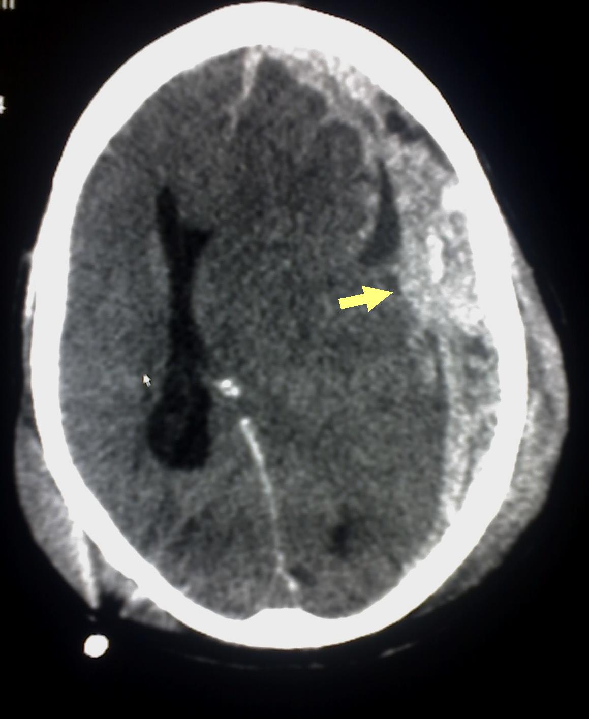
No individual history and physical examination findings can eliminate the possibility of intracranial injury in head trauma. Skull x-rays are only used to assess for foreign bodies, gunshots, or stab wounds. CT head is advocated for all patients with moderate to severe head injury (see Image. Subdural Hematoma Detected on Head CT). For patients at low risk for intracranial injuries, 2 externally validated rules to obtain a head CT scan after TBI are utilized.[18][19]

New Orleans Criteria

  • Headache
  • Vomiting (any)
  • Age > 60 years
  • Drug or alcohol intoxication
  • Seizure
  • Trauma visible above clavicles
  • Short-term memory deficits

Canadian CT Head Rule

  • Dangerous mechanism of injury
  • Vomiting 2 or more times
  • Age > 65 years
  • GCS score < 15, 2 hours post-injury
  • Any sign of basal skull fracture
  • Possible open or depressed skull fracture
  • Amnesia for events 30 minutes before injury

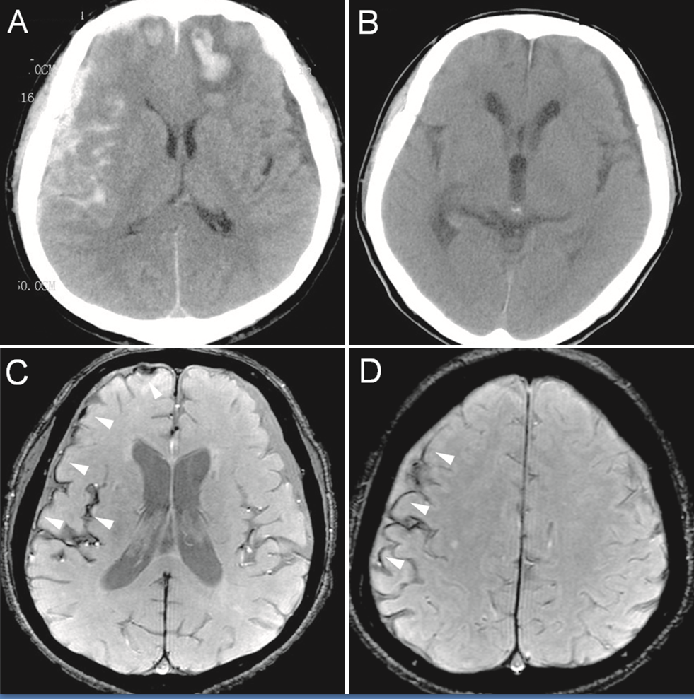
CT images sequentially reveal effacement of the ipsilateral lateral horn with a displacement of septum pellucidum, progressive compression/obliteration of the basal cisterns, and truncated brainstem with duret hemorrhages eventually leading to a 'white cerebellar' sign (see Image. CT and Enhanced Gradient Echo T2 Star-Weighted Angiography (ESWAN) of the Brain).[17] Marshall CT classification does not detail the subtypes of intracranial lesions but was utilized by the IMPACT for outcome prediction.[3] Rotterdam and Helsinki CT score individual CT characteristics separately.[3] 

Treatment / Management

The prime mandate in the management of head trauma involves preventing secondary brain insults. The algorithm in the management involves:

  • Securing airway and maintaining ventilation
  • Maintaining adequate cerebral perfusion pressure
  • Preventing hypoxia, hypotension, hypercapnia, or hypocapnia.
  • Evaluating and managing raised ICP
  • Obtain urgent neurosurgical consultation for traumatic intracranial lesions
  • Identifying and treating other life-threatening injuries or conditions (if they exist).[1][20][21]

Airway, Breathing, and Circulation

Identify any condition which might compromise the airway, such as pneumothorax. For sedation, consider using short-acting agents that have a minimal effect on blood pressure or intracranial pressure (ICP). Etomidate or propofol are used for induction and vecuronium or rocuronium as muscle relaxants. Consider endotracheal intubation in the following situations:

  • Inadequate ventilation or gas exchange, such as hypercarbia, hypoxia, or apnea
  • Severe head injury 
  • Inability to protect the airway
  • Severely agitated patient

The cervical spine should be maintained in line during intubation. Nasotracheal intubation should be avoided in facial trauma or basilar skull fracture patients. The targets should maintain oxygen saturation>90%, PaO>60 mm Hg, and PCO2 at 35 to 45 mm Hg of mercury.

Avoid hypotension. Normal blood pressure may not be adequate to maintain adequate flow and CPP if ICP is elevated. The target is maintaining systolic blood pressure >90 mm Hg and mean arterial pressure (MAP) >80 mm Hg. Isolated head trauma usually does not cause hypotension. Look for another cause if the patient is in hypovolemic shock. 

Mild Head Trauma

The majority of head trauma is mild. These patients can be discharged following a routine neurological examination due to minimal risk of developing an intracranial lesion.

Consider observing for at least 4 to 6 hours if no imaging was obtained. Consider hospitalization if these other risk factors are present:

  • Bleeding disorder
  • Patients taking anticoagulation therapy or antiplatelet therapy
  • Previous neurosurgical procedure 

Provide strict return precautions for patients discharged without imaging. A study of metabolomics and microRNAs can help predict the need for CT among these cohorts.[13][22][13] CENTER-TBI compared 6 biomarkers (S100B, glial fibrillary acidic protein (GFAP), ubiquitin carboxy-terminal hydrolase L1 (UCH-L1), neuron-specific enolase [NSE], neurofilament light [NfL], and total tau) were compared with 4 clinical decision rules (the Canadian CT head rule, the CT in Head injury Patients, NICE, and New Orleans criteria).[13] GFAP outperformed other biomarkers in dichotomizing the need for CT images among cohorts with mild head injury in the study.[13]

Scandinavian Neurotrauma Committee advocates CT imaging among cohorts with minimal head injury presenting with the following clinical characteristics:

  • Loss of consciousness and repeated vomiting alongside increased S100B assay
  • Seizures, neurological deficits, and clinical evidence of depressed or skull base fractures
  • Patients on therapeutic anticoagulation or having bleeding disorders.
  • Age >65 years and on antiplatelet medications.

This minimizes health costs, radiation hazards, and emergency overcrowding without compromising patients' safety.[23]

Moderate to Severe Head Trauma

Prehospital and ED care should prioritize:

  • Stabilization of physiological parameters as per the principles of Advanced Trauma Life Support.
  • Rapid correction of impaired coagulation
  • Noninvasive techniques to monitor ICP
  • Emergent surgical evacuation of mass lesions or decompressive craniectomy and
  • Temporary measures to counteract ICP[24]

Current recommendations include:

  • Optimizing Oxygenation (>90%), blood pressure (SBP >110 mm Hg), and maintaining euthermia. The CENTER TBI core study reported 5% hypoxia and 10% hypotension compared to approx 20% of both hypoxia and hypotension in IMPACT studies.[13] Prehospital guidelines of the Brain Trauma Foundation recommend intubation with a GCS of 8 or less.[13]Collaborative European NeuroTrauma Effectiveness Research in Traumatic Brain Injury CENTER-TBI advocates in-hospital intubation with a GCS of 10 or lower.[13] A preference for euthermia may relate to coagulopathy and infection with hypothermia, as well as increased metabolic demand and energy failure with hyperthermia.
  • Supplemental oxygen to all patients in the prehospital environment
  • Maintain end-tidal CO2 reading between 35 to 45 mm Hg
  • Pupil and GCS assessment every 30 mins
  • Prophylactic administration of hyperosmolar therapy in the absence of signs of herniation and prehospital tranexamic acid is discouraged.[25]

The central dogma in managing raised ICP is preventing secondary insults. CT and MRI images provide only provide a single-point snapshot picture. Ventricular catheters represent a "global" ICP and are the gold standard.[16] Parenchymal monitors, though easier to insert in cases of midline shift or malignant brain swelling, cannot be re-calibrated in vivo, only measure a localized pressure, and have a high propensity for drift issues in long-term usage. The current noninvasive techniques such as pulsatility index from transcranial Doppler (TCD), tympanic membrane displacement (otoacoustic emissions), near-infrared spectroscopy, and optic nerve sheath diameter (ONSD) assessments are not accurate enough to replace traditional invasive techniques and have significant inter-rater variability.[16] 

The ICP pressure waves reveal Lundberg A (plateau waves peaking to 50 mm Hg and persisting for 5 to 20 minutes) and B waves (rhythmic oscillations occurring every 1 to 2 minutes peaking up to 20 to 30 mm Hg) with progressive increment in intracranial pressure. ICP waveform will have a dicrotic wave (P2) larger than the percussion (P1) and the tidal wave (P3).[17] Higher RAP (correlation coefficient (R) between the pulse pulsation amplitude and mean ICP) values connote compromised brain compliance. RAP gradually falls below zero as the brain's ICP increases, exhausting the autoregulatory capacity.[16] The current standards of care while managing severe TBI are the application of multimodal monitoring (MMM), Pressure-Reactivity index, and adherence to ICP-guided therapy.[16][26] Level IIA evidence supports the role of decompressive hemicraniectomy, and level IIB evidence supports the role of ICP monitoring.[17] Brain Trauma Foundation guidelines advocate ICP monitoring:

  1. Severe TBI [Glasgow coma scale (GCS) of 3 to 8] with abnormal CT images or
  2. At least 2 criteria are age over 40, systolic blood pressure under 90 mm Hg, or abnormal posturing.[17]

Other ICP Monitoring Recommendations (Level III Recommendations) include:

  • Patients with an initial normal CT scan or with minor changes in CT images but later show features of neurologic worsening or progression on the repeat scan
  • Evidence of brain swelling, eg, compressed or absent basal cisterns
  • Extensive bifrontal contusions independent of the neurological condition
  • When sedation interruption to check neurological function is not justified, eg, respiratory failure from lung contusions and flail chest
  • When the neurological examination is not reliable, eg, maxillofacial trauma or spinal cord injury.[16]

World Society of Emergency Surgery strongly recommends following guidelines for the 'Hub and Spike' model of managing severe head injury:

  • Telemedicine to facilitate the early transfer of radio images
  • Hemodynamic stabilization before referral
  • Clear therapeutic communications and collaborations between personnel at the multispeciality level
  • Transfer via trained and certified health team with serial neurological monitoring (check only pupils without interrupting sedation among cohorts with features of raised intracranial pressure
  • Pupillpmetry study or ocular ultrasound study for optic nerve sheath diameter assessment should not unnecessarily delay the transfer of patients.
  • The patient should be sedated, intubated, and on mechanical ventilation during transfer, with the head of the bed elevated to 30° to 45°
  • Provision of cardiorespiratory and invasive ABP monitoring, keeping SAP >110 and MAP >90 mm Hg
  • Platelet count>75.000/mm3, PT/aPTT value<1.5, the standard control with early reversal of anticoagulant/antiplatelet agents
  • Aim for normothermia, Hb level >7 g/d, SpO2 >94%, PaCO2 of 35 to 38 mm Hg, serum Na level of 140 to 145 mEq/L
  • Increased sedation, osmotherapy, and short-term hyperventilation are justified for patients with brain herniation awaiting emergent neurosurgery.[27]

Proposed tiers in the management of refractory intracranial hypertension include:

  1. Evacuation of hematoma harbingering or causing cerebral herniation
  2. Physiological neuroprotection
  3. Sedation, analgesics, and ventilation
  4. CSF drainage
  5. Osmotherapy
  6. Hyperventilation
  7. Hypothermia
  8. Barbiturate coma
  9. Decompressive hemicraniectomy.[17]

Level of Evidence of Brain Trauma Foundation guidelines for severe head injury:

  • Level IIA for decompressive craniectomy (not less than 12 x 15 cm or 15 cm diameter) for diffuse cerebral injury with ICP above 20 mm Hg and refractory to first-tier therapies
  • Level IIB against the use of prophylactic hypothermia
  • Hyperosmolar therapy is not currently recommended
  • Level III for early (first 12 hours of injury) and continuous CSF drainage in patients with GCS<6  
  • Level IIB for prophylactic temporary hyperventilation after 24 hours of injury with SjO2 or BtpO2 measurements
  • Level IIB against barbiturate burst suppression for prophylaxis of raised ICP
  • Level I against the use of steroids
  • Level IIA to meet basal caloric replacement at least by the fifth day and at most by the seventh day
  • Level IIB recommendation for transgastric jejunal feeding to reduce ventilator-associated pneumonia
  • Level IIA evidence that early tracheostomy reduces days in mechanical ventilation. Oral care does not reduce ventilator-associated pneumonia but may predict acute respiratory distress syndrome risk.
  • Level III evidence supporting antimicrobial-impregnated ventricular catheters
  • Level III evidence for deep vein thrombosis prophylaxis with LMWH or low-dose unfractionated heparin alongside mechanical prophylaxis
  • Level IIA evidence for prevention of early post-traumatic seizures (within 7 d of injury) only.[28]

Pertinent Studies and Ongoing Trials

Benchmark Evidence from South American Trials: The Treatment of Intracranial Pressure (BEST TRIP) trial treatment revealed no significant differences in clinical outcomes among cohorts managed on an ICP-based management algorithm compared to those with serial CTs and clinical examination alone.[16]

Prognosis

The outcomes after head trauma depend on many factors. Different factors govern the prognosis of the patient with head trauma, which includes the age of the patient, presenting Glasgow coma scale, anisocoria, duration of onset of herniation, associated polytrauma, concurrent hypoxia or hypotension, type of lesion (extradural or subdural hemorrhage), and Marshall and Rotterdam CT scores.[17] The post-resuscitation GCS score is one of the most variable governing outcomes. Patients with a GCS of less than 8 at presentation have high mortality. International Mission for Prognosis And Clinical Trial (IMPACT), TRACK-TBI, and CENTER-TBI studies have also formulated prognostic scoring systems.[3][29] 25% of outcome variance in head trauma is governed by genomics inheritable.[13]

A newer armamentarium in patient management involves monitoring partial pressure of brain tissue oxygen (PbtO2) and cerebral metabolism using microdialysis and ICP and cerebral perfusion pressure (CPP) centered management.[30] Early and continuum rehabilitation has a pivotal role in long-term clinical outcomes.[13] Precision medicine through interdisciplinary collaboration and a holistic approach should be promoted.[13] Machine learning also aids in predicting adverse outcomes as well as mortality.[31][32][33]

Complications

Complications include the following:

  • Neurological deficits
  • Cerebrospinal fluid (CSF) leaks
  • Pneumocephalus[34]
  • Vasospasm
  • Neurovascular injuries
  • Hydrocephalus
  • Seizures
  • Stunned myocardium syndrome
  • Neurogenic pulmonary edema
  • Malignant cerebral edema
  • Dyselectrolytemia-cerebral salt wasting syndrome (CSW) and syndrome of inappropriate antidiuretic hormone syndrome (SIADH)
  • Endocrinopathies such as hypopituitarism[35]
  • Paroxysmal sympathetic hyperactivity[36]
  • Central nervous system (CNS) infection[37]
  • Deep vein thrombosis 
  • Spasticity[38]
  • Depression and post-traumatic stress disorder (PTSD)[39]
  • Dependency
  • Dementia[40]
  • Chronic traumatic encephalopathy (especially in military personnel and athletes related to contact sports[41]
  • Mortality. 

Deterrence and Patient Education

Deterrence and patient education are significant when head trauma has occurred. The gravity of head injuries cannot be understated, as they can result in long-term cognitive impairment, mental health issues, and diminished quality of life. Therefore, educating patients about the importance of taking preventative measures, such as wearing helmets while playing sports or engaging in other high-risk activities, is vital. Patients should also be aware of the warning signs and symptoms of head injuries and seek immediate medical attention if they experience any symptoms. 90% to 95% of patients undergoing CT imaging are negative.[13] However, CT head imaging alone does not permanently exclude neuronal damage.[13] By emphasizing the significance of deterrence and patient education, we can reduce the incidence of head trauma and ensure patients receive prompt and appropriate care when needed. 

Enhancing Healthcare Team Outcomes

Head trauma is a significant public health problem that accounts for thousands of admissions annually and costs the healthcare system billions of dollars. Most patients with head trauma are seen in the emergency department; the head injury is often associated with other organ injuries. Most patients require admission and monitoring in an intensive care unit (ICU) setting. The outcome of these patients depends on the severity of the head trauma, initial GCS score, and any other organ injury. Data indicate that those patients with an initial GCS of 8 or less have a mortality rate of 30% within 2 weeks of the damage. Other negative prognostic factors include advanced age, elevated intracranial pressure, and the presence of a gross neurologic deficit on presentation. ICU nurses play a vital role in managing these patients, from providing primary medical care, monitoring DVT and ulcer prophylaxis, monitoring the patient for complications, and reporting concerns to the team. The dietitian manages the nutrition, and physical therapists provide bedside exercises to prevent muscle wasting.

Patients with a GCS of less than 9 often require mechanical ventilation, tracheostomy, and a feeding tube. With prolonged hospital stays, they are prone to pressure ulcers, aspiration, sepsis, failure to thrive, and deep vein thrombus. Patients who are deemed brain dead are assessed by the team, including specialists from the end of life care. Recovery in most patients can take months or years. Those discharged may have residual deficits in executive function or neurological deficits. Some require speech, occupational, and physical therapy for months. In addition, the social worker should assess the home environment to ensure it is safe and offers amenities for the disabled person. Only through such a team approach can the morbidity of head trauma be lowered.

Many young people still lead a lifestyle that predisposes them to head injury. Young people still drink and drive, text while driving, abuse alcohol and illicit drugs, and are often involved in high-risk sporting activities, which makes them susceptible to head trauma.[42][43] The cornerstone in preventing head trauma is public awareness in following traffic rules, wearing helmets and seat belts, and avoiding drunk driving.[17] Stringent resuscitation, early specialist management, timely intervention for primary insult, minimizing secondary insults, and target-driven care (ICP reduction below 20 mm Hg and CPP of around 60 mm Hg) are the cornerstones of management.[17] A patient-centered 'care bundle' through a holistic team approach is paramount.[17] Long-term improvement should be optimized through vocational intervention programs (VIP) and cognitive rehabilitation strategies.[44][45] The transition should be focused on 'precision medicine.'[3] The gaps in patient care while transitioning from hospital to community setup should be minimized.[46]


(Click Image to Enlarge)
<p>Subdural Hematoma Detected on Head CT

Subdural Hematoma Detected on Head CT. Abusive head trauma or shaken baby syndrome may cause subdural hematoma (arrow), indicating bleeding between the dura mater of the meninges and the brain, as detected on the head CT of a patient.


Glitzy queen00, (Public Domain), via Wikimedia Commons. 


(Click Image to Enlarge)
Original description: Figure 2
Original description: Figure 2. CT and enhanced gradient echo T2 star-weighted angiography (ESWAN) images of the brain of a 54-year-old man who experienced a traumatic brain injury. An axial head CT image displays right frontotemporal SAH (Fisher grade 4) with bilateral frontal contusions and intracerebral hematoma (A). A follow-up CT image 26 weeks after the brain injury indicates that the hemorrhages were completely resolved and the lateral ventricles were mildly enlarged (B). A follow-up MRI (1.5T) image was obtained 26 weeks following the head injury (C,D). The axial ESWAN image displays a rim of hypointensity (arrowheads), with hemosiderin deposits forming along the cerebral convexity (C, D).
Contributed by Hongwei Zhao, Jin Wang, Zhonglie Lu, Qingjie Wu, Haijuan Lv, Hu Liu, Xiangyang Gong
Details

Author

Faizan Shaikh

Editor:

Muhammad Waseem

Updated:

2/24/2024 2:48:16 PM

References


[1]

Brommeland T, Helseth E, Aarhus M, Moen KG, Dyrskog S, Bergholt B, Olivecrona Z, Jeppesen E. Best practice guidelines for blunt cerebrovascular injury (BCVI). Scandinavian journal of trauma, resuscitation and emergency medicine. 2018 Oct 29:26(1):90. doi: 10.1186/s13049-018-0559-1. Epub 2018 Oct 29     [PubMed PMID: 30373641]

Level 1 (high-level) evidence

[2]

Ng SY, Lee AYW. Traumatic Brain Injuries: Pathophysiology and Potential Therapeutic Targets. Frontiers in cellular neuroscience. 2019:13():528. doi: 10.3389/fncel.2019.00528. Epub 2019 Nov 27     [PubMed PMID: 31827423]


[3]

Wilson MH, Ashworth E, Hutchinson PJ, British Neurotrauma Group. A proposed novel traumatic brain injury classification system - an overview and inter-rater reliability validation on behalf of the Society of British Neurological Surgeons. British journal of neurosurgery. 2022 Oct:36(5):633-638. doi: 10.1080/02688697.2022.2090509. Epub 2022 Jun 30     [PubMed PMID: 35770478]

Level 1 (high-level) evidence

[4]

Schweitzer AD, Niogi SN, Whitlow CT, Tsiouris AJ. Traumatic Brain Injury: Imaging Patterns and Complications. Radiographics : a review publication of the Radiological Society of North America, Inc. 2019 Oct:39(6):1571-1595. doi: 10.1148/rg.2019190076. Epub     [PubMed PMID: 31589576]


[5]

Wang R, Yang DX, Ding J, Guo Y, Ding WH, Tian HL, Yuan F. Classification, risk factors, and outcomes of patients with progressive hemorrhagic injury after traumatic brain injury. BMC neurology. 2023 Feb 13:23(1):68. doi: 10.1186/s12883-023-03112-x. Epub 2023 Feb 13     [PubMed PMID: 36782124]


[6]

Portaro S, Naro A, Cimino V, Maresca G, Corallo F, Morabito R, Calabrò RS. Risk factors of transient global amnesia: Three case reports. Medicine. 2018 Oct:97(41):e12723. doi: 10.1097/MD.0000000000012723. Epub     [PubMed PMID: 30313071]

Level 3 (low-level) evidence

[7]

Salehpour F, Bazzazi AM, Aghazadeh J, Hasanloei AV, Pasban K, Mirzaei F, Naseri Alavi SA. What do You Expect from Patients with Severe Head Trauma? Asian journal of neurosurgery. 2018 Jul-Sep:13(3):660-663. doi: 10.4103/ajns.AJNS_260_16. Epub     [PubMed PMID: 30283522]


[8]

Alao T, Munakomi S, Waseem M. Penetrating Head Trauma. StatPearls. 2024 Jan:():     [PubMed PMID: 29083824]


[9]

Dewan MC, Rattani A, Gupta S, Baticulon RE, Hung YC, Punchak M, Agrawal A, Adeleye AO, Shrime MG, Rubiano AM, Rosenfeld JV, Park KB. Estimating the global incidence of traumatic brain injury. Journal of neurosurgery. 2018 Apr 27:130(4):1080-1097. doi: 10.3171/2017.10.JNS17352. Epub 2018 Apr 27     [PubMed PMID: 29701556]


[10]

Mohammadifard M, Ghaemi K, Hanif H, Sharifzadeh G, Haghparast M. Marshall and Rotterdam Computed Tomography scores in predicting early deaths after brain trauma. European journal of translational myology. 2018 Jul 10:28(3):7542. doi: 10.4081/ejtm.2018.7542. Epub 2018 Jul 16     [PubMed PMID: 30344974]


[11]

Lalwani S, Hasan F, Khurana S, Mathur P. Epidemiological trends of fatal pediatric trauma: A single-center study. Medicine. 2018 Sep:97(39):e12280. doi: 10.1097/MD.0000000000012280. Epub     [PubMed PMID: 30278499]

Level 2 (mid-level) evidence

[12]

Schneider ALC, Wang D, Ling G, Gottesman RF, Selvin E. Prevalence of Self-Reported Head Injury in the United States. The New England journal of medicine. 2018 Sep 20:379(12):1176-1178. doi: 10.1056/NEJMc1808550. Epub     [PubMed PMID: 30231228]


[13]

Maas AIR, Menon DK, Manley GT, Abrams M, Åkerlund C, Andelic N, Aries M, Bashford T, Bell MJ, Bodien YG, Brett BL, Büki A, Chesnut RM, Citerio G, Clark D, Clasby B, Cooper DJ, Czeiter E, Czosnyka M, Dams-O'Connor K, De Keyser V, Diaz-Arrastia R, Ercole A, van Essen TA, Falvey É, Ferguson AR, Figaji A, Fitzgerald M, Foreman B, Gantner D, Gao G, Giacino J, Gravesteijn B, Guiza F, Gupta D, Gurnell M, Haagsma JA, Hammond FM, Hawryluk G, Hutchinson P, van der Jagt M, Jain S, Jain S, Jiang JY, Kent H, Kolias A, Kompanje EJO, Lecky F, Lingsma HF, Maegele M, Majdan M, Markowitz A, McCrea M, Meyfroidt G, Mikolić A, Mondello S, Mukherjee P, Nelson D, Nelson LD, Newcombe V, Okonkwo D, Orešič M, Peul W, Pisică D, Polinder S, Ponsford J, Puybasset L, Raj R, Robba C, Røe C, Rosand J, Schueler P, Sharp DJ, Smielewski P, Stein MB, von Steinbüchel N, Stewart W, Steyerberg EW, Stocchetti N, Temkin N, Tenovuo O, Theadom A, Thomas I, Espin AT, Turgeon AF, Unterberg A, Van Praag D, van Veen E, Verheyden J, Vyvere TV, Wang KKW, Wiegers EJA, Williams WH, Wilson L, Wisniewski SR, Younsi A, Yue JK, Yuh EL, Zeiler FA, Zeldovich M, Zemek R, InTBIR Participants and Investigators. Traumatic brain injury: progress and challenges in prevention, clinical care, and research. The Lancet. Neurology. 2022 Nov:21(11):1004-1060. doi: 10.1016/S1474-4422(22)00309-X. Epub 2022 Sep 29     [PubMed PMID: 36183712]


[14]

GBD 2016 Traumatic Brain Injury and Spinal Cord Injury Collaborators. Global, regional, and national burden of traumatic brain injury and spinal cord injury, 1990-2016: a systematic analysis for the Global Burden of Disease Study 2016. The Lancet. Neurology. 2019 Jan:18(1):56-87. doi: 10.1016/S1474-4422(18)30415-0. Epub 2018 Nov 26     [PubMed PMID: 30497965]

Level 1 (high-level) evidence

[15]

Munakomi S, Cherian I. Newer insights to pathogenesis of traumatic brain injury. Asian journal of neurosurgery. 2017 Jul-Sep:12(3):362-364. doi: 10.4103/1793-5482.180882. Epub     [PubMed PMID: 28761509]


[16]

Munakomi S, Das JM. Intracranial Pressure Monitoring. StatPearls. 2024 Jan:():     [PubMed PMID: 31194438]


[17]

Munakomi S, Das JM. Brain Herniation. StatPearls. 2024 Jan:():     [PubMed PMID: 31194403]


[18]

Pavlović T, Milošević M, Trtica S, Budinčević H. Value of Head CT Scan in the Emergency Department in Patients with Vertigo without Focal Neurological Abnormalities. Open access Macedonian journal of medical sciences. 2018 Sep 25:6(9):1664-1667. doi: 10.3889/oamjms.2018.340. Epub 2018 Sep 24     [PubMed PMID: 30337984]


[19]

Hajiaghamemar M, Lan IS, Christian CW, Coats B, Margulies SS. Infant skull fracture risk for low height falls. International journal of legal medicine. 2019 May:133(3):847-862. doi: 10.1007/s00414-018-1918-1. Epub 2018 Sep 7     [PubMed PMID: 30194647]


[20]

Jacquet C, Boetto S, Sevely A, Sol JC, Chaix Y, Cheuret E. Monitoring Criteria of Intracranial Lesions in Children Post Mild or Moderate Head Trauma. Neuropediatrics. 2018 Dec:49(6):385-391. doi: 10.1055/s-0038-1668138. Epub 2018 Sep 17     [PubMed PMID: 30223286]


[21]

Bayley MT, Lamontagne ME, Kua A, Marshall S, Marier-Deschênes P, Allaire AS, Kagan C, Truchon C, Janzen S, Teasell R, Swaine B. Unique Features of the INESSS-ONF Rehabilitation Guidelines for Moderate to Severe Traumatic Brain Injury: Responding to Users' Needs. The Journal of head trauma rehabilitation. 2018 Sep/Oct:33(5):296-305. doi: 10.1097/HTR.0000000000000428. Epub     [PubMed PMID: 30188459]


[22]

Fedoruk RP, Lee CH, Banoei MM, Winston BW. Metabolomics in severe traumatic brain injury: a scoping review. BMC neuroscience. 2023 Oct 16:24(1):54. doi: 10.1186/s12868-023-00824-1. Epub 2023 Oct 16     [PubMed PMID: 37845610]

Level 2 (mid-level) evidence

[23]

Vedin T, Karlsson M, Edelhamre M, Clausen L, Svensson S, Bergenheim M, Larsson PA. A proposed amendment to the current guidelines for mild traumatic brain injury: reducing computerized tomographies while maintaining safety. European journal of trauma and emergency surgery : official publication of the European Trauma Society. 2021 Oct:47(5):1451-1459. doi: 10.1007/s00068-019-01145-x. Epub 2019 May 14     [PubMed PMID: 31089789]


[24]

Hossain I, Rostami E, Marklund N. The management of severe traumatic brain injury in the initial postinjury hours - current evidence and controversies. Current opinion in critical care. 2023 Dec 1:29(6):650-658. doi: 10.1097/MCC.0000000000001094. Epub 2023 Oct 11     [PubMed PMID: 37851061]

Level 3 (low-level) evidence

[25]

Hawryluk GWJ, Lulla A, Bell R, Jagoda A, Mangat HS, Bobrow BJ, Ghajar J. Guidelines for Prehospital Management of Traumatic Brain Injury 3rd Edition: Executive Summary. Neurosurgery. 2023 Dec 1:93(6):e159-e169. doi: 10.1227/neu.0000000000002672. Epub 2023 Sep 26     [PubMed PMID: 37750693]


[26]

Taccone FS, Sterchele ED, Piagnerelli M. Multimodal neuromonitoring in traumatic brain injury patients: the search for the Holy Graal. Critical care (London, England). 2023 Oct 16:27(1):396. doi: 10.1186/s13054-023-04679-0. Epub 2023 Oct 16     [PubMed PMID: 37845770]


[27]

Picetti E, Catena F, Abu-Zidan F, Ansaloni L, Armonda RA, Bala M, Balogh ZJ, Bertuccio A, Biffl WL, Bouzat P, Buki A, Cerasti D, Chesnut RM, Citerio G, Coccolini F, Coimbra R, Coniglio C, Fainardi E, Gupta D, Gurney JM, Hawryluk GWJ, Helbok R, Hutchinson PJA, Iaccarino C, Kolias A, Maier RW, Martin MJ, Meyfroidt G, Okonkwo DO, Rasulo F, Rizoli S, Rubiano A, Sahuquillo J, Sams VG, Servadei F, Sharma D, Shutter L, Stahel PF, Taccone FS, Udy A, Zoerle T, Agnoletti V, Bravi F, De Simone B, Kluger Y, Martino C, Moore EE, Sartelli M, Weber D, Robba C. Early management of isolated severe traumatic brain injury patients in a hospital without neurosurgical capabilities: a consensus and clinical recommendations of the World Society of Emergency Surgery (WSES). World journal of emergency surgery : WJES. 2023 Jan 9:18(1):5. doi: 10.1186/s13017-022-00468-2. Epub 2023 Jan 9     [PubMed PMID: 36624517]

Level 3 (low-level) evidence

[28]

Carney N, Totten AM, O'Reilly C, Ullman JS, Hawryluk GW, Bell MJ, Bratton SL, Chesnut R, Harris OA, Kissoon N, Rubiano AM, Shutter L, Tasker RC, Vavilala MS, Wilberger J, Wright DW, Ghajar J. Guidelines for the Management of Severe Traumatic Brain Injury, Fourth Edition. Neurosurgery. 2017 Jan 1:80(1):6-15. doi: 10.1227/NEU.0000000000001432. Epub     [PubMed PMID: 27654000]


[29]

Steyerberg EW, Mushkudiani N, Perel P, Butcher I, Lu J, McHugh GS, Murray GD, Marmarou A, Roberts I, Habbema JD, Maas AI. Predicting outcome after traumatic brain injury: development and international validation of prognostic scores based on admission characteristics. PLoS medicine. 2008 Aug 5:5(8):e165; discussion e165. doi: 10.1371/journal.pmed.0050165. Epub     [PubMed PMID: 18684008]

Level 1 (high-level) evidence

[30]

Stein KY, Froese L, Sekhon M, Griesdale D, Thelin EP, Raj R, Tas J, Aries M, Gallagher C, Bernard F, Gomez A, Kramer AH, Zeiler FA. Intracranial Pressure-Derived Cerebrovascular Reactivity Indices and Their Critical Thresholds: A Canadian High Resolution-Traumatic Brain Injury Validation Study. Journal of neurotrauma. 2024 Apr:41(7-8):910-923. doi: 10.1089/neu.2023.0374. Epub 2023 Nov 22     [PubMed PMID: 37861325]

Level 1 (high-level) evidence

[31]

Mekkodathil A, El-Menyar A, Naduvilekandy M, Rizoli S, Al-Thani H. Machine Learning Approach for the Prediction of In-Hospital Mortality in Traumatic Brain Injury Using Bio-Clinical Markers at Presentation to the Emergency Department. Diagnostics (Basel, Switzerland). 2023 Aug 5:13(15):. doi: 10.3390/diagnostics13152605. Epub 2023 Aug 5     [PubMed PMID: 37568968]


[32]

Courville E, Kazim SF, Vellek J, Tarawneh O, Stack J, Roster K, Roy J, Schmidt M, Bowers C. Machine learning algorithms for predicting outcomes of traumatic brain injury: A systematic review and meta-analysis. Surgical neurology international. 2023:14():262. doi: 10.25259/SNI_312_2023. Epub 2023 Jul 28     [PubMed PMID: 37560584]

Level 1 (high-level) evidence

[33]

Terabe ML, Massago M, Iora PH, Hernandes Rocha TA, de Souza JVP, Huo L, Massago M, Senda DM, Kobayashi EM, Vissoci JR, Staton CA, de Andrade L. Applicability of machine learning technique in the screening of patients with mild traumatic brain injury. PloS one. 2023:18(8):e0290721. doi: 10.1371/journal.pone.0290721. Epub 2023 Aug 24     [PubMed PMID: 37616279]


[34]

Das JM, Bajaj J. Pneumocephalus. StatPearls. 2024 Jan:():     [PubMed PMID: 30571033]


[35]

Tahara S, Otsuka F, Endo T. Recognition and Practice of Hypopituitarism After Traumatic Brain Injury and Subarachnoid Hemorrhage in Japan: A Survey. Neurology and therapy. 2024 Feb:13(1):39-51. doi: 10.1007/s40120-023-00553-x. Epub 2023 Oct 24     [PubMed PMID: 37874463]

Level 3 (low-level) evidence

[36]

Salasky VR, Chowdhury SH, Chen LK, Almeida E, Kong X, Armahizer M, Pajoumand M, Schrank GM, Rabinowitz RP, Schwartzbauer G, Hu P, Badjatia N, Podell JE. Overlapping Physiologic Signs of Sepsis and Paroxysmal Sympathetic Hyperactivity After Traumatic Brain Injury: Exploring A Clinical Conundrum. Neurocritical care. 2023 Oct 26:():. doi: 10.1007/s12028-023-01862-7. Epub 2023 Oct 26     [PubMed PMID: 37884690]


[37]

Zhao W, Guo S, Xu Z, Wang Y, Kou Y, Tian S, Qi Y, Pang J, Zhou W, Wang N, Liu J, Zhai Y, Ji P, Jiao Y, Fan C, Chao M, Fan Z, Qu Y, Wang L. Nomogram for Predicting Central Nervous System Infection Following Traumatic Brain Injury in the Elderly. World neurosurgery. 2024 Mar:183():e28-e43. doi: 10.1016/j.wneu.2023.10.088. Epub 2023 Oct 23     [PubMed PMID: 37879436]

Level 2 (mid-level) evidence

[38]

Tani J, Wen YT, Hu CJ, Sung JY. Current and Potential Pharmacologic Therapies for Traumatic Brain Injury. Pharmaceuticals (Basel, Switzerland). 2022 Jul 6:15(7):. doi: 10.3390/ph15070838. Epub 2022 Jul 6     [PubMed PMID: 35890136]


[39]

Carmichael J, Ponsford J, Gould KR, Spitz G. Characterizing depression after traumatic brain injury using a symptom-oriented approach. Journal of affective disorders. 2024 Jan 15:345():455-466. doi: 10.1016/j.jad.2023.10.130. Epub 2023 Oct 24     [PubMed PMID: 37879410]


[40]

Graham NS, Sharp DJ. Dementia after traumatic brain injury. BMJ (Clinical research ed.). 2023 Oct 19:383():2065. doi: 10.1136/bmj.p2065. Epub 2023 Oct 19     [PubMed PMID: 37857435]


[41]

Munakomi S, Puckett Y. Repetitive Head Injury Syndrome. StatPearls. 2024 Jan:():     [PubMed PMID: 31082057]


[42]

Fitzpatrick S, Leach P. Neurosurgical aspects of abusive head trauma management in children: a review for the training neurosurgeon. British journal of neurosurgery. 2019 Feb:33(1):47-50. doi: 10.1080/02688697.2018.1529295. Epub 2018 Oct 24     [PubMed PMID: 30353746]


[43]

Hussain E. Traumatic Brain Injury in the Pediatric Intensive Care Unit. Pediatric annals. 2018 Jul 1:47(7):e274-e279. doi: 10.3928/19382359-20180619-01. Epub     [PubMed PMID: 30001441]


[44]

Simpson GK, McRae P, Gates TM, Daher M, Johnston D, Cameron ID. A vocational intervention that enhances return to work after severe acquired brain injury: A pragmatic trial. Annals of physical and rehabilitation medicine. 2023 Nov:66(8):101787. doi: 10.1016/j.rehab.2023.101787. Epub 2023 Oct 25     [PubMed PMID: 37890426]


[45]

Fleming J, Hamilton C, Ownsworth T, Doig E, Swan S, Holmes E, Griffin J, Shum DHK. The perspectives of participants with traumatic brain injury on prospective memory rehabilitation incorporating compensatory and metacognitive skills training. Patient education and counseling. 2024 Jan:118():108023. doi: 10.1016/j.pec.2023.108023. Epub 2023 Oct 20     [PubMed PMID: 37866073]

Level 3 (low-level) evidence

[46]

Wiles MD, Braganza M, Edwards H, Krause E, Jackson J, Tait F. Management of traumatic brain injury in the non-neurosurgical intensive care unit: a narrative review of current evidence. Anaesthesia. 2023 Apr:78(4):510-520. doi: 10.1111/anae.15898. Epub 2023 Jan 12     [PubMed PMID: 36633447]

Level 3 (low-level) evidence