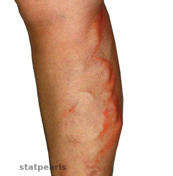
Septic Thrombophlebitis

Earn CME/CE in your profession:


Continuing Education Activity

Suppurative (septic) thrombophlebitis describes thrombosis in a vein that occurs in the setting of inflammation and infection. This condition is characterized by the presence of a thrombus that is associated with inflammation and pus formation (suppuration) both in the venous wall and surrounding the vessel. Although this process can occur in both superficial and deep vessels throughout the body, it is seen most commonly in venous structures, including peripheral veins, pelvic veins, superior vena cava, internal jugular vein, portal veins or dural sinuses. This activity describes the evaluation and management of septic thrombophlebitis and highlights the role of the interprofessional team in improving care for affected patients.

Objectives:

  • Identify the etiology of septic thrombophlebitis.
  • Explain the evaluation of septic thrombophlebitis.
  • Outline the treatment options for septic thrombophlebitis.
  • Describe interprofessional team strategies for enhancing care coordination and communication to enhance the management of patients with septic thrombophlebitis and improve outcomes.

Introduction

Septic or suppurative thrombophlebitis (STP) is defined by the presence of an endovascular thrombus in the setting of associated bacterial or fungal infection. Venous infections can arise from an associated intravenous catheter, skin breakdown, or by invasion from adjacent nonvascular structures. Although this process can occur in both superficial and deep vessels of the body, it is seen most commonly in venous structures (e.g., peripheral veins, pelvic veins, superior vena cava, internal jugular vein, portal veins or dural sinuses). The patient presentation may vary from a mild infection of the superficial veins to a severe, systemic infection. Once STP is present, several complications related to the hematogenous bacterial spread and septic thromboembolism may develop, such as endocarditis, septic arthritis, and even septic uveitis. The morbidity and mortality of STP if not treated, is high, although the risk of significant morbidity and mortality depends on the location of the thrombus.[1][2][3][4][5]

Etiology

Even though phlebitis can occur without an underlying infection, simply as a result of catheter-related mechanical or chemical irritation, an infection should always be in the differential diagnosis. Septic thrombophlebitis can occur spontaneously; however, the majority of cases with superficial, peripheral vein involvement result from breaks in the skin, as with venipuncture done for phlebotomy, or intravenous injection. In some instances, intravenous catheters, such as peripherally inserted central catheters (PICC) lines can be the initiating culprit in peripheral vein as well as deep veins such as superior vena cava (SVC). With inferior vena cava or SVC, STP occurs most often as a consequence of an indwelling catheter. [6][7][8]

Other conditions such as Lemierre syndrome, pylephlebitis, STP of the dural sinuses, and pelvis veins often result from an invading infection from a near non-vascular structure. For example, STP of the pelvic veins may result from endometritis, pelvic inflammatory disease, intra-abdominal infections, abdominal surgery, childbirth, or septic abortion. Lemierre syndrome, which is thrombophlebitis of the internal jugular vein, is most often caused by pharyngitis but may occur secondary to a dental infection.  Dural sinus STP may result from an ear, nose, or throat infection, such as mastoiditis, otitis media, or even meningitis.[4][6][7][8]

Epidemiology

Individuals at extremes of age, such as neonates and the elderly appear to be the most vulnerable to STP. This is likely attributable to undeveloped host defenses in neonates, and a decline in immunologic function as well as additional comorbid disease in the elderly. [9][10][11] Patients with active malignancy, who have an increased risk for developing deep vein thrombosis (DVT) due to their impaired fibrinolytic systems along with their severely immunocompromised state also develop STP. In these patients, STP may be difficult to diagnose, as they often have more than one source of bacteremia.[12][13]  Others, such as burned patients, those on long term steroid regimen or intravenous drug users, also have a higher incidence of developing STP.[14][15][16][17]

The current incidence of catheter-associated (peripheral) STP is estimated at 0.5 cases of bloodstream infections per 1000 days of a peripherally inserted intravenous device. For non-tunneled, non-medicated, central venous catheters, the incidence is estimated at 2.7 per 1000 intravenous device days. Approximately 4.2% of burn patients have been reported to experience peripheral STP.[14][15][18][15]

Deep (non-catheter associated) STP is seen much less common; the exact incidence, therefore, not yet described. However, in the case of pelvic STP, which is seen most frequently in women of childbearing age, younger than 20 years old, the frequency has been reported to vary from 1:500 to 1:2000 in cesarean deliveries, while the frequency for vaginal deliveries has been reported to be 1:9000. Other risk factors associated with pelvic STP are the black race and multiple gestations.[19] 

Pylephlebitis, which is STP of the portal vein, has been cited for having a mortality rate of up to 25% despite the use of antibiotics and an incidence of 2.7 per 100,000 person-years.[20]

Other rare conditions such as Lemierre syndrome, which commonly occur in healthy, young adults have been cited to occur at an incidence of 14.4 cases per million per year in patients between 15-24 years old. The mortality rate reported in a 3-year prospective study was 9%.[21]

Prior to the widespread use of antibiotics, STP was the main cause of cerebral sinus thrombosis and was nearly universally fatal. However, it is now considered rare. Despite this, the mortality is still reported to be approximately 30%  for STP of the cavernous sinus and 78% for STP of the superior sagittal sinus, with less than 50% achieving full recovery. This condition is also more common in children than in adults.[22][23][22] 

Pathophysiology

While the exact pathophysiologic mechanism of STP is not fully understood, it is believed that STP initially begins by a disruption of the endovascular wall, which leads to a thrombus formation. This disruption of the vein wall can be due to an endovascular device, such as a peripheral line or central line. However, it can also be from a preexisting hypercoagulable state, such as those in patients with cancer. Once the thrombus is present, invasion of a microorganism may ensue from skin breakdown or an adjacent structure. Once an organism has entered the vein, it proliferates and can further add to the thrombus formation and inflammation.[24]

The most common organism involved in STP is Staphylococcus aureus. However, others have been implicated, such as viral, parasitic, and fungal organisms, and even polymicrobial. Other common organisms are those of the streptococcal species, anaerobes as well as gram-negative organisms.[22][25]

Other conditions, such as Lemierre syndrome, occur from bacterial invasion from a nearby structure. Lemierre syndrome refers to thrombophlebitis of the internal jugular vein. It occurs most commonly as a result of bacterial pharyngitis that may be associated with a peritonsillar abscess, which may then rupture and spreads to surrounding tissues and venous structures. There is characteristic bacterial involvement of the carotid sheath with associated bacteremia.[26] Lemierre's syndrome is often caused by the anaerobic organism Fusobacterium necrophorum.[27][4]

Infection in any part of the middle third of the face, such as the nose, periorbital regions, tonsils, or soft palate, poses the greatest risk of dural venous sinus STP. These structures drain directly into the cavernous sinus, through the ophthalmic veins, facial veins, and the pterygoid plexus, and thus infection can spread via this route. Dental infections, meningitis, and sinusitis have also been reported to be associated with dural sinus STP through the direct spread of infection. [23][22] 

Pylephlebitis is a rare form of STP that can be associated with diverticulitis (most commonly), pancreatitis, appendicitis, inflammatory bowel disease, hemorrhoidal disease as well as any intra-abdominal infection that involves structures draining into the portal vein. It is believed to start as an infection of the neighboring areas drained by the portal system, that can progress and hematogenously spread to the liver and bacteremia.[20][28][4]

Pelvic and ovarian vein STP occurs most commonly in the setting of childbirth and within the first three weeks post-partum due to local spread of uterine infection, such as endometritis or chorioamnionitis. The hypercoagulable state that occurs in the setting of pregnancy contributes to thrombus formation, and damage to the iliofemoral vessels during childbirth further promotes this process.[19] Other than its association with the postpartum period, Pelvic STP has also been associated with pelvic inflammatory disease as well as intrauterine device insertion. [29][30]

History and Physical

Patients with STP may present with a range of symptoms from mild swelling and pain of a superficial vein to septic shock, depending on the length of time and location of the infected thrombus.

As the majority of cases are due to peripheral vein involvement, those patients may present with a fever higher than 100.4 F along with erythema and tenderness along the course of the vein as well as purulent drainage at the catheter insertion site. Swelling of the extremity as well as frank purulent material from the vein has been reported.[31]

Some patients may have associated hypoxemia, respiratory distress, and chest pain due to septic emboli to the lungs. Septic pulmonary embolism is rare but has been reported and should be considered in a patient with an extrapulmonary source of infection in the setting of bacteremia and an associated thrombus.[32]

In the case of jugular vein STP, the patients may present with fever, localized throat or neck pain, dysphagia, trismus, neck swelling or induration over the jugular vein, along with the sternocleidomastoid muscle, or over the angle of the jaw. Additionally, ulcerations, a pseudomembrane, or erythema may be visualized in the oropharynx of a patient with jugular vein STP. Lemierre's syndrome can also present with post septal cellulitis, sinusitis, or dental pain from a dental infection.[27]

Most patients presenting with pylephlebitis exhibit a fever with abdominal pain; however, nausea, vomiting, and jaundice is rare. Some may present with hypotension, tachycardia, and severe sepsis. Septic emboli may additionally travel to joints or bones, resulting in septic arthritis or osteomyelitis - in which case the patient may complain of joint pains or body aches (e.g., in addition to fever, malaise, and night sweats).[4]

Patients with STP of the pelvic veins can present in one of two ways. With ovarian vein thrombophlebitis, there is usually fever and abdominal pain localized to the side of the affected vessel, with tenderness present. The pain may be described as radiating to the groin or the upper abdomen and as constant pain.[4][33]  On the other hand, a patient with deep septic pelvic thrombophlebitis may have a more subtle presentation. They will often not look ill between fever spikes, and abdominal pain or tenderness is often not present. Patients often exhibit only a fever postpartum and fail to respond to antibiotics. It is known as "enigmatic fever" for this reason.[4][34]

Evaluation

Septic thrombophlebitis should be suspected in patients who continue to spike fevers despite 72 hours of adequate antibiotic coverage, especially in the setting of an endovascular catheter.[35] [36][37]

The best available test remains a contrast-enhanced computed tomography (CT) scan. The CT scan allows for the evaluation of any filling defects within a vessel that may potentially contain a clot and may additionally demonstrate any surrounding inflammation. Diagnosis is then made based on this radiographic evidence of thrombosis, taken together with results from blood cultures or cultures of purulent material obtained from a suspected site (e.g., tip culture in the case of catheter-associated thrombophlebitis, or Gram stain and culture of purulent material from a soft-tissue site). Tip cultures from both peripheral and central sites should be sent for comparison, if available. Diagnosis can be microbiologic with culture results together with radiographic evidence of disease. [38]

If CT scanning is unavailable, magnetic resonance imaging (MRI) may also be used for the diagnosis of most cases of STP. MRI combined with MR venography is the most sensitive, non-invasive test for evaluating the dural sinuses.[22]

Ultrasound may be useful in some cases of STP (e.g., if there is an abscess present very close to the involved vessel). It can also be diagnostic if a thrombus is revealed in the setting of positive blood culture. In the cases of pelvic or dural vein thrombophlebitis, however, ultrasound will not provide an adequate study (due to poor penetration). [4]

Additional laboratory studies may include a complete blood count, blood chemistries, hepatic enzymes, liver function tests, international normalized ratio/prothrombin time, and if dural sinuses are involved, consider obtaining cerebrospinal fluid cultures.[22] 

Treatment / Management

The treatment of STP depends on the source of infection, the organisms involved, the structures being affected, and the individual patient's physiology. The main goals of treatment include removal of the source of infection, such as peripheral or central intravenous catheters, broad-spectrum intravenous antibiotic administration, possible anticoagulation (although this remains controversial), and evaluation by a surgical team for possible surgical intervention. [39][40]

If there is frank purulence, then the infected vessel may need to be excised; this is possible in the periphery but not so when the central vessels are involved.

The role of anticoagulation is controversial. Some authors recommend anticoagulation to prevent the propagation of the thrombus, while others favor anticoagulation only if there is an extension of the thrombus; however, there are no controlled studies up to date. Anticoagulation should be held in patients who are thrombocytopenic, with a platelet count of less than 50,000/microL due to a high risk of bleeding. [41][42][41]

Differential Diagnosis

Other conditions may mimic septic thrombophlebitis. In patients with cancer, it is often difficult to distinguish STP from the common deep venous thrombosis (DVT) that cancer patients experience due to hypercoagulation. Cancer patients are often bacteremic from other sources due to their severe immunocompromise, and there is no good way to differentiate the septic patient, who has an indwelling catheter with a thrombus, from the septic patient who has septic thrombophlebitis. Some have looked at the role of  fluorine-18 fluorodeoxyglucose positron emission tomography (FDG-PET) and found it to be specific for the diagnosis of STP in this population of patients with concurrent malignancy.[12][13] 

Cerebral venous thrombosis can be mimicked by pre septal cellulitis or orbital cellulitis or by ophthalmologic migraines, which can present with 3rd nerve palsy, decreased vision, eye pain, ophthalmoplegia, and proptosis.[22]

Prognosis

Septic thrombophlebitis mortality has drastically decreased over the last century due to early antibiotic use and improved diagnostics. Some conditions, such as dural sinus thrombosis, still carry high morbidity and mortality despite advances.[22][4] 

Complications

  • Emboli
  • Sepsis
  • Vessel thrombosis

Consultations

  • Vascular surgeon: in some cases excision of the vein may be required to remove the source of infection.
  • Interventional radiology
  • Infectious disease

Enhancing Healthcare Team Outcomes

Because STP can occur in any vessel, the condition is best managed by an interprofessional team that includes a surgeon, an interventional radiologist, and an infectious disease specialist. Nurses are often the first professionals to note the condition because it is often linked to peripheral vein cannulation. Once the condition is diagnosed, immediate treatment is necessary to prevent metastatic foci of infection in the systemic circulation. Mortality rates between 3 and 78% have been reported depending on the location of the vein and the extent of infection. Infections associated with candida have the highest mortality. When the dural sinuses are involved, the mortality can exceed 70%. Today, with better imaging and improved diagnosis, the mortality rates have dropped, but any delay in treatment is associated with high morbidity and mortality. Pharmacists, particularly those specialized in infectious disease, are involved in helping the team in selecting medications, with the review of medications dosing, and checking for drug-drug interactions. Once discharged, patients often need to follow up to ensure that they have not developed endocarditis or recurrent infection; nursing can provide the needed monitoring and report any issues to the clinician staff. To lessen the mortality, peripheral vein cannulation should be preferred over central vein cannulation. close communication between nurses and other clinicians is vital if one wants to improve the outcomes.[43][10][22] [Level 5]


(Click Image to Enlarge)
<p>Migratory Thrombophlebitis</p>

Migratory Thrombophlebitis


Contributed by S Bhimji, MD

Details

Author

Demis N. Lipe

Author

Lisa A. Foris

Editor:

Kevin C. King

Updated:

7/4/2023 12:21:56 AM

References


[1]

Losno RA, Vidal-Sicart S, Grau JM. Multiple pyomyositis secondary to septic thrombophlebitis. Medicina clinica. 2019 Jun 21:152(12):515-516. doi: 10.1016/j.medcli.2018.07.020. Epub 2018 Oct 16     [PubMed PMID: 30340839]


[2]

Paker M, Cohen JT, Moed N, Shleizerman L, Masalha M, Ashkenazi D, Mazzawi S. Facial vein thrombophlebitis: A case report and literature review. International journal of pediatric otorhinolaryngology. 2018 Oct:113():298-301. doi: 10.1016/j.ijporl.2018.08.015. Epub 2018 Aug 16     [PubMed PMID: 30174005]

Level 3 (low-level) evidence

[3]

Nemakayala DR, P Rai M, Kavuturu S, Rayamajhi S. Atypical Presentation of Lemierre's Syndrome Causing Septic Shock and Acute Respiratory Distress Syndrome. Case reports in infectious diseases. 2018:2018():5469053. doi: 10.1155/2018/5469053. Epub 2018 Jul 2     [PubMed PMID: 30057835]

Level 3 (low-level) evidence

[4]

Chirinos JA, Garcia J, Alcaide ML, Toledo G, Baracco GJ, Lichtstein DM. Septic thrombophlebitis: diagnosis and management. American journal of cardiovascular drugs : drugs, devices, and other interventions. 2006:6(1):9-14     [PubMed PMID: 16489845]


[5]

Giquello JA, Dupoiron D, Lemarié C, Lorimier G, Granry JC. Septic uveitis revealing internal jugular vein thrombophlebitis. The Lancet. Infectious diseases. 2011 Apr:11(4):332. doi: 10.1016/S1473-3099(11)70030-5. Epub     [PubMed PMID: 21453874]


[6]

Alabraba E, Manu N, Fairclough G, Sutton R. Acute parotitis due to MRSA causing Lemierre's syndrome. Oxford medical case reports. 2018 May:2018(5):omx056. doi: 10.1093/omcr/omx056. Epub 2018 May 31     [PubMed PMID: 29942528]

Level 3 (low-level) evidence

[7]

Ho VT, Rothenberg KA, McFarland G, Tran K, Aalami OO. Septic Pulmonary Emboli From Peripheral Suppurative Thrombophlebitis: A Case Report and Literature Review. Vascular and endovascular surgery. 2018 Nov:52(8):633-635. doi: 10.1177/1538574418779469. Epub 2018 Jun 18     [PubMed PMID: 29909751]

Level 3 (low-level) evidence

[8]

De Smet K,Claus PE,Alliet G,Simpelaere A,Desmet G, Lemierre's syndrome: a case study with a short review of literature. Acta clinica Belgica. 2018 May 21     [PubMed PMID: 29783881]

Level 3 (low-level) evidence

[9]

San-Juan R, Viedma E, Chaves F, Lalueza A, Fortún J, Loza E, Pujol M, Ardanuy C, Morales I, de Cueto M, Resino-Foz E, Morales-Cartagena A, Rico A, Romero MP, Orellana MÁ, López-Medrano F, Fernández-Ruiz M, Aguado JM. High MICs for Vancomycin and Daptomycin and Complicated Catheter-Related Bloodstream Infections with Methicillin-Sensitive Staphylococcus aureus. Emerging infectious diseases. 2016 Jun:22(6):1057-66. doi: 10.3201/eid2206.151709. Epub     [PubMed PMID: 27192097]


[10]

Kim M, Kwon H, Hong SK, Han Y, Park H, Choi JY, Kwon TW, Cho YP. Surgical treatment of central venous catheter related septic deep venous thrombosis. European journal of vascular and endovascular surgery : the official journal of the European Society for Vascular Surgery. 2015 Jun:49(6):670-675. doi: 10.1016/j.ejvs.2015.01.023. Epub 2015 Mar 14     [PubMed PMID: 25784507]


[11]

Nasr DM, Brinjikji W, Cloft HJ, Saposnik G, Rabinstein AA. Mortality in cerebral venous thrombosis: results from the national inpatient sample database. Cerebrovascular diseases (Basel, Switzerland). 2013:35(1):40-4. doi: 10.1159/000343653. Epub 2013 Feb 14     [PubMed PMID: 23428995]


[12]

Miceli M, Atoui R, Walker R, Mahfouz T, Mirza N, Diaz J, Tricot G, Barlogie B, Anaissie E. Diagnosis of deep septic thrombophlebitis in cancer patients by fluorine-18 fluorodeoxyglucose positron emission tomography scanning: a preliminary report. Journal of clinical oncology : official journal of the American Society of Clinical Oncology. 2004 May 15:22(10):1949-56     [PubMed PMID: 15143089]


[13]

Miceli M, Atoui R, Thertulien R, Barlogie B, Anaissie E, Walker R, Jones-Jackson L. Deep septic thrombophlebitis: an unrecognized cause of relapsing bacteremia in patients with cancer. Journal of clinical oncology : official journal of the American Society of Clinical Oncology. 2004 Apr 15:22(8):1529-31     [PubMed PMID: 15084634]


[14]

Pruitt BA Jr, McManus WF, Kim SH, Treat RC. Diagnosis and treatment of cannula-related intravenous sepsis in burn patients. Annals of surgery. 1980 May:191(5):546-54     [PubMed PMID: 7369818]


[15]

Gillespie P, Siddiqui H, Clarke J. Cannula related suppurative thrombophlebitis in the burned patient. Burns : journal of the International Society for Burn Injuries. 2000 Mar:26(2):200-4     [PubMed PMID: 10716366]


[16]

Strinden WD, Helgerson RB, Maki DG. Candida septic thrombosis of the great central veins associated with central catheters. Clinical features and management. Annals of surgery. 1985 Nov:202(5):653-8     [PubMed PMID: 4051612]


[17]

Baker CC, Petersen SR, Sheldon GF. Septic phlebitis: a neglected disease. American journal of surgery. 1979 Jul:138(1):97-103     [PubMed PMID: 464215]


[18]

Maki DG, Kluger DM, Crnich CJ. The risk of bloodstream infection in adults with different intravascular devices: a systematic review of 200 published prospective studies. Mayo Clinic proceedings. 2006 Sep:81(9):1159-71     [PubMed PMID: 16970212]

Level 1 (high-level) evidence

[19]

Dotters-Katz SK, Smid MC, Grace MR, Thompson JL, Heine RP, Manuck T. Risk Factors for Postpartum Septic Pelvic Thrombophlebitis: A Multicenter Cohort. American journal of perinatology. 2017 Sep:34(11):1148-1151. doi: 10.1055/s-0037-1604245. Epub 2017 Jul 13     [PubMed PMID: 28704844]


[20]

Choudhry AJ, Baghdadi YM, Amr MA, Alzghari MJ, Jenkins DH, Zielinski MD. Pylephlebitis: a Review of 95 Cases. Journal of gastrointestinal surgery : official journal of the Society for Surgery of the Alimentary Tract. 2016 Mar:20(3):656-61. doi: 10.1007/s11605-015-2875-3. Epub 2015 Jul 10     [PubMed PMID: 26160320]

Level 3 (low-level) evidence

[21]

Hagelskjaer Kristensen L, Prag J. Lemierre's syndrome and other disseminated Fusobacterium necrophorum infections in Denmark: a prospective epidemiological and clinical survey. European journal of clinical microbiology & infectious diseases : official publication of the European Society of Clinical Microbiology. 2008 Sep:27(9):779-89. doi: 10.1007/s10096-008-0496-4. Epub 2008 Mar 11     [PubMed PMID: 18330604]

Level 2 (mid-level) evidence

[22]

Khatri IA, Wasay M. Septic cerebral venous sinus thrombosis. Journal of the neurological sciences. 2016 Mar 15:362():221-7. doi: 10.1016/j.jns.2016.01.035. Epub 2016 Jan 19     [PubMed PMID: 26944152]


[23]

Southwick FS, Richardson EP Jr, Swartz MN. Septic thrombosis of the dural venous sinuses. Medicine. 1986 Mar:65(2):82-106     [PubMed PMID: 3512953]


[24]

Tagalakis V, Kahn SR, Libman M, Blostein M. The epidemiology of peripheral vein infusion thrombophlebitis: a critical review. The American journal of medicine. 2002 Aug 1:113(2):146-51     [PubMed PMID: 12133753]


[25]

Wilson Dib R, Chaftari AM, Hachem RY, Yuan Y, Dandachi D, Raad II. Catheter-Related Staphylococcus aureus Bacteremia and Septic Thrombosis: The Role of Anticoagulation Therapy and Duration of Intravenous Antibiotic Therapy. Open forum infectious diseases. 2018 Oct:5(10):ofy249. doi: 10.1093/ofid/ofy249. Epub 2018 Oct 1     [PubMed PMID: 30377625]


[26]

Sinave CP, Hardy GJ, Fardy PW. The Lemierre syndrome: suppurative thrombophlebitis of the internal jugular vein secondary to oropharyngeal infection. Medicine. 1989 Mar:68(2):85-94     [PubMed PMID: 2646510]


[27]

Chirinos JA, Lichtstein DM, Garcia J, Tamariz LJ. The evolution of Lemierre syndrome: report of 2 cases and review of the literature. Medicine. 2002 Nov:81(6):458-65     [PubMed PMID: 12441902]

Level 3 (low-level) evidence

[28]

Plemmons RM, Dooley DP, Longfield RN. Septic thrombophlebitis of the portal vein (pylephlebitis): diagnosis and management in the modern era. Clinical infectious diseases : an official publication of the Infectious Diseases Society of America. 1995 Nov:21(5):1114-20     [PubMed PMID: 8589130]


[29]

Yamamoto S, Okamoto K, Okugawa S, Moriya K. Fusobacterium necrophorum septic pelvic thrombophlebitis after intrauterine device insertion. International journal of gynaecology and obstetrics: the official organ of the International Federation of Gynaecology and Obstetrics. 2019 Apr:145(1):122-123. doi: 10.1002/ijgo.12760. Epub 2019 Feb 7     [PubMed PMID: 30648745]


[30]

Revzin MV, Mathur M, Dave HB, Macer ML, Spektor M. Pelvic Inflammatory Disease: Multimodality Imaging Approach with Clinical-Pathologic Correlation. Radiographics : a review publication of the Radiological Society of North America, Inc. 2016 Sep-Oct:36(5):1579-96. doi: 10.1148/rg.2016150202. Epub     [PubMed PMID: 27618331]


[31]

Stein JM, Pruitt BA Jr. Suppurative thrombophlebitis. A lethal iatrogenic disease. The New England journal of medicine. 1970 Jun 25:282(26):1452-5     [PubMed PMID: 5419294]


[32]

Brenes JA, Goswami U, Williams DN. The association of septic thrombophlebitis with septic pulmonary embolism in adults. The open respiratory medicine journal. 2012:6():14-9. doi: 10.2174/1874306401206010014. Epub 2012 May 8     [PubMed PMID: 22611460]


[33]

Witlin AG, Sibai BM. Postpartum ovarian vein thrombosis after vaginal delivery: a report of 11 cases. Obstetrics and gynecology. 1995 May:85(5 Pt 1):775-80     [PubMed PMID: 7724112]

Level 3 (low-level) evidence

[34]

Dunn LJ,Van Voorhis LW, Enigmatic fever and pelvic thrombophlebitis. Response to anticoagulants. The New England journal of medicine. 1967 Feb 2     [PubMed PMID: 6016063]


[35]

O'Neill JA Jr, Pruitt BA Jr, Foley FD, Moncrief JA. Suppurative thrombophlebitis--a lethal complication of intravenous therapy. The Journal of trauma. 1968 Mar:8(2):256-67     [PubMed PMID: 4868813]


[36]

Rebelo J, Nayan S, Choong K, Fulford M, Chan A, Sommer DD. To anticoagulate? Controversy in the management of thrombotic complications of head & neck infections. International journal of pediatric otorhinolaryngology. 2016 Sep:88():129-35. doi: 10.1016/j.ijporl.2016.06.013. Epub 2016 Jun 6     [PubMed PMID: 27497400]

Level 3 (low-level) evidence

[37]

Hagan IG, Burney K. Radiology of recreational drug abuse. Radiographics : a review publication of the Radiological Society of North America, Inc. 2007 Jul-Aug:27(4):919-40     [PubMed PMID: 17620459]


[38]

Andes DR,Urban AW,Acher CW,Maki DG, Septic thrombosis of the basilic, axillary, and subclavian veins caused by a peripherally inserted central venous catheter. The American journal of medicine. 1998 Nov     [PubMed PMID: 9831430]


[39]

Mimoz O, Rayeh F, Debaene B. [Catheter-related infection in intensive care. Physiopathology, diagnosis, treatment and prevention]. Annales francaises d'anesthesie et de reanimation. 2001 Jun:20(6):520-36     [PubMed PMID: 11471500]


[40]

Noël-Savina E, Paleiron N, Le Gal G, Descourt R. [Septic pulmonary embolism after removal of a venous access device for septic thrombophlebitis]. Journal des maladies vasculaires. 2012 Jun:37(3):146-9. doi: 10.1016/j.jmv.2012.02.003. Epub 2012 Apr 7     [PubMed PMID: 22483563]


[41]

Roche JP, Hansen MR. The role of anticoagulation in the management of pediatric temporal bone septic thrombophlebitis. The Laryngoscope. 2016 May:126(5):1027-8. doi: 10.1002/lary.25755. Epub 2015 Dec 15     [PubMed PMID: 26666669]


[42]

Bhatia K,Jones NS, Septic cavernous sinus thrombosis secondary to sinusitis: are anticoagulants indicated? A review of the literature. The Journal of laryngology and otology. 2002 Sep     [PubMed PMID: 12437798]


[43]

Cupit-Link MC, Nageswara Rao A, Warad DM, Rodriguez V. Lemierre Syndrome: A Retrospective Study of the Role of Anticoagulation and Thrombosis Outcomes. Acta haematologica. 2017:137(2):59-65. doi: 10.1159/000452855. Epub 2016 Dec 23     [PubMed PMID: 28006761]

Level 2 (mid-level) evidence