Continuing Education Activity
The forearm is a vital structure in the human body that is essential for completing activities of daily living. It is designed to help maximize versatility by allowing pronation and supination of the hand. Forearm fractures can lead to significant short-term and long-term disability, particularly if treated incorrectly. The Monteggia fracture is a fracture of the proximal ulna associated with dislocation of the radial head. Monteggia fractures can be difficult to diagnose, and debilitating complications can occur if proper management is not instituted. Technological advances in radiography and fracture research have helped to better define, classify, and guide operative management.
Objectives:
- Identify common physical exam findings of a Monteggia fracture.
- Describe the typical imaging findings associated with a Monteggia fracture.
- Review the injuries commonly seen in association with a Monteggia fracture.
- Explain strategies to enhance communication and collaboration between medical, radiologic, and surgical teams to improve care for patients with Monteggia fractures.
Introduction
The forearm is a vital structure in the human body that is essential for completing activities of daily living. It is designed to help maximize versatility by allowing pronation and supination of the hand. Forearm fractures can lead to significant short-term and long-term disability, particularly if treated incorrectly. Originally described by Giovanni Battista Monteggia in 1814, the Monteggia fracture is a fracture of the proximal ulna associated with a dislocation of the radial head. Technological advances in radiography and fracture research have helped to better define, classify, and guide operative management. [1][2][3][4]Monteggia fractures remain difficult to diagnose clinically, and debilitating complications can occur if proper management is not initiated.
Etiology
Monteggia fractures most commonly result from a direct blow to the forearm with the elbow extended and forearm in hyperpronation. The energy from the ulnar fracture gets transmitted along the interosseous membrane leading to rupture of the proximal quadrate and annular ligaments, disrupting the radiocapitellar joint. In conjunction with the bimodal distribution, diaphyseal forearm fractures in young males are commonly due to high-energy trauma, for example, falls from height, sports injuries, motor vehicle accidents, and fractures in elderly females are due to low-energy trauma such as a ground level fall.[5][6][7]
Epidemiology
Monteggia fractures account for approximately 1% to 2% of all forearm fractures. Distal forearm fractures are far more frequent than midshaft forearm fractures, which occur in about 1 to 10 per 10,000 people per year. The most significant risk factors for midshaft forearm fractures include sports (football and wrestling), osteoporosis, and post-menopausal phase. These risk factors correlate with a bimodal occurrence with the highest incidence occurring in young males (10:10,000) and elderly females (5:10,000).
Pathophysiology
The osseous forearm is composed of the radius and ulna bones. The proximal radial head articulates with the capitellum of the humerus (radiocapitellar joint), rotating within the annular ligament during pronation and supination. Distally, the radius connects with the scaphoid and lunate bones of the wrist. The ulnar head supplements the triangular fibrocartilage complex (TFCC) at the wrist. Proximally, the ulna consists of the coronoid and olecranon. The alignment and stability of the radius and ulna originate from three ligamentous structures: the interosseous membrane, the annular ligament, and the TFCC. The interosseous membrane is responsible for distributing axial load force to the forearm, 60% to the radiocapitellar joint and 40% to the ulnohumeral joint. The radiocapitellar joint primarily stabilizes the proximal forearm while the TFCC predominantly supports the distal forearm.
Classification System
In 1967, Dr. Jose Luis Babo classified Monteggia fractures into four types. These types depend on the direction of the radial head dislocation.[8]
Type I
- The proximal ulna is fractured and radial head dislocation directed anteriorly.
- Most common type in children accounting for 70% of cases, 15% of cases in adults.
- Mechanism of Injury: (1) direct blow to posterior elbow, (2) hyper-pronated force on an outstretched arm, (3) contracted biceps resists forearm extension causing dislocation and followed by impact leading to ulna fracture.
Type II
- Both the ulnar shaft fracture and radial head dislocation are directed posteriorly
- Mechanism of Injury: Axial load directed up the forearm with a slightly flexed elbow.
- Most common type in adults accounting for approximately 80% of cases.
- Associated with an instability of the ulnohumeral joint and high rates of radial head fracture and posterior interosseous nerve injury.
Type III
- Ulnar fracture with a radial head dislocation directed laterally.
- Mechanism of Injury: Varus force on an extended elbow leads to a greenstick fracture of the ulna.
- More frequently seen in children.
Type IV
- Fractures of the ulnar and radial shafts with an anterior radial head dislocation
- Rarest type and poorly understood mechanism.
History and Physical
Patients with diaphyseal forearm fractures usually complain of pain at the site of injury. An examination should begin with visual inspection paying close attention to the skin and soft tissue for visible bony deformities, muscle contusions, skin lacerations, tendon damage and neurovascular deficits. It is imperative to identify wounds overlying fracture sites (i.e., open fracture), which requires immediate surgical intervention. Gentle palpation should be performed identifying deformities and focal tenderness. Examination of the proximal and distal joint should be performed to identify concomitant injuries. Avoid probing open wounds. High mechanism crush injuries warrant a detailed neurovascular exam with repeat serial exams looking for signs of acute compartment syndrome. Inquire about numbness, weakness, paresthesias, and radiating pain. Although nerve injury is less common, examination of the radial and median nerve distribution is essential in identifying nerve damage. Ulnar nerve injury is rare.
Evaluation
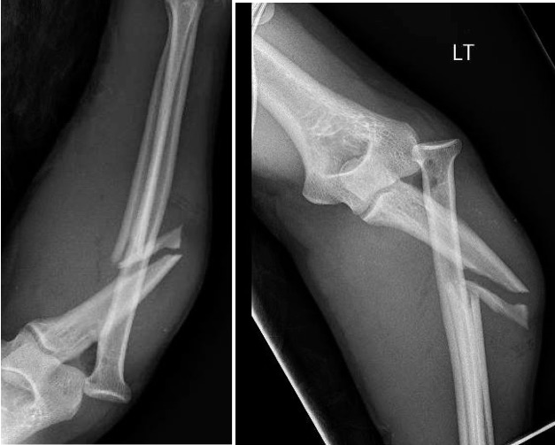
If a forearm fracture and/or dislocation are suspected, radiographs are warranted. An anteroposterior and lateral view will usually identify the injury. An additional oblique view may help better classify the injury. Additional radiographs of the distal wrist and proximal elbow should be obtained with any suspicion of coexistent injury.[8][9][10]
Advanced imaging is not usually indicated on initial assessment. For pre-operative planning computed tomography (CT) may be utilized to evaluate for non-union and magnetic resonance imaging (MRI) can help assess for TFCC tears and interosseous membrane disruption.
Treatment / Management
All Monteggia fractures are considered unstable and require intervention. Emergent orthopedic consultation is essential for open fractures and vascular compromise. Urgent orthopedic consultation is indicated for neurologic deficits without vascular compromise.[11][12][13]
Initial management for a suspected fracture includes rest, ice, immobilization, and elevation. In most circumstances, closed reduction should be attempted. If the annular ligament is trapped within the joint, reduction may be unobtainable. Patients with a Monteggia fracture should be placed in a sugar-tong splint with urgent referral to an orthopedist.
Pediatrics
Children usually have better overall outcomes than adults. This is thought to be due to multiple influences, including the remodeling ability of small angle deformities, shorter healing time, and overall solidity of Monteggia fractures in children. Management is determined by the characteristics of the ulna fracture. Non-operative management is successful in this population if the ulna has undergone a plastic deformation (bending or bowing without fracture) or an incomplete fracture (greenstick). This fracture should be treated with a closed reduction and splinted with the elbow flexed at approximately 110-degrees in full supination for 6 weeks. Complete ulnar fractures will require operative management. Short, oblique fractures should be stabilized with elastic intramedullary titanium nail fixation. Comminuted or long oblique ulna fractures are fixed by ORIF using plates and screws.
Adults
Operative management is crucial for the majority of adult Monteggia fractures. Adults are more prone to the persistent angulation and shortening despite closed reduction techniques. The most common operative repair is an ORIF. In most cases, a single compression plate is placed with approximately six cortical screws anchored proximally and distally. The radial head dislocation usually reduces easily after the ulna fracture is realigned. After surgery, the extremity is placed in a long-arm splint with full supination and elbow flexion around 100-degrees for Babo types 1, 3, and 4 fractures. For Babo type 2 fractures the elbow should be splinted at 70-degrees.
As with all fractures, the length of recovery depends on multiple variables including the severity of the injury, intended use of the extremity, and the individual’s ability to heal. Rehabilitation usually begins at 2 weeks after surgical fixation. The goal of rehabilitation is the return of full range of motion and fine motor skills with the absence of pain. Total return to activity depends upon the severity of the injury as well as the patient’s intended use of their upper extremities. Athletes and manual workers may require more prolonged rehabilitation. Typical return to full activity in patients with low physical demands can occur after 8 to 12 weeks. Patients with high demand activity (athletes and manual workers) may require up to 12 to 16 weeks of rehab. Surgical hardware is usually left in permanently, with less than 10% of patients needing hardware removal.
Differential Diagnosis
- Emergent management of hand dislocation
Pearls and Other Issues
Monteggia injuries are easily missed, particularly the radial head dislocation, as the ulnar shaft fracture can be distracting. To better recognize subtle dislocations, a line can be drawn through the radial shaft and head. This line should pass through the middle third of the capitellum. If it does not, a dislocation should be suspected.
The complexity of Monteggia fractures leads to a variety of outcomes. Children usually fair better than their adult counterparts. Difficulty in management and outcomes increases with Babo type 2 fractures and when associated with other comorbid fractures (radial head and coronoid process fractures).
Nerve injuries can occur from a laceration or entrapment, with radial and median nerve injuries being the most common. The most common associated motor deficit is a posterior interosseous nerve palsy. Higher incidence is seen with Babo type 2 fractures causing a radial head contusion and/or compression against the supinator muscle. Ulnar nerve injuries are rare. Nerve injuries rarely require treatment, and the majority of patients have complete resolution of symptoms in 9 to 12 weeks.
Malunion and nonunion occur in approximately 2% to 10% of cases, which is higher than the average forearm fracture nonunion rate (2%). Other notable complications include acute compartment syndrome (pre-surgical and post-surgical), radioulnar synostosis (1% to 6%), elbow stiffness from protracted immobilization (adults), myositis ossificans, ulnohumeral osteoarthritis, and wound infections (0% to 3%).
Enhancing Healthcare Team Outcomes
Patients with Monteggia fractures frequently present to the emergency room, but the diagnosis of these fractures is not simple. When the diagnosis is missed, these fractures carry very high morbidity. Hence the emergency department physician, nurse practitioner or nurse provider should refer these patients to an orthopedic surgeon ASAP. Initial management for a suspected fracture includes rest, ice, immobilization, and elevation. In most circumstances, closed reduction should be attempted. If the annular ligament is trapped within the joint, reduction may be unobtainable. Patients with a Monteggia fracture should be placed in a sugar-tong splint with urgent referral to an orthopedist.
All Monteggia fractures are considered unstable and require intervention. Emergent orthopedic consultation is essential for open fractures and vascular compromise. Urgent orthopedic consultation is indicated for neurologic deficits without vascular compromise.
Even after treatment, the outcomes of patients with Monteggia fractures are guarded. Chronic pain and limited range of motion are common complaints.[14] (Level V)