Continuing Education Activity
Sialolithiasis is the most frequent cause of salivary gland swelling, affecting the major salivary glands: parotid, submandibular, and sublingual glands. The condition predominantly affects individuals of 30 to 60 years old and is more common in males. The most frequent symptom is cyclical gland swelling and pain associated with meals. Initial management is conservative, with various minimally invasive surgical options available for persistent cases. This activity reviews the presentation, evaluation, and treatment of sialolithiasis and highlights the role of the interprofessional team in evaluating and treating this condition.
Objectives:
Identify the etiology of sialolithiasis.
Review the evaluation of sialolithiasis.
Outline the treatment and management options available for sialolithiasis.
Summarize interprofessional team strategies for improving care coordination and communication to improve clinical care and outcomes for patients with sialolithiasis.
Introduction
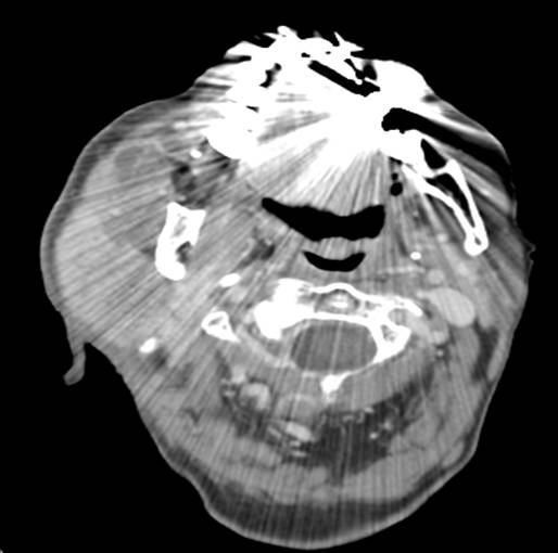
Sialolithiasis is a benign condition involving the formation of stones within the ducts of the major salivary glands: parotid, submandibular, and sublingual glands (see Image. Neck CT, Sialolithiasis in Right Parotid Duct). It is the most frequent cause of salivary gland swelling, with a reported incidence of 1 in 10000 to 1 in 30000.[1]In some cases, sialoliths can obstruct the salivary ducts, leading to inflammation, superimposed bacterial infection termed sialadenitis, or in rare cases, abscess formation. There are various presenting symptoms, with the most common being cyclical postprandial swelling of the affected gland and decreased salivary flow.[2]
In the case of larger salivary stones involving the distal submandibular duct (Wharton’s duct), diagnosis is often possible based on physical examination. Cases involving smaller stones within the distal submandibular duct or parotid duct (Stenson’s duct) were historically diagnosed utilizing conventional radiography, sialography, and digital subtraction sialography. Modern diagnostic techniques involve point-of-care ultrasound, computed tomography (CT), magnetic resonance imaging (MRI), and direct visualization with sialoendoscopy.[2][3] A variety of treatment options exist for sialolithiasis, including sialogogues, direct massage of distal stones out of the duct, and other procedures including interventional sialography, lithotripsy, sialoendoscopy, and surgery.[3][4]
Etiology
The etiology of salivary stones formation remains elusive, and research into etiologic factors is still limited due to the relatively rare incidence of the disease, which makes extensive studies difficult. Factors that are believed to affect salivary stones formation are divided into two major groups: anatomical, affecting saliva formation or flow (i.e., duct stenosis or inflammation), and compositional (i.e., increased calcium content or altered enzyme function).
Research examining the geographic distribution of hard water and salivary calculi formation demonstrated no correlation with an increased incidence of salivary stones in areas of increased water hardness.[5] Studies examining the effects of hypercalcemia using animal models demonstrated no evidence of increased salivary stones with hypercalcemia.[3] Additional factors such as decreased fluid intake and pharmacologic side effects resulting in decreased salivary production (i.e., diuretic use) remain under study. In recent years tobacco smoking has been discussed as a potential risk factor for the formation of salivary stones. Tobacco may induce inflammation within the salivary ducts and decrease the production of salivary amylase.[1][3]
Epidemiology
The incidence of sialolithiasis is estimated at 1 in 10000 to 1 in 30000 individuals.[1] The primary age of diagnosis is between 30 and 60 years old, with a higher incidence in males. Approximately 85% of sialoliths occur within the submandibular gland, making it the most common location for sialolithiasis. One of the reasons is that the submandibular duct ascends towards its opening in the oral cavity, resulting in a stagnant flow of saliva. Additionally, the submandibular gland produces predominately mucinous saliva, which is more viscous than the secretions created by the parotid gland, resulting in a more stagnant flow of secretions. The submandibular gland also produces more alkaline saliva, which predisposes to precipitation of inorganic salts (i.e., calcium and phosphate), further leading to salivary stone formation. Approximately 15% of salivary stones occur within the parotid gland, and less than 5% occur within the sublingual and minor salivary glands.[2][6]
Pathophysiology
The exact pathogenesis of sialolithiasis is not well understood, but two dominant theories were suggested. One of them proposes that there are multiple internal microcalculi within salivary gland secretory granules. When these microcalculi get secreted into the salivary ducts, they may act as a nidus for the formation of larger calculi, ultimately forming a sialolith.[7] The second hypothesis suggests bacteria or food debris within the oral cavity enter the distal submandibular or parotid ducts. Over time, this organic substrate may act as a nidus for the formation of larger calculi.[8]
Histopathology
Histologically, sialoliths are composed of varying ratios of organic and inorganic materials within an inner core. The inner core is considered the initial sialolith, which subsequently enlarges from the deposition of additional organic and inorganic material, forming the outer lamellae (layers). Inorganic materials include hydroxyapatite, whitlockite, and octacalcium phosphate, with hydroxyapatite being the most prevalent. The exact ratios of inorganic material found within a sialolith depend on the chemical environment in which it forms. Organic materials found within sialoliths include glycoproteins, cellular debris, bacteria, and mucopolysaccharides.[3][7] Studies utilizing scanning electron microscopy (SEM) and polymerase chain reaction (PCR) have demonstrated bacteria within sialoliths, predominantly the Streptococcus genus.[9][10] Bacteria are not present in all sialoliths suggesting they are not necessary for their formation.
History and Physical
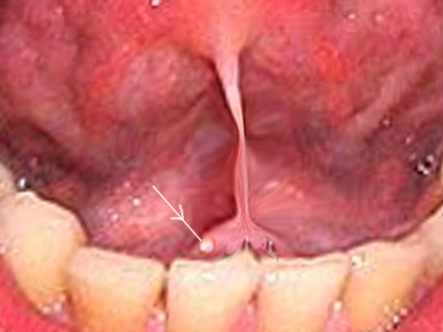
Clinical diagnosis of sialolithiasis can be challenging as patients may not be symptomatic unless the stone obstructs the salivary ducts, termed sialadenitis. Patients with obstructing stones will commonly present a history of unilateral salivary gland swelling and acute onset of pain that worsens with meals.[4] The physical exam will demonstrate asymmetric swelling of the affected salivary gland. Approximately 60% of parotid stones and 30% of submandibular stones will be located distally in their respective ducts. If large enough, these stones may be visually apparent on examination of the oral cavity (see Image. Submandibular Stone). Salivary stones typically demonstrate an oval or round shape and a white or yellow color on visual inspection. If not visually apparent, the stones are usually palpable along the anatomic course of the affected salivary duct or gland.[11][12][13]
Salivary stones’ size and weight vary. Sialoliths’ diameter ranges between 2.1 and 10 mm, and only 7.6% exceeds 15 mm in diameter.[14] Submandibular stones are usually larger than parotid ones.[15] Sialoliths weigh 300 mg on average, ranging between 1 mg to 5 gr.[14]
Evaluation
Conventional radiographs using occlusal views were the initial diagnostic choice in the past. These can detect large radiopaque ductal stones relatively well, but they miss smaller and parenchymal stones. Additionally, only around 80% of stones are radioopaque, which leads to many stones not being diagnosed. While conventional radiography can still be useful in the initial workup of salivary stones, modern imaging techniques using ultrasound or CT scans can accurately diagnose the sialoliths and provide their exact location.[3]
Sialography is traditionally regarded as the gold standard for diagnosing sialolithiasis as it allows excellent visualization of the salivary ducts and underlying ductal pathology. In this technique, contrast is injected via a small needle, enabling radiographic visualization, acting as a sialogogue, and allowing passage of smaller stones. Disadvantages include radiation exposure and risk of contrast reaction.[3][16]
Non-contrast computed tomography (NCCT) is a widely used modality for the evaluation of sialolithiasis. It is a valid tool for diagnosing sialolithiasis when it is big enough or when the radiographic slices are done every millimeter.[3] Advantages include excellent sensitivity and specificity for calcified stones, fast acquisition time, and widespread availability. Disadvantages include radiation exposure and limited evaluation of the ductal system or underlying pathology (i.e., obstructing masses). The administration of intravenous contrast has been used only as an adjunct to NCCT due to concerns small vessels may simulate calcifications resulting in false-positive results. A recent study in the American Journal of Neuroradiology comparing contrast-enhanced CT (CECT) and NCCT demonstrated excellent sensitivity and specificity with no false-positive results. Benefits of initial contrast-enhanced CT include better evaluation of the ductal system, improved soft-tissue contrast in assessing salivary masses and decreased radiation dose (compared to dual-phase NCCT with CECT).[17]
MR Sialography demonstrates similar sensitivity and specificity to NCCT for the evaluation of sialolithiasis. Benefits of MR include excellent visualization of the salivary ducts without the need for contrast and no radiation exposure. Disadvantages include cost, image acquisition time, and limited availability compared to NCCT and Ultrasonography.[3][16]
Sialendoscopy allows for direct visualization of salivary stones and the salivary ducts, thus providing excellent sensitivity and specificity. In addition to aiding in the diagnosis of sialolithiasis, clinicians increasingly use sialoendoscopy for therapy and stone removal, given the advancements in endoscopic technology.[3] Research has shown sialoendoscopy to be a safe and effective alternative to conventional open surgical techniques with a more favorable complication profile.[18] Additionally, sialoendoscopy is performable in the outpatient setting utilizing local anesthesia.
Ultrasonography provides a non-invasive method of imaging sialolithiasis. The usual appearance of sialoliths on ultrasound is a hyperechoic focus with posterior shadowing. Benefits to ultrasonography include no radiation exposure, real-time image interpretation, and widespread availability. Ultrasound is often available at the point of care and can provide rapid diagnostic information. Ultrasound is highly operator-dependent, revealing a wide range of reported sensitivity and specificity. Reported sensitivities are best for stones greater than 2 to 3 mm in size. Recent research has demonstrated sensitivity and specificity for stones greater than 3 mm in size comparable to conventional NCCT and sialoendoscopy.[19]
Treatment / Management
Management of sialolithiasis should begin with conservative measures, including massaging the salivary gland, nonsteroidal anti-inflammatory drugs (NSAIDs), and sialogogues. Signs of infection, including cervical adenopathy, purulent discharge from the salivary ducts, or erythema surrounding the salivary ducts, indicate the need for antibiotic therapy.[4][11]
Further treatment is dictated based on the sialolith's size, number, and location if conservative management is unsuccessful.
Mobile submandibular stones measuring less than 5 mm located within the distal duct should initially undergo management with endoscopy. Impacted submandibular stones within the distal duct and stones larger than 5 mm should have treatment with transoral duct slitting. Stones of 5 to 7 mm within the proximal duct or hilar region should receive initial treatment endoscopically. If this is unsuccessful or the stone becomes impacted, the next step is a transoral surgical approach.
External shockwave lithotripsy (ESWL) is an option for not palpable stones or visualized under endoscopy. Intraparenchymal stones can be extracted endoscopically if they are 5 to 7 mm and visualized. Larger stones require treatment with transoral slitting. ESWL is indicated for smaller stones that are not palpable and not visualized endoscopically. ESWL is generally unsuccessful for stones larger than 7 to 10 mm. Surgical excision of the submandibular gland should be a last resort.[11][20][21][22]
Salivary stones within the parotid duct that measure less than 7 mm and are mobile require endoscopic removal. If endoscopic management is unsuccessful or the stones have become impacted, external shockwave lithotripsy is considered the most appropriate second-line therapy with subsequent endoscopic removal of fragmented stones. Treatment of salivary stones that do not respond to external shockwave lithotripsy is with a combined transcutaneous and endoscopic approach (assuming the stone is visible under endoscopy). Surgical excision of the parotid gland should be a last resort.[11][21][22]
Differential Diagnosis
The differential diagnosis for salivary gland swelling includes:
- Sialolithiasis
- Sialadenitis (inflammatory or infectious)
- Neoplasm
However, the clinical presentation can vary, and the differential diagnosis for the pathology of the oral cavity and face can be extensive and relies heavily on physical exam findings and clinical history. Conditions that can have a similar presentation include:
- Cellulitis
- Poor dentition and dental abscess formation
- Infection of the buccal or masticator space
- Herpes zoster
- Neoplasm
Prognosis
Sialolithiasis has an excellent prognosis, and the majority of patients can be managed conservatively with sialogogues and NSAIDs. The minimally invasive procedures discussed above have excellent success rates with minimal morbidity compared to traditional surgical techniques. Sialadenectomy for the treatment of sialolithiasis is rarely necessary with modern treatment techniques.[21]
Complications
The primary complications of sialolithiasis are the development of sialadenitis, acute or chronic, and atrophy of the affected salivary gland. Obstruction of the salivary glands by a sialolith blocks the flow of saliva resulting in swelling and pain. Additionally, this blockage of flow prevents the removal of bacteria and debris from the salivary duct, resulting in bacterial infection. If the obstruction is chronic, the blocked flow of saliva will damage the salivary glands' acinar cells, resulting in local inflammation. Without proper treatment, it can result in permanent fibrosis of the gland and atrophy.[7]
Deterrence and Patient Education
Patients should be informed that sialolithiasis has an excellent prognosis and resolve with conservative management in most cases. Patients require education on common initial symptoms such as glandular swelling and pain with meals that suggest they have formed a new sialolith. While sialolithiasis is usually idiopathic, the formation of stones can be secondary to an obstructing process such as ductal stenosis or neoplasm. Patients should be educated about the need to inform their clinician of recurrent or worsening symptoms that would indicate a need for a more advanced imaging workup or referral to a head and neck specialist.
Enhancing Healthcare Team Outcomes
Sialolithiasis is the most common benign cause of salivary gland swelling; however, it remains a relatively rare diagnosis with an incidence of 1 in 10000 to 1 in 30000. The disease commonly gets diagnosed in the primary care or emergency medicine setting, where initial patient contact is mostly with nurses, nurse practitioners, physician assistants, primary care physicians, and emergency medicine physicians. Given the oral nature of the disease, some patients may initially present to their dentist for evaluation of symptoms. Diagnostic radiologists are often involved in the initial workup for imaging recommendations and interpretation. The potential involvement of all these disciplines is why an interprofessional team approach is necessary for the diagnosis and management of this condition.
An interprofessional approach and open communication among providers can aid in ensuring patients receive appropriate initial management resulting in a quicker resolution of symptoms. Communication among the radiologist and primary care provider can help timely referral to an otolaryngologist when a large sialolith is likely to fail conservative measures. Additionally, patients should be educated to inform their provider if symptoms are not improving or are worsening, indicating failure of conservative measures and the potential need for referral to an otolaryngologist for advanced interventions. Well-informed nursing staff can assist in the management and provide patients with assurance that with modern multimodal treatment algorithms, success rates approach 100%. Gland removal is rarely necessary, especially with interprofessional team collaboration.[21] [Level 5]