Continuing Education Activity
Balanoposthitis is a common disease process involving the glans penis and prepuce. It affects up to 12% to 20% of pediatric and adult males. This activity reviews the evaluation and management of balanoposthitis as well as strategies for the healthcare team to improve outcomes.
Objectives:
- Identify the etiology and epidemiology of balanoposthitis.
- Describe the appropriate history, physical, and evaluation of balanoposthitis.
- Review the treatment and management options available for balanoposthitis.
- Outline interprofessional team strategies for improving patient care and compliance.
Introduction
Balanoposthitis is an inflammation that affects both the glans penis and prepuce. Many etiologies exist, but it can generally classify as either infectious, irritant, or traumatic in origin.
Balanitis refers to inflammation of the glans penis, while posthitis refers to inflammation of the prepuce. At birth, the prepuce, commonly referred to as the foreskin, is attached by adhesions to the glans. These nonpathological adhesions are responsible for physiologic phimosis or decreased retraction of the foreskin. Hsieh et al. found that 17.1% of first-grade boys had physiologic phimosis that decreased to 1.2% by seventh grade in a cohort of healthy children in Taiwan.[1] In contrast, pathologic phimosis refers to an inability to retract the foreskin secondary to scarring of the prepuce.[2]
Etiology
The most common etiology of balanoposthitis is poor hygiene, commonly referred to as nonspecific balanoposthitis. Other etiologies include inflammatory skin diseases, infection, trauma, and cancer. Candidal infection is prevalent in children and can be associated with diaper rash. Other infectious causes include aerobic bacteria such as Staphylococcus aureus and Group A Streptococcus, anaerobic bacteria, and viruses such as human papillomavirus. Some inflammatory etiologies include contact dermatitis, reactive arthritis, and lichen sclerosus.[3]
Epidemiology
Balanoposthitis is a relatively common condition affecting both pediatric patients and adults. Prevalence in males of all ages is between 12% to 20%.[4] Pediatric patients most commonly present around ages 2 to 5 years; this is likely due to physiologic phimosis and hygiene habits. In adults, uncircumcised males with diabetes mellitus are at the highest risk, with a prevalence of around 35%.[5][6] Meta-analysis has shown that circumcision can decrease the prevalence of inflammatory conditions of the glans penis by 68%.[4]
Pathophysiology
Pathophysiologic processes can vary widely depending on etiology. These can be irritant, allergic, infectious, autoimmune-mediated, and secondary to trauma or malignancy. The majority of cases commence with moisture such as urine, sweat, or smegma (physiologic secretion from genital sebaceous glands) becoming trapped within the preputial space secondary to adhesions and poor hygiene, which in turn creates a nidus for bacteria and fungi. Balanoposthitis can also be commonly provoked by irritants and allergens, causing non-specific inflammation leading to erythema and pruritis.
History and Physical
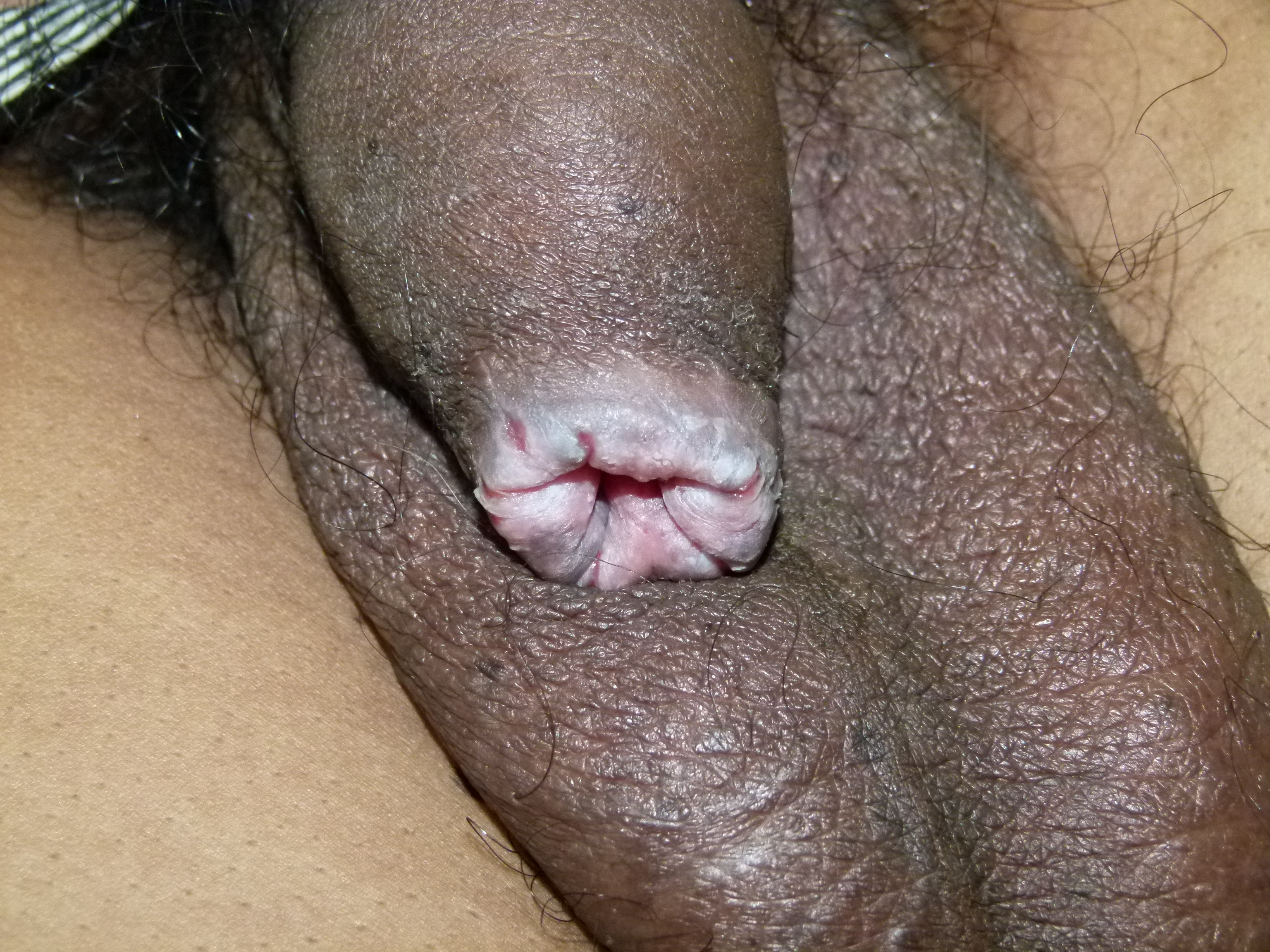
Balanoposthitis can present as penile pain, pruritus, discharge, erythema, rash, or inconsolable crying. By definition, this only presents in uncircumcised boys. It can be more common in patients with poor hygiene. Depending on the etiology and disease progression, it may also occur with or without phimosis, scarring of the prepuce, inability to void, ulcers, other lesions, and rashes.
Evaluation
A thorough history and physical exam are sufficient in most cases of balanoposthitis for diagnosis and establishing a course of treatment. It is essential to evaluate the duration of symptoms, hygiene habits, infectious exposures, potential allergens, and sexual practices. The examination should focus on the presence of phimotic discharge, urethral discharge, urinary retention, erythema, edema, tenderness, scarring, inguinal lymphadenitis, testicular edema, or tenderness.
Physiologic preputial smegma can be confused with discharge. Discharge is more exudative in appearance, may have a foul order, and may be associated with erythema and tenderness. Penile ulcers, versicles, urethral discharge, and other lesions point toward sexually transmitted infections as etiology and indicate the need for appropriate tests.
Children in which sexual abuse is not suspected and in the absence of lesions or discharge are manageable empirically, and further testing is not required. Otherwise, guidance for further evaluation should be by other symptoms and a history of exposures.
If a sexually transmitted disease is suspected, cultures and swab for nucleic acid amplification test (NAAT) for gonorrhea and chlamydia could provide a diagnosis. In adults and pediatric patients, whether suspecting sexual abuse or not, culture for Group A beta-hemolytic Streptococcus may be indicated.
In new cases of balanoposthitis, especially candidal etiology outside of the younger child period, evaluation for diabetes may be necessary as this may be the first sign of diabetes.[5]
Although most cases will respond to hygiene improvements or empiric treatment, patients with symptoms that recur or are refractory to treatment after 4 weeks may warrant biopsy to investigate etiology and appropriate treatment further.
Treatment / Management
Upon presentation, if phimosis is severe enough to cause urinary obstruction, the patient should be urgently catheterized and seen by a specialist. If unable to place a catheter, the patient may require more invasive interventions such as a dorsal slit.[7] Circumcision can be deferred until preputial edema has subsided.[7]
Nonspecific Balanoposthitis - is the most common etiology in children. This condition is usually due to poor hygiene. There should be no ulcers or lesions, and although some preputial discharge can be present on a physical exam, there should be no urethral drainage. Assessment is by milking the urethra from the base of the penis to the glans. Treatment involves gentle cleaning of the area 2 to 3 times per day. If physiologic phimosis is present, forceful prepuce retraction should be avoided. In patients able to retract the foreskin, clean the area with gentle use of cotton swab. After commencing a proper hygiene routine, symptoms usually resolve within five days.
Irritant Balanoposthitis - usually presents as mild erythema with or without pruritus. It is more common in patients presenting with atopic dermatitis. This condition may be due to frequent or aggressive washing with soap.[3] Most of these cases can be appropriately managed by avoiding strong soaps and applying emollients such as vaseline multiple times daily. Potential allergens such as latex condom use, lubricants, detergents used in underwear should be investigated and avoided. Hydrocortisone 1% is an option, applying a thin layer of cream BID for 1 to 2 weeks.
Candidal infection - a common cause of balanoposthitis in children may present in association with or secondary to diaper dermatitis. Its classic description is an erythematous rash with satellite lesions that is tender to palpation. In adults, it can present in association with diabetes mellitus, immunosuppressive disorders, or the use of broad-spectrum antibiotics. This condition can have topical treatment with seven days of miconazole 0.25% cream applied on each diaper change. Nystatin cream 100000 u/gram TID for two weeks is also an option.
Bacterial Balanoposthitis - should be suspected in the presence of intense erythema along with transudative or exudative preputial discharge.
Common aerobic bacteria are Streptococcus pyogenes and Staphylococcus aureus. Mild cases can receive therapy with topical antibiotics such as mupirocin 2% cream TID for 7 to 14 days. In severe cases or when phimosis prevents topical treatment, therapy should commence with oral antibiotics such as cephalexin or erythromycin for one week.[8] Cases with concomitant Group A Streptococcal pharyngitis is treatable as pharyngitis with a beta-lactam.
Sexually transmitted infections - If present, urethral drainage when milking the penis from the base should prompt evaluation and treatment for sexually transmitted infections such as gonorrhea and chlamydia.[9] Neisseria gonorrhea or Chlamydia trachomatis can receive treatment with a single dose of ceftriaxone 250 mg IM and a single dose of azithromycin 1g PO. A painless ulcer may indicate syphilis infection, which can be treated with Penicillin G 50000U/kg IM once. In children, sexual abuse evaluation should also merit consideration in the presence of urethral discharge.
Anaerobic bacterial infection - should be suspected in cases with erythema, edema, and foul-smelling exudate. Mild cases can be treated with topical metronidazole, while more severe cases should have treatment with oral antibiotics such as oral metronidazole for seven days.
Circinate Balanitis - is described as pale macules with white margins that may coalesce, which can occur in isolation or association with reactive arthritis. Treatment includes management of any underlying conditions and the use of topical steroids such as hydrocortisone 1%, applying a thin layer BID for 1 to 2 weeks.
Viral Balanoposthitis - includes herpes simplex virus and human papillomavirus. Herpes simplex presents as an erythematous base with overlying vesicles that may rupture. A first episode is treated with oral acyclovir for 7 to 10 days, whereas for recurrent episodes, 5-day treatment is appropriate. Human papillomavirus can present as diffuse erythema, and treatment includes podophyllotoxin 0.5% gel BID for three days repeated weekly for up to 4 weeks.
Fixed drug eruptions - described as round, erythematous patches that turn darker with or without associated edema and vesicles that appear after using medications including tetracyclines, phenolphthalein, phenacetin, NSAIDs, barbiturates, and sulfa-drugs.[10] These lesions most commonly occur in the genitals or oral mucosa. Fixed drug eruptions resolve upon discontinuing the medication and return shortly on the same location if that medication is resumed. Hydrocortisone 1% can be used, applying a thin layer of cream BID for 1 to 2 weeks.
Differential Diagnosis
- Diaper dermatitis
- Psoriasis
- Discoid (nummular) eczema
- Lichen planus
- Circinate balanitis
- Lichen sclerosus, also known as balanitis xerotica obliterans
- Fixed drug eruption
- Human papillomavirus
- Squamous cell carcinoma
- Reactive arthritis
Prognosis
The majority of patients without a clear infectious etiology will respond to changes in hygiene and empiric therapy with emollients within one to two weeks. About one in ten of those patient’s symptoms, however, will recur needing further evaluation and more targeted management.[3]
Complications
Patients with symptoms that recur or are refractory to treatment after four weeks, present with associated pathologic phimosis or urinary obstruction, should be referred to urology. A biopsy may be warranted to investigate etiology further.
Refractory cases may represent cancerous or precancerous lesions, including balanitis xerotica obliterans or squamous cell carcinoma.[11][12] Clinicians may miss these without histopathologic examination.[13] Circumcision or 1 cm wedge biopsy of the affected area is necessary to obtain a pathologic diagnosis and histologic grade.[12] These may require reconstructive surgery, radiation, and chemotherapy, depending on the stage.
Deterrence and Patient Education
The clinician and other healthcare staff must provide guidance to patients regarding appropriate preputial hygiene; this can be preventative and therapeutic in the majority of balanoposthitis cases. Proper preputial hygiene involves gentle cleaning of the area 2 to 3 times per day. If physiologic phimosis is present, forceful prepuce retraction should be avoided. In patients able to retract the foreskin, the area can be cleaned with gentle use of cotton swab. Avoidance of strong or scented soaps is also a recommendation as they can be irritating. Investigating other possible irritants and providing guidance on how to avoid them can also significantly improve symptoms.
Pearls and Other Issues
Prevention
Balanoposthitis can be prevented by first establishing proper hygiene habits, including washing area routinely, avoiding forceful retraction, forceful scrubbing, or cleaning under the prepuce in young boys with physiologic phimosis and avoiding strong soaps.
Another way to prevent balanoposthitis is circumcision. Although controversy exists regarding recommendations for circumcising neonates, research ahs show it to prevent penile dermatosis, urinary tract infections, penile cancer, and sexually transmitted infections such as HIV and syphilis.[12][14]
Conflicting literature exists regarding whether circumcision prevents balanitis compared to balanoposthitis in uncircumcised patients.[14][15] However, meta-analysis data has shown that inflammatory conditions of the glans penis have a 3.1 times higher prevalence in uncircumcised males.[16]
Avoiding high-risk sexual behavior may help prevent balanoposthitis by decreasing the risk of sexually transmitted infections such as syphilis, herpes simplex virus, and human papillomavirus.
Disposition/referral
The majority of patients without a clear infectious etiology will respond to changes in hygiene and empiric therapy with emollients within one to two weeks. About one in ten of those patient’s symptoms, however, will recur needing further evaluation and more targeted management. [3] Patients with symptoms that recur or are refractory to treatment after four weeks, present with associated pathologic phimosis or urinary obstruction, should be referred to urology. A biopsy may be warranted to investigate etiology further.
Refractory cases may represent cancerous or precancerous lesions, including balanitis xerotica obliterans, squamous cell carcinoma.[11][12] These may be missed often without histopathologic examination. [13] Circumcision or 1 cm wedge biopsy of the affected area should be performed to obtain a pathologic diagnosis and histologic grade. [12] These may require reconstructive surgery, radiation, and chemotherapy, depending on the stage.
Enhancing Healthcare Team Outcomes
An interprofessional approach to healthcare when manging balanoposthitis can have a positive impact on patient outcomes and safety. A strong network of communication is imperative to have a strong healthcare team. Detailed documentation can improve communication and avoid redundant testing, unnecessary antibiotics, and loss of follow up. Communication with nursing can help nurses further clarify patient education, prescriptions, and the importance of follow up. If available, in house pharmacy can be involved to verify proper dosing and mode of administration for patients as well as compatibility with other medications. In refractory or urgent cases, early urology involvement is recommended to avoid complications. Pharmacists review prescriptions, check for drug interactions, and counsel patients and their families.
It is essential to also think of the patient as part of the interprofessional healthcare team. Providing the patient with a clear definition of the diagnosis, possible complications, importance of close follow up, and detailed home management instructions can also have a positive impact on compliance and patient care. [Level 5]